
Higginsarctus signeae, Hansen & Kristensen, 2021
|
publication ID |
https://doi.org/ 10.5852/ejt.2021.762.1461 |
|
publication LSID |
lsid:zoobank.org:pub:43F5C871-A651-47FB-B0A8-29C41EEEEBDD |
|
DOI |
https://doi.org/10.5281/zenodo.5218541 |
|
persistent identifier |
https://treatment.plazi.org/id/4F3D0D8B-BC13-4B98-9DD5-A5DA3A3BD817 |
|
taxon LSID |
lsid:zoobank.org:act:4F3D0D8B-BC13-4B98-9DD5-A5DA3A3BD817 |
|
treatment provided by |
Felipe |
|
scientific name |
Higginsarctus signeae |
| status |
gen. et sp. nov. |
Higginsarctus signeae View in CoL gen. et sp. nov.
urn:lsid:zoobank.org:act:4F3D0D8B-BC13-4B98-9DD5-A5DA3A3BD817
Figs 2–7 View Fig View Fig View Fig View Fig 5 View Fig View Fig
Florarctinae nov. gen. 1 et nov. sp. 1 – Hansen et al. 2001. — Hansen 2005.
Diagnosis (characters uniquely defining the taxon are written in bold)
Characterized by flat, rectangular secondary clavae with slightly protruding contours. Each clava inserted on a small elevation of the ventral cuticle. Unilobed, rectangular antero-lateral alae with weakly undulating distal margins without indentations. Unilobed, rectangular medio-lateral alae with weakly undulating distal margins without indentations. Antero-lateral alae and medio-lateral alae similar in size. Bilobed postero-lateral alae with a medial arched identation. Postero-lateral alae larger than antero-lateral alae and medio-lateral alae. Quadrilobed, rectangular caudal ala with 3 arched indentations, 1 medial and 2 lateral. Lateral lobes of the caudal ala nearly rectangular. Leg sense organs I–III with similar length. Genital stoup present. Anus inserted on ventral folium.
Etymology
The new species is dedicated to Signe G. Hansen, the daughter of the first author.
Material examined
Holotype FAROE ISLANDS • ♀; North Atlantic Ocean , Faroe Bank; 61°23.18′ N, 08°35.13′ W; depth 273 m; 1 Apr. 1992; R.M. Kristensen leg.; BIOFAR Station 788; coarse shell gravel; NHMD-293899 . GoogleMaps
Allotype FAROE ISLANDS • ♂; same collection data as for holotype; 61°00′ N, 08°13′ W; depth 120 m; 19 Sep. 1998; R.M. Kristensen leg.; BIOFAR Station 2013; coarse shell gravel; NHMD-293900 . GoogleMaps
Paratypes FAROE ISLANDS • 1 ♀; same collection data as for holotype; 61°12.33′ N, 08°28.23′ W; depth 148 m; 1 Apr. 1992; R.M. Kristensen leg.; BIOFAR Station 786; fine shell sand; NHMD-293898 GoogleMaps • 1 ♀; same collection data as for holotype; 61°00′ N, 08°13′ W; depth 120 m; 19 Sep. 1998; R.M. Kristensen leg.; BIOFAR Station 2013; coarse shell gravel; NHMD-293901 GoogleMaps • 1 ♀; same collection data as for preceding; used for SEM; NHMD-293902 GoogleMaps .
Type locality
Faroe Bank, North Atlantic Ocean (between 61°00′ N, 08°13′ W and 61°23.18′ N, 08°35.13′ W; depth range, 120–273 m).
Description
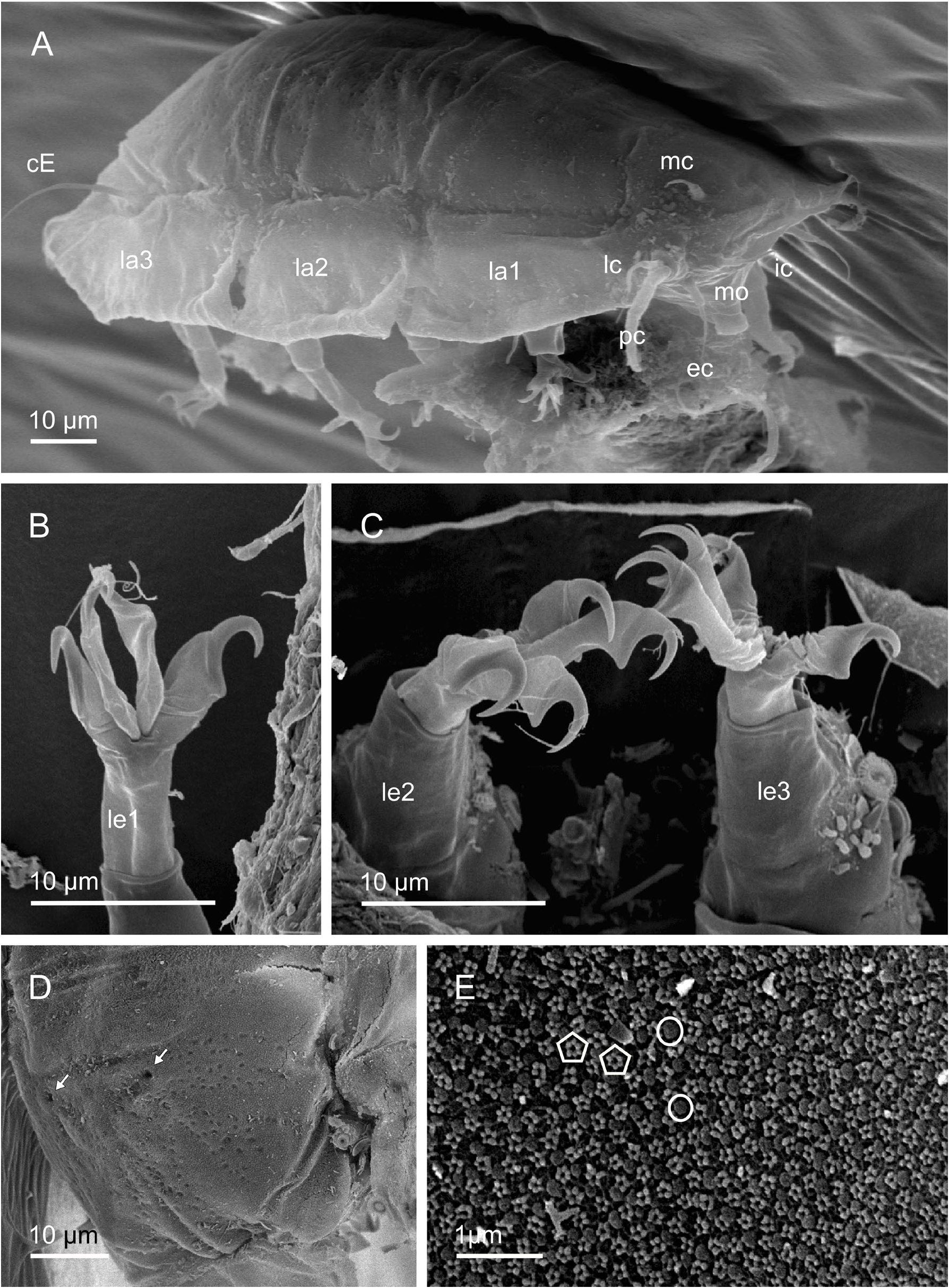
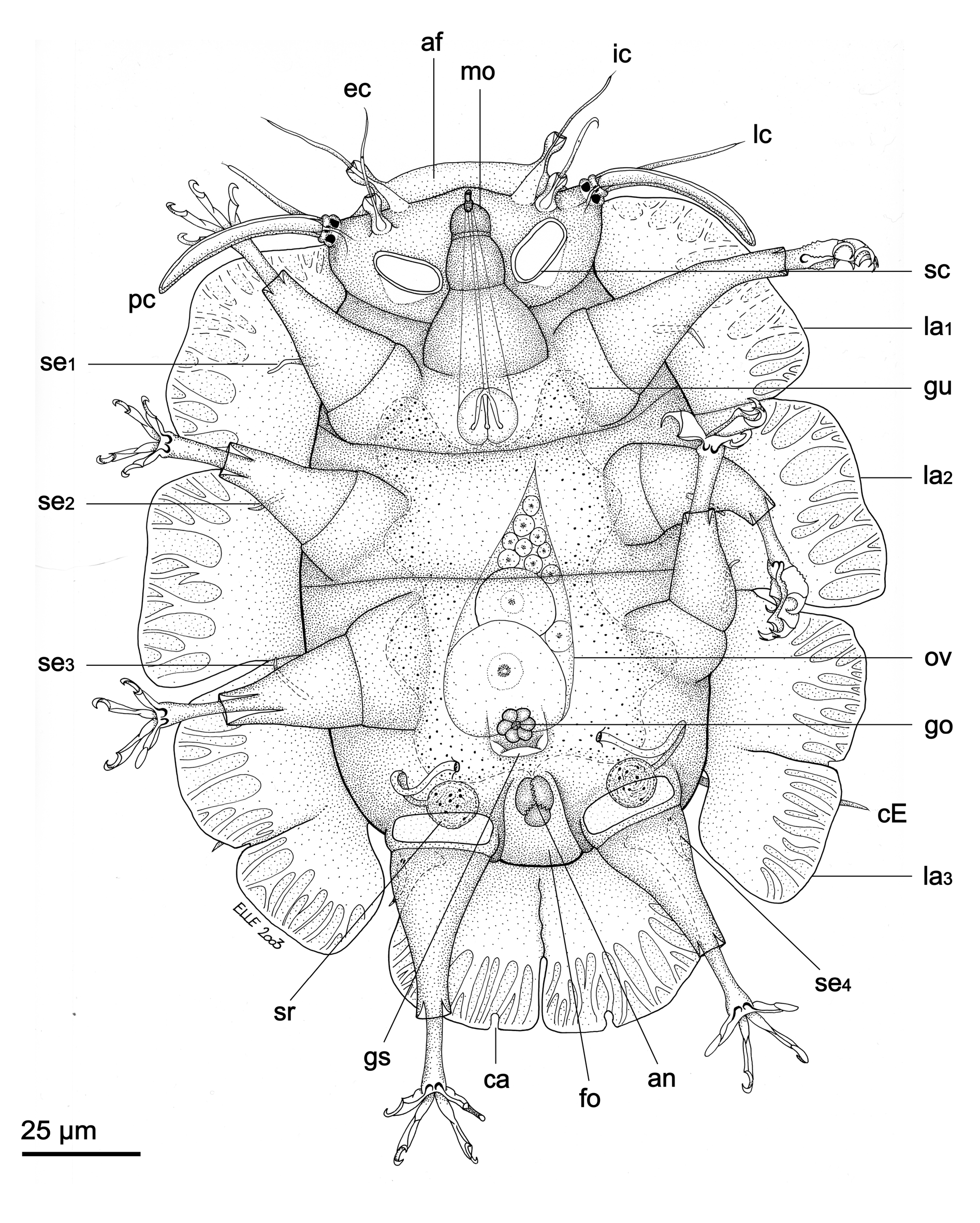
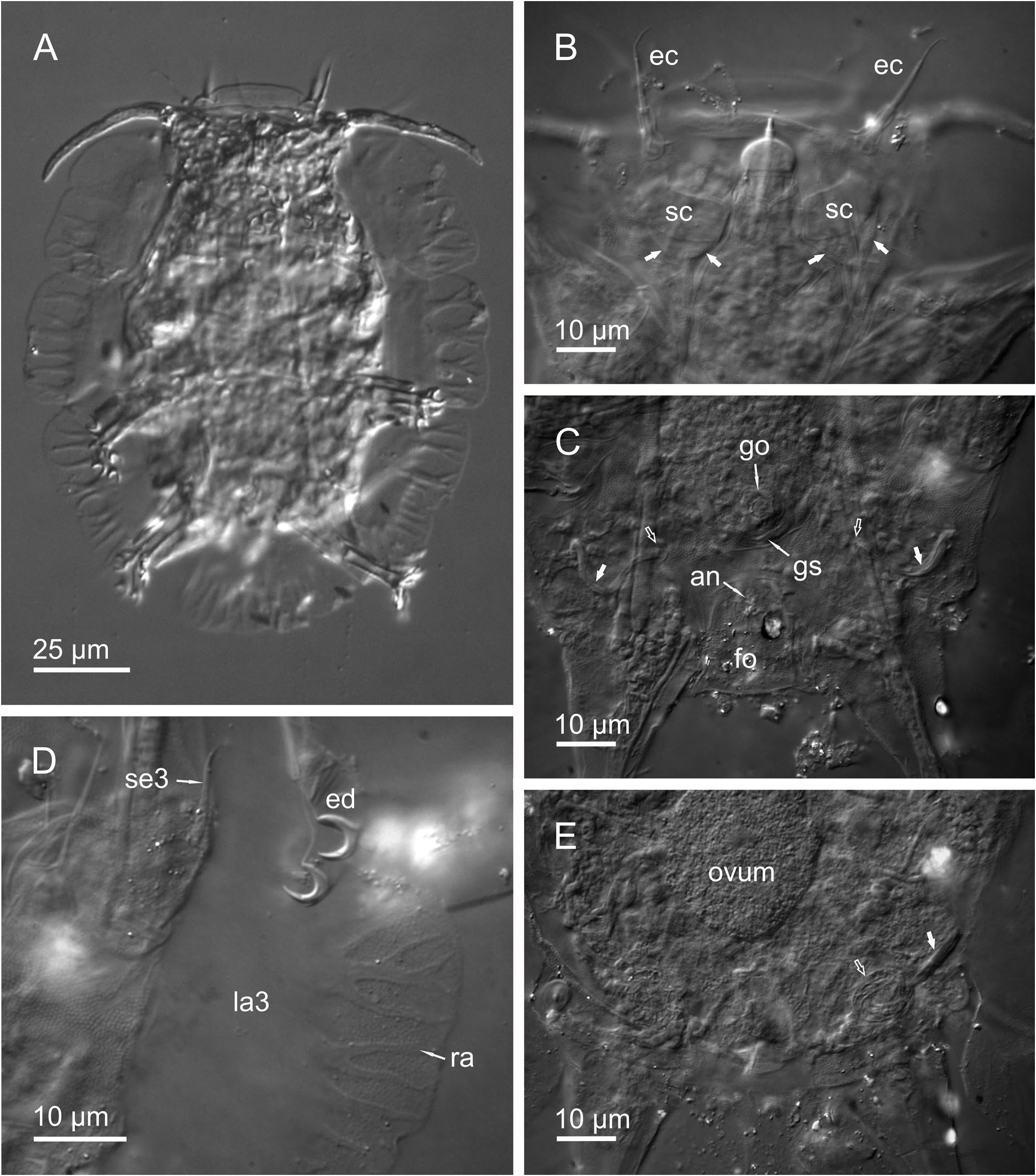
HABITUS. The holotypic female ( Figs 2 View Fig , 3B–E View Fig ) is 147 µm long from the anterior margin of the head to the posterior margin of the body. The body is ovoid, being broadest (94 µm) at the level between the second and third pair of legs. It is highly convex dorsally, and flattened ventrally. The anterior margin of the head taper off, into a thin membrane-like structure (the ala anterior; van der Land 1968), which is different from the alae of the body. It is here referred to as the frontal ala ( Fig. 2 View Fig ). The dorsal cuticle has four transverse inter-segmental folds: one anterior to the first pair of legs, two between the first and second pair of legs and one between the second and third pair of legs. The SEM studies of a paratypic female shows two large pores mid-dorsally, at the level of the third pair of legs ( Fig. 5D View Fig 5 ). A deep longitudinal furrow extends mid-dorsally from the insertion of the caudal ala and halfway to the dorsal pores. Also only visible by SEM (in LM the dorsal cuticle appears smooth with small pillars inside the epicuticle), the dorsal cuticle is remarkably sculptured with numerous small knob-like structures of two different types: one type which is organized in rosettes of five, and another type which is larger, with a more spongy appearance, occurring separately. The two types are evenly distributed in the cuticle ( Fig. 5E View Fig 5 ). This kind of dorsal cuticular structure has never been observed in any tardigrade species before. The ventral cuticle is perfectly smooth with small pillars inside the epicuticle.
ALAE. Eight alae, which are all clearly separated from each other, are present ( Fig. 2 View Fig ): frontal ala, a pair of antero-lateral alae, a pair of medio-lateral alae, a pair of postero-lateral alae and a single caudal ala (see also paratypes Figs 4A View Fig , 5A View Fig 5 ). The antero-lateral alae and medio-lateral alae are all similar in size and shape being unilobed and rectangular with weakly undulating distal margins without indentations. The postero-lateral alae each have a medial arched indentation in the distal margin, dividing the ala into two lobes of equal size. The caudal ala is narrowed at the insertion on the body and has an overall rectangular shape with a deep medial, arched indentation and a pair of lateral, arched indentations dividing the ala into four lobes. The medial lobes are small and rounded whereas the lateral lobes are larger and more rectangular. The pillars in the alae are visible as closely spaced dots. The proximal halve of the lateral and caudal alae is internally supported by continuous procuticle which sends out branching processes (ramuli) into the distal halve of the alae. The branching processes all start at the same level, thus creating a clear dividing line between continuous procuticle and branching procuticle, giving the alae a very characteristic appearance ( Figs 2 View Fig , 3D View Fig ).
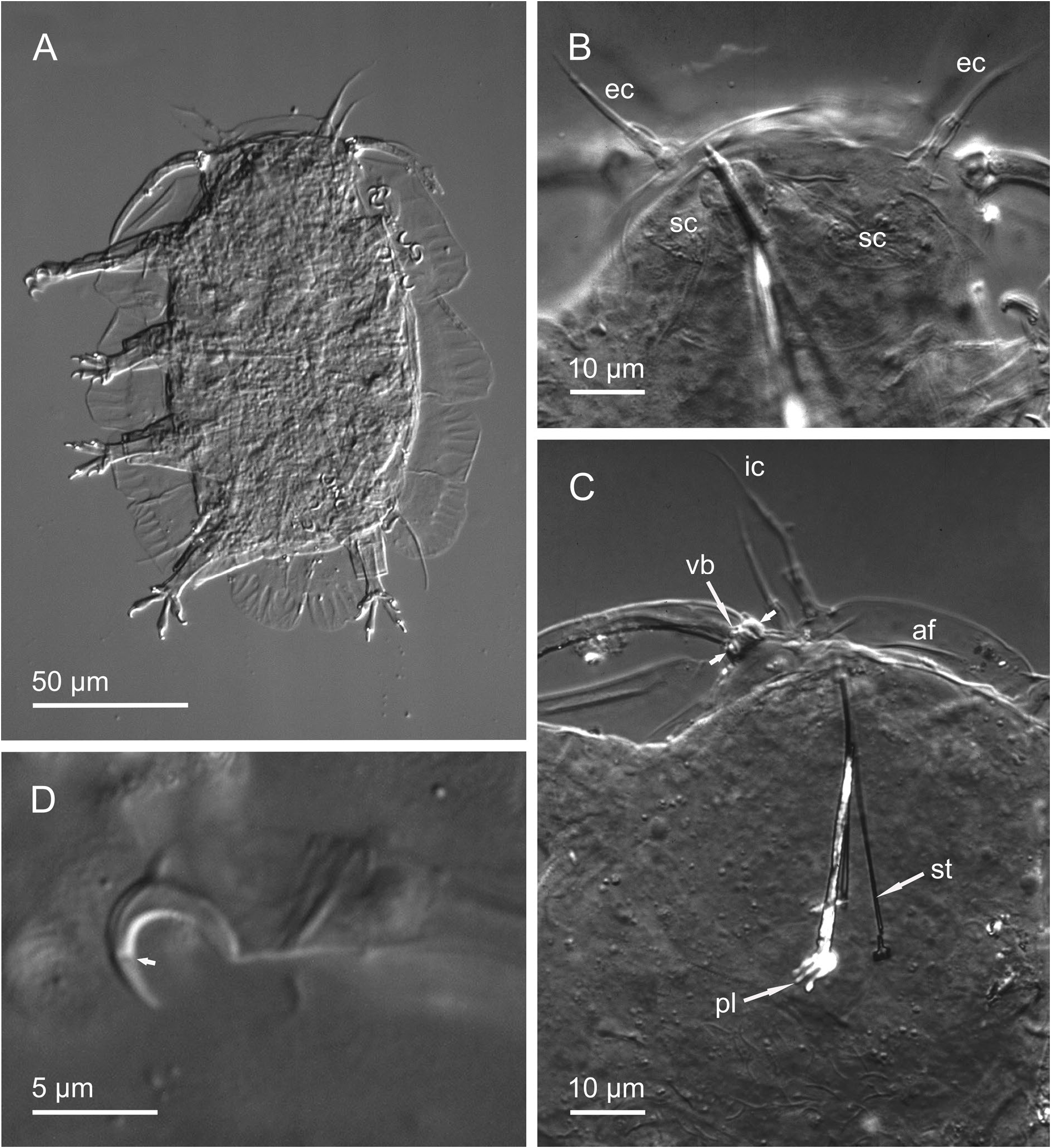
SENSORY ORGANS. The head is well defined from the body by a constriction and a complete set of sense organs is present. All the cephalic cirri consist of an hourglass-shaped scapus, a long tubular portion and a protruding flagellum. As in most other species of Florarctinae the scapus of each cirrus appears somewhat outsized, enveloping the internal sensory structures rather than lining them. The internal cirri (35 µm) emerge from the frontal ala at the anterior margin of the head. The external cirri (29 µm) are inserted ventrally and the median cirrus mid-dorsally. The primary clava is relatively long (43 µm), slightly curved and non-flexible ( Fig. 2 View Fig and paratypes Figs 3A View Fig , 7A View Fig ). A van der Land’s body is visible inside its base. Primary clava and lateral cirrus (34 µm) arise on the same cirrophore, and a common membrane (extended margin of cirrophore) surrounds the base of primary clava and lateral cirrus. A very large and thick cuticular ring supports the cirrophore internally, and is probably functioning as an anchor of the primary clava (see paratype Fig. 7C View Fig ). The secondary clavae are large, rectangular flat sacs (8 µm × 15 µm) flanking the mouth cone ( Fig. 3B View Fig ). Each clava is inserted on a small elevation of the ventral cuticle. In SEM of a paratypic female, the contours of the secondary clavae are recognizable, protruding slightly from the ventral cuticle ( Fig. 4B View Fig ). The leg I sense organ (11 µm) is an unsegmented spine with a slightly swollen base and a terminal tube. The sense organs of leg II (8 µm) and III (8 µm) are unsegmented tapering spines. The fourth leg sense organ (12 µm) is an elongate papilla with a basal van der Land’s body and a terminal pore. The cirrus E (46 µm) has a prominent cirrophorus, scapus and a long tapering flagellum. The scapus has a highly complex composition. The outer lining is composed of small pellets organized in regular rings. At least 33 rings are recognisable by LM and SEM, however the pellets are recognizable as separate units, only by using SEM-technique ( Fig. 4F–G View Fig ).
LEGS, DIGITS AND CLAWS. The legs consist of coxa, femur, tibia and tarsus as found in all species of Florarctinae . A rectangular folium-like structure is weakly evident ventrally on each coxa of leg pair IV ( Fig. 2 View Fig ). Whether or not this is a true folium awaits the analysis of additional material. The external digits are supported by internal hook-shaped peduncles. The external claw is simple and with a calcar. The internal claw has an accessory spine, but no calcar. All the claws are of the same size, however the external claws are thicker basally and the internal claws have an almost straight portion dorsally ( Fig. 3D View Fig and paratype Fig. 5B–C View Fig 5 ). An internal partition is evident as a small notch in each claw, dividing the claw in a basal portion and a distal portion ( Fig. 3D View Fig and paratype Fig. 7D View Fig ).
BUCCO- PHARYNGEAL APPARATUS. The mouth cone is large and consists of three parts; a large basal cone, a middle telescopic cylinder and a terminal, very refractive cupola, through which the distal part of the stylet sheaths, protrude ( Fig. 3B View Fig and paratype Fig. 7B View Fig ). The buccal tube is 41 µm long and thin and has a small refractive bulb anterior to the placoids. The stylets are 44 µm long and very thin, each with a small furca ( Fig. 2 View Fig and paratype Fig. 7C View Fig ). The placoids are short, thick and strongly curved. Each placoid has a droplet-shaped terminal swelling ( Fig. 2 View Fig and paratype Fig. 7C View Fig ).
REPRODUCTIVE SYSTEM. Consists of a single ovary bearing numerous small oocytes and three larger ova. The ovary is 58 µm long and is attached dorsally, at the level of the second pair of legs. The gonopore consists of a rosette with six large cells. Posterior to the rosette, the cuticle forms what appears in LM to be a broad fold. The SEM studies of a paratypic female shows that the cuticle is in fact forming a kind of basin or cup-like structure ( Fig. 4E View Fig ). It is here referred to as a ‘genital stoup’. The two cuticular seminal receptacles each consist of a spheroid vesicle and an S-shaped genital duct ( Figs 2 View Fig , 3C, E View Fig ). The cuticle is slightly elevated at each duct opening but does not form a true papilla. The vesicles are filled with spermatozoa. The anus is situated on a large rectangular folium ( Figs 2 View Fig , 3C View Fig ), and is closed by a threelobed cuticular system consisting of two large lateral lobes and a smaller posterior lobe.
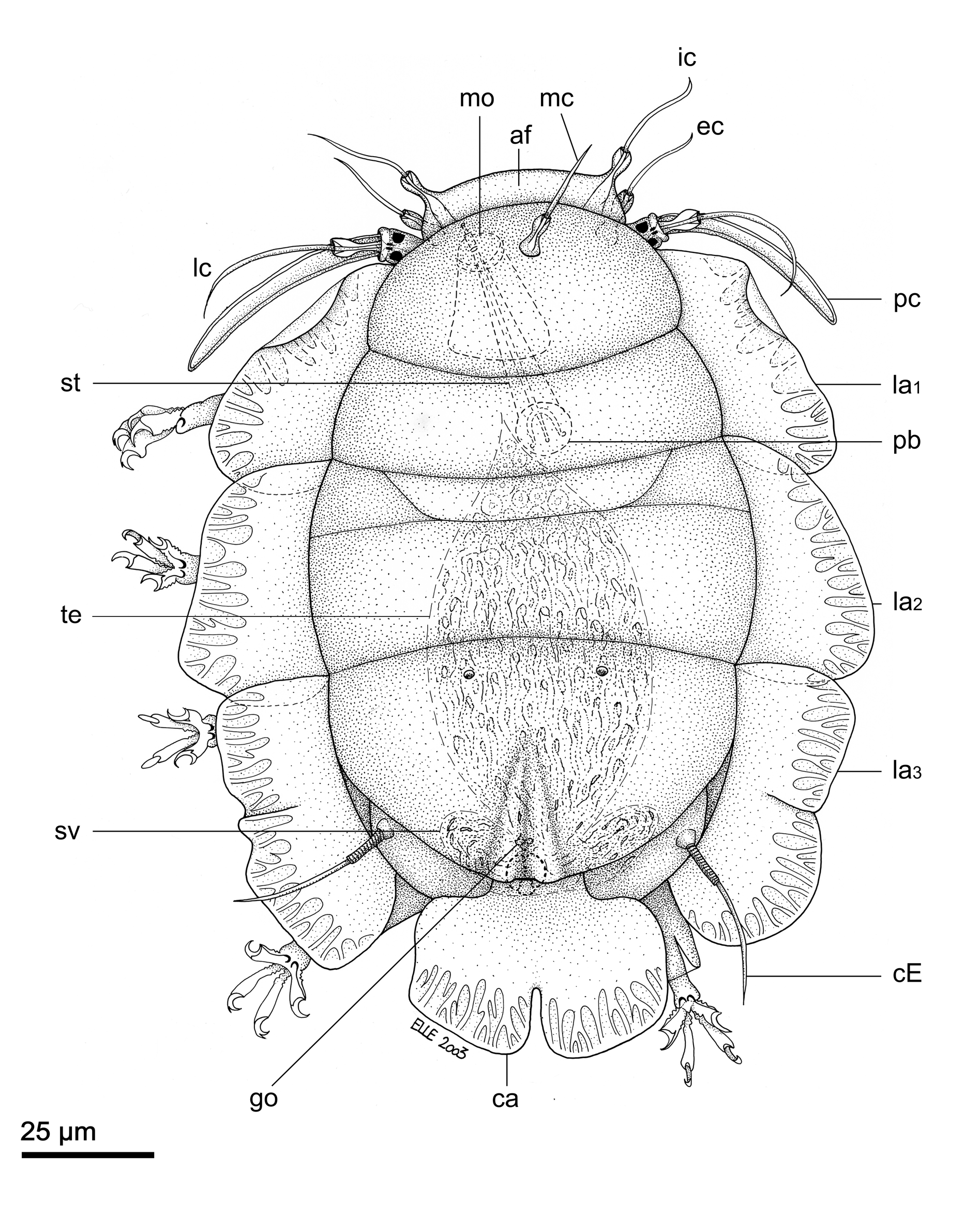
Allotypic male ( Figs 6–7 View Fig View Fig )
No strong secondary sexual dimorphism is observed in Higginsarctus signeae gen. et sp. nov. The male is a little smaller (134 µm) than the female, and the primary clavae a little longer (46 µm). Also the secondary clavae have a slightly different shape, being more oval than rectangular ( Fig. 7B View Fig ). Bacterial vesicles are present just beneath the secondary clavae. The male gonopore is a small triangular papilla with two large pores (openings of the gonoducts), and is situated very close to the anus. The testis is large (87 µm), extending to the pharyngeal bulb, at the level of the first pair of legs. Two small lateral seminal vesicles are present.
Paratypes The paratype NHMD-293898 is a juvenile female without a fully developed rosette gonopore. It measures 113 µm in body length, 66 µm in width and the primary clavae are 42 µm. The other paratype NHMD-29902 is a fully mature female with a body length of 142 µm, body width of 90 µm and the
primary clavae are 49 µm.
Ecology and distribution
Known only from the type locality.

No known copyright restrictions apply. See Agosti, D., Egloff, W., 2009. Taxonomic information exchange and copyright: the Plazi approach. BMC Research Notes 2009, 2:53 for further explanation.
|
Kingdom |
|
|
Phylum |
|
|
Class |
|
|
Order |
|
|
Family |
|
|
Genus |
Higginsarctus signeae
| Hansen, Jesper G. & Kristensen, Reinhardt M. 2021 |
Florarctinae
| Hansen & Kristensen 2021 |