
Higginsarctus laurae, Hansen & Kristensen, 2021
|
publication ID |
https://doi.org/ 10.5852/ejt.2021.762.1461 |
|
publication LSID |
lsid:zoobank.org:pub:43F5C871-A651-47FB-B0A8-29C41EEEEBDD |
|
DOI |
https://doi.org/10.5281/zenodo.5213166 |
|
persistent identifier |
https://treatment.plazi.org/id/E4929598-4B3D-4932-A6FE-9B32DC74CF0D |
|
taxon LSID |
lsid:zoobank.org:act:E4929598-4B3D-4932-A6FE-9B32DC74CF0D |
|
treatment provided by |
Felipe |
|
scientific name |
Higginsarctus laurae |
| status |
gen. et sp. nov. |
Higginsarctus laurae View in CoL gen. et sp. nov.
urn:lsid:zoobank.org:act:E4929598-4B3D-4932-A6FE-9B32DC74CF0D
Figs 14–16 View Fig View Fig View Fig
Diagnosis (characters uniquely defining the taxon are written in bold)
Characterized by large, spherical secondary clavae. Trilobed antero-lateral alae each with pair of medial pointed indentations. Antero-lateral alae larger than postero-lateral alae. Bilobed mediolateral alae with medial pointed indentations. Medio-lateral alae smaller than postero-lateral alae. Bilobed postero-lateral alae with medial pointed identations. Bilobed, round caudal ala with deep
medial pointed indentations. Leg sense organs I–III with similar length. Genital stoup present. Ventral cuticle with folia.
Etymology
The new species is dedicated to Laura Pavesi in acknowledgement of her continuous enthusiasm and effort to services the tardigrade collection at NHMD.
Material examined
Holotype FRANCE • ♀; North Atlantic Ocean , English Channel , Bay of Morlaix, Roscoff; 48°43′ N, 03°54′ W; depth 20–30 m; 15 Mar. 1982; R.M. Kristensen leg.; Dentalium sand; NHMD-293905 . GoogleMaps
Paratypes FRANCE • 6 ♀♀; same collection data as for holotype; NHMD-293906 to NHMD-203011 GoogleMaps .
Description
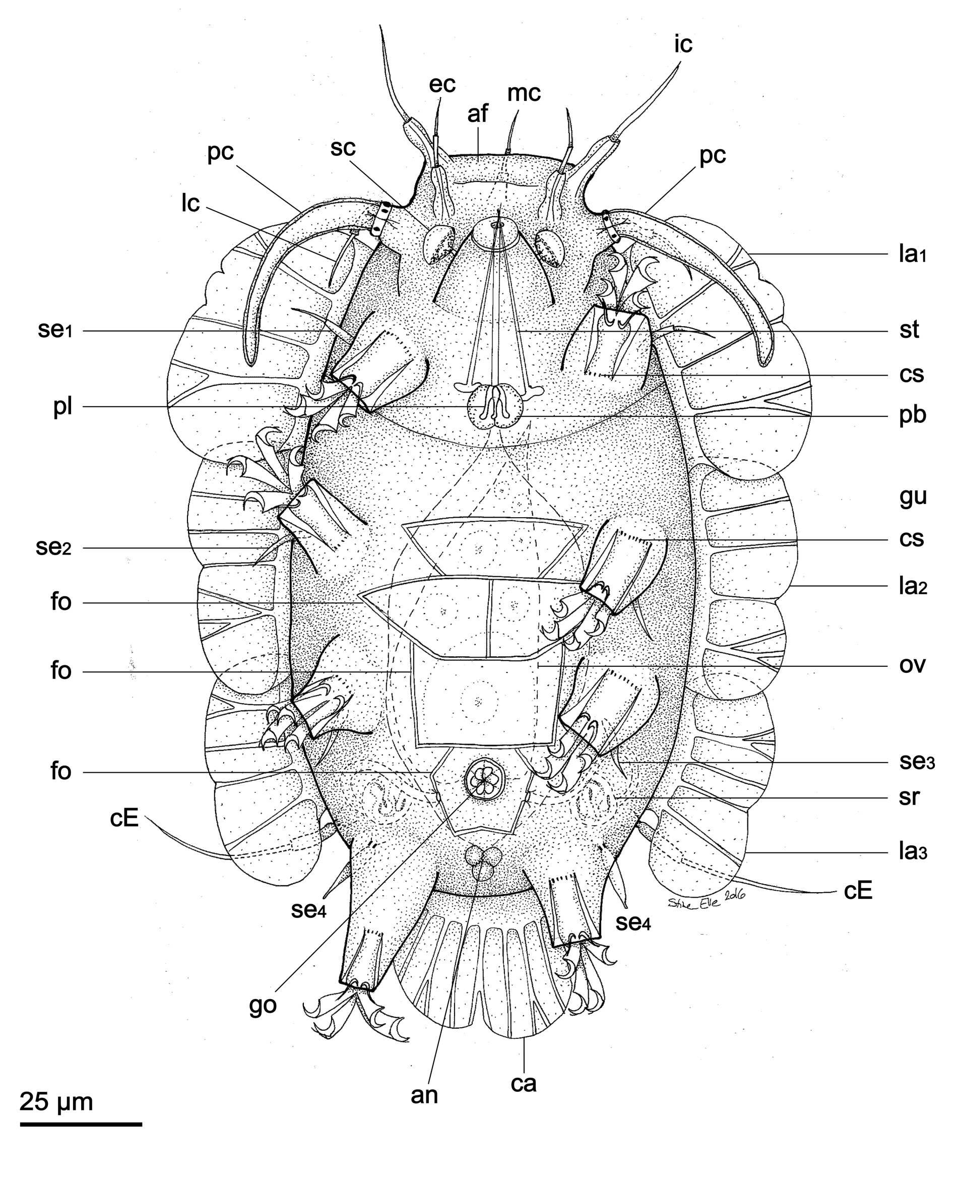
HABITUS. The holotypic female ( Figs 14–15 View Fig View Fig ) is 147 µm long from the anterior margin of the head to the posterior margin of the body. The body is ovoid, being broadest (83 µm) at the level between the second and third pair of legs. The dorsal cuticle has four transverse inter-segmental folds: one anterior to the first pair of legs, two between the first and second pair of legs and one between the second and third pair of legs. Five folia in the form of leaf-like structures ( Fig. 14 View Fig and paratype Fig. 16C View Fig ) are present on the ventral cuticle in the area between the legs, extending from the second pair of legs to the anus.
ALAE. Typical for the genus, eight alae, which are all clearly separated from each other, are present: frontal ala, a pair of antero-lateral alae, a pair of medio-lateral alae, a pair of postero-lateral alae and a single caudal ala ( Figs 14 View Fig , 15A, D View Fig and paratype Fig. 16A View Fig ). The antero-lateral alae which are larger than the medio-lateral alae, each have a medial pair of pointed indentations, dividing the ala into a small medial lobe and two larger lateral lobes. The medio-lateral alae which are larger than the postero-lateral alae, each have a medial pointed indentation dividing the ala into two lobes of equal size. The posterolateral alae each have a medial pointed indentation dividing the ala into two lobes of equal size. The caudal ala has an overall round shape with a deep medial, pointed indentation dividing the ala into two lobes ( Fig. 15D–E View Fig ). As in all species of the new genus, the proximal halve of the lateral and caudal alae is internally supported by continuous procuticle which sends out branching processes (ramuli) into the distal halve of the alae ( Figs 14 View Fig , 15A, D View Fig ).
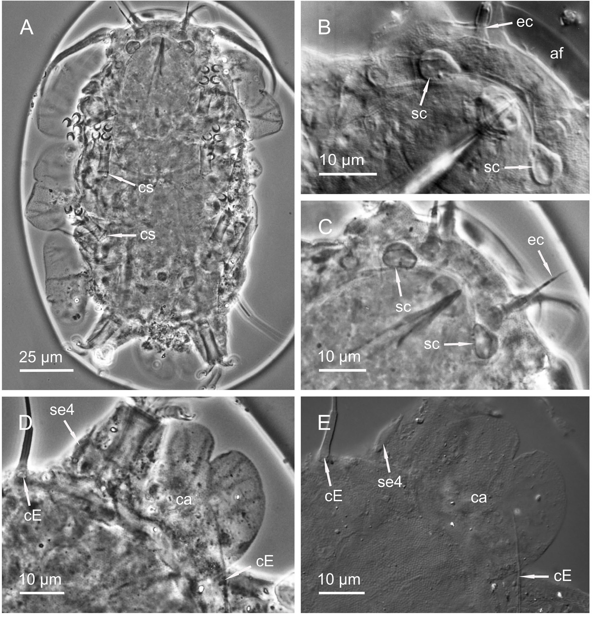
SENSORY ORGANS. The head is well defined from the body by a constriction and a complete set of sense organs is present ( Fig. 14 View Fig ). All the cephalic cirri consist of an hourglass-shaped scapus, a long tubular portion and a protruding flagellum. As in most other species of Florarctinae the scapus of each cirrus appears somewhat outsized, enveloping the internal sensory structures rather than lining them. The internal cirri (35 µm) emerge from the frontal ala at the anterior margin of the head. The external cirri (26 µm) are inserted ventrally and the median cirrus (18 µm) mid-dorsally. Typical for the genus, the primary clava (47 µm) is slightly curved and non-flexible ( Figs 14 View Fig , 15A View Fig ). A van der Land’s body is visible inside its base. Primary clava and lateral cirrus (27 µm) arise on the same cirrophore, and a common membrane (extended margin of cirrophore) surrounds the base of primary clava and lateral cirrus. A very large and thick cuticular ring supports the cirrophore internally. The secondary clavae are large, spherical papillae (8 µm × 5 µm) flanking the mouth cone ( Fig. 15B–C View Fig and paratype Fig. 16B View Fig ). A small refractive organ is visible inside the base of each clavae. The leg I sense organ (15 µm) is a slightly segmented spine with a long swollen base and a terminal tube. The sense organs of leg II (14 µm) and III (14 µm) are unsegmented tapering spines. The fourth leg sense organ (18 µm) is an elongate papilla with a basal van der Land’s body and a terminal pore ( Fig. 15D–E View Fig ). The cirrus E (45 µm) has a prominent cirrophorus, scapus and a long tapering flagellum ( Fig. 15D–E View Fig )
LEGS, DIGITS AND CLAWS. The legs consist of coxa, femur, tibia and tarsus as found in all species of Florarctinae . A row of numerous cuticular stripes of unknown function marks the transition from femur to tibia ( Figs 14 View Fig , 15A View Fig ). The external digits are supported by internal hook-shaped peduncles. The external claw is simple and with a calcar. The internal claw has an accessory spine, but no calcar. All the claws are of the same size, however the external claws are thicker basally and the internal claws have an almost straight portion dorsally. An internal partition is evident as a small notch in each claw, dividing the claw in a basal portion and a distal portion.
BUCCO- PHARYNGEAL APPARATUS. The mouth cone ( Figs 14 View Fig , 15B–C View Fig and paratype Fig. 16B View Fig ) is large with a terminal, very refractive cupola, through which the distal part of the stylets protrudes. The buccal tube is 35 µm long and thin and has a small refractive bulb anterior to the placoids. The stylets are 36 µm long and very thin, each with a well-developed furca. The placoids are short, thick and slightly curved (paratype Fig. 16D View Fig ). Each placoid has a droplet-shaped terminal swelling.
REPRODUCTIVE SYSTEM. Consists of a single ovary bearing numerous small oocytes and a single larger ovum. The ovary is 83 µm long and is attached dorsally, at the level of the first pair of legs. The gonopore is inserted on a large folium and consists of a rosette with six large cells ( Fig. 14 View Fig and paratype Fig. 16C View Fig ). The two cuticular seminal receptacles each consist of a spheroid vesicle and an S-shaped genital duct ( Fig. 14 View Fig ). The cuticle is slightly elevated at each duct opening but does not form a true papilla. The anus is a trilobed cuticular system consisting of two large lateral lobes and a smaller posterior lobe.
Paratypes
The 6 paratypes NHMD-293906 to NHMD-293911 are all mature females with a body length ranging from 118 µm to 139 µm, body width from 60 µm to 73 µm and primary clava length from 39 µm to 42 µm.
Ecology and distribution
Known only from the type locality.

No known copyright restrictions apply. See Agosti, D., Egloff, W., 2009. Taxonomic information exchange and copyright: the Plazi approach. BMC Research Notes 2009, 2:53 for further explanation.
|
Kingdom |
|
|
Phylum |
|
|
Class |
|
|
Order |
|
|
Family |
|
|
Genus |