
Gastrops willistoni Cresson, 1914
|
publication ID |
https://doi.org/ 10.11646/zootaxa.4869.1.7 |
|
publication LSID |
lsid:zoobank.org:pub:8E922358-3363-463E-A03F-A59A43E12DBB |
|
DOI |
https://doi.org/10.5281/zenodo.4418210 |
|
persistent identifier |
https://treatment.plazi.org/id/277D87E8-9B6F-FF88-FF36-1580FD68F808 |
|
treatment provided by |
Plazi |
|
scientific name |
Gastrops willistoni Cresson |
| status |
|
Gastrops willistoni Cresson View in CoL
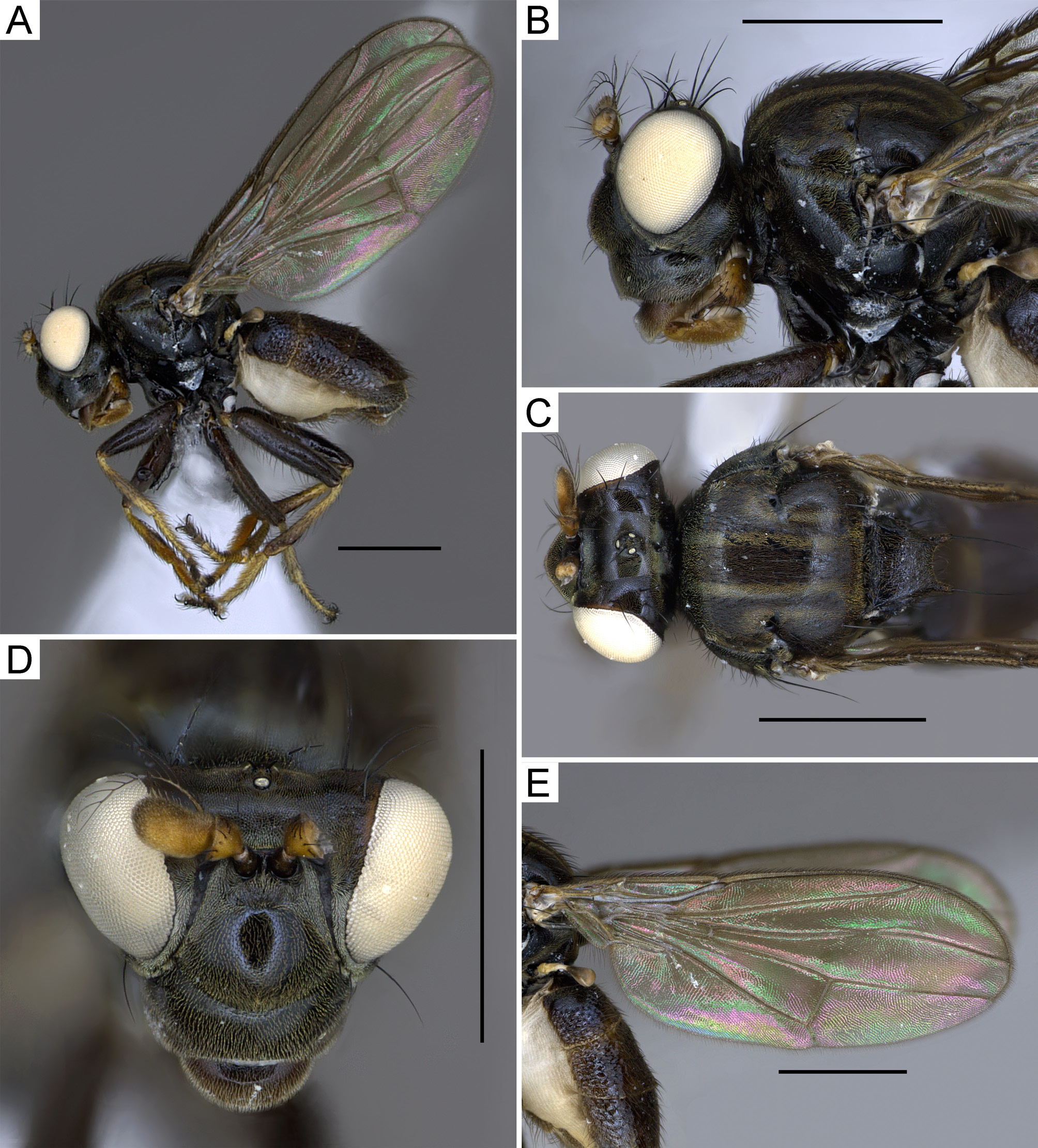
Diagnosis adult. Medium-size species, body length 3.6–4.5 mm (n=5); general color dark brown ( Fig. 3 View FIGURE 3 ); pedicel and postpedicel yellowish brown; arista with 12–14 rays; head sparsely golden tomentose ( Fig. 3B, C, D View FIGURE 3 ); mesonotum golden tomentose with two (sometimes four) greyish stripes; scutellum flattened with one pair of small apical tubercles bearing marginal setae, without lateral tubercles ( Fig. 3B, C View FIGURE 3 ); wings hyaline without infuscate pattern ( Fig. 3E View FIGURE 3 ); femora black; tibiae brown; tarsi reddish brown ( Fig. 3A View FIGURE 3 ) ( Lizarralde de Grosso 1998; Zatwarnicki & Hagenlund 2017).
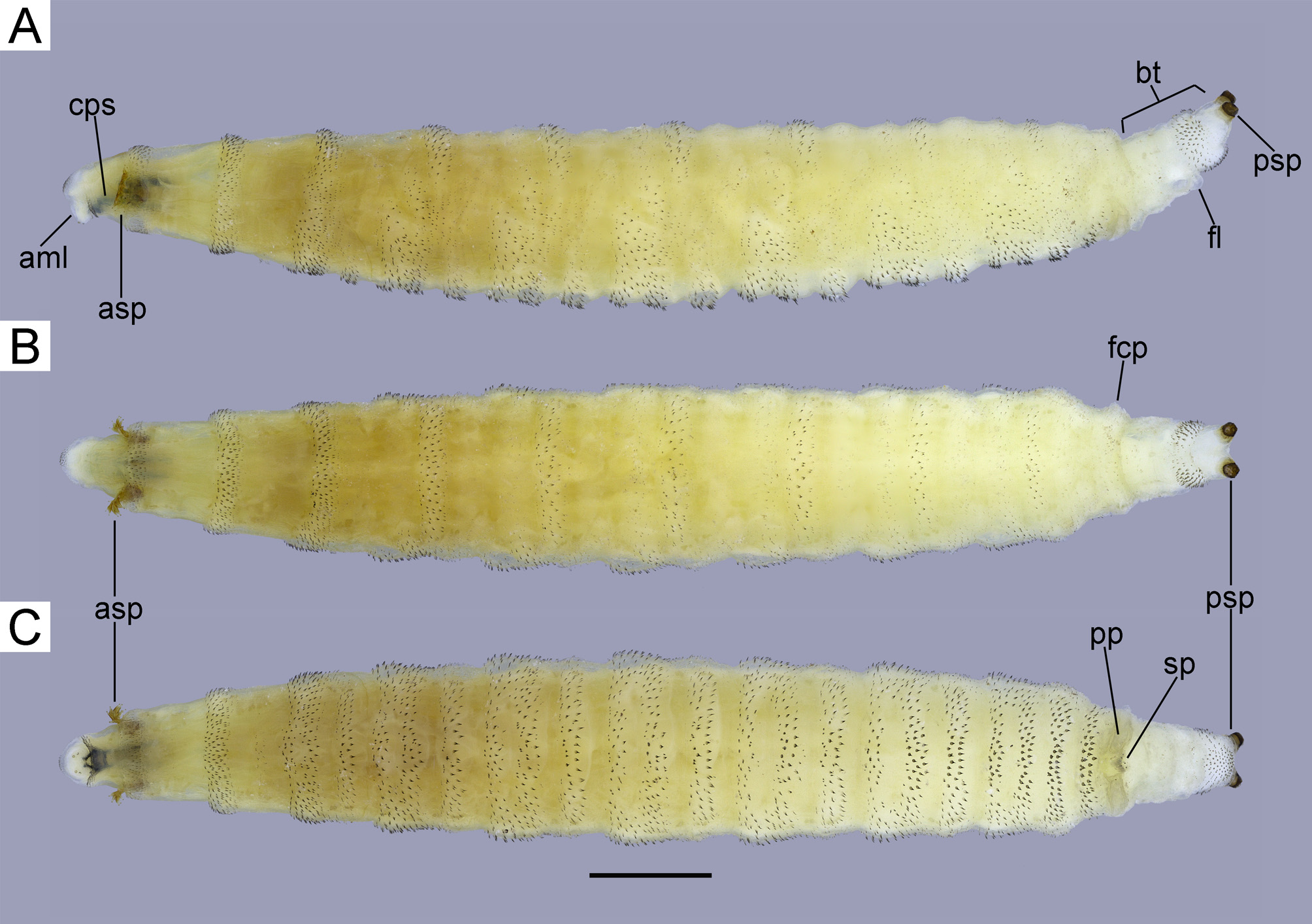
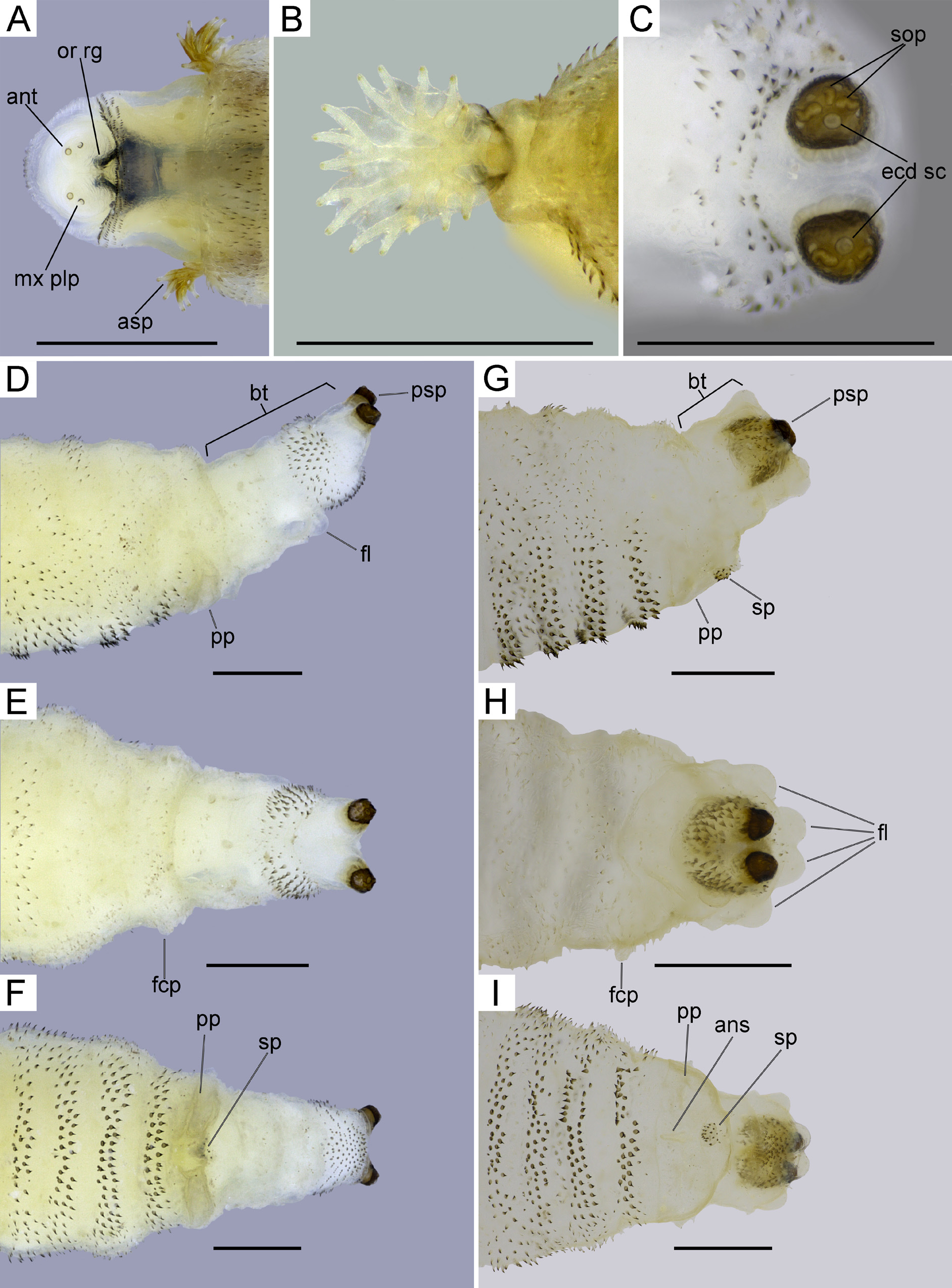
Description of immature stages. Third instar larva ( Figs. 4 View FIGURE 4 , 5 View FIGURE 5 ): main body length 6.11–9.66 mm (n=10); breathing tube length: 0.68–1.05 mm (evaginated n=5) and 0.28–0.67 mm (invaginated n=5); maximum width in dorsal view 0.87–1.57 mm (n=10); whitish and yellowish to dark yellow (after fixation) except anterior spiracles yellowish, posterior spiracular caps dark brown and cephalopharyngeal skeleton black; shape fusiform, thoracic segments telescoping anteriorly, caudal segment retractile, invaginated in most preserved specimens, directed upward and bifurcate distally; anterior segments somewhat retracted and invaginated or completely evaginated in preserved specimens; segment 1 (pseudocephalic) projected anteriorly in two small lobes (antennomaxillary lobes), each lobe with minute antenna directed anteriorly (anteroventrally in invaginated specimens); maxillary sensory papilla ventral to each antenna, slightly smaller, directed anteroventrally (ventrally in invaginated specimens); oral ridges beneath maxillary papillae in direction to oral region with 3–4 clusters of minute dark brown spinules arranged in short rows ( Figs. 4A View FIGURE 4 , 5A View FIGURE 5 , 6 View FIGURE 6 A–C); segment 2 (prothoracic) with anterior ventral half (postoral border) with a cluster of 10–12 rows of dark brown spinules, disappearing laterally, dorsal anterior margin (approx. 1/3 of segment length) with a cluster of 8–9 rows of dark brown spinules diminishing in size posteriorly, remainder of segment glabrous; anterior spiracles whitish or yellowish (brown in some preserved specimens) ( Figs. 4 View FIGURE 4 A–C, 5A, B) present at posterior border, retractile, almost completely invaginated and enclosed under anterior margin of mesothoracic segment in the majority of preserved specimens (only a few ones completely evaginated), fan-shaped in lateral view with basal trunk branching in smaller 11–18 twigs (finger-like ramifications) connected by a thin lamellae almost until tip ( Fig 5B View FIGURE 5 ); segment 3 (mesothoracic) with spinule band completely encircling anterior margin, composed of 4–6 rows of irregularly arranged stout spinules, mid-laterally with a very slight prominence that continues ventrally, remainder of segment with somewhat sparse and almost translucent spinules, except posterior margin (approx. 1/4 of segment length) which is bare; segment 4 (metathoracic) with spinule band of 4–6 rows completely encircling anterior margin, mid-ventrally with a belt of 3–4 rows of spinules diminishing laterally, posterior margin with a band of spinules (6–7 rows) that completely encircles posterior margin, this posterior band expands laterally and obliquely, slightly directing forward, ventrally it is divided in two bands by a central bare spot (fusiform-ellipsoid shape) with 2–3 rows of spinules anteriorly and 3–4 posteriorly to this ventral bare spot, mid-dorsal and lateral parts of segment covered by translucent sparse spinules, except ventrally between anterior band, ventral belt and posterior band of spinules, bare; segment 5 (first abdominal) with ventral belt of spinules diminishing laterally and directed forward, meeting the posterior spinule band from segment 4, posterior spinule band from segment 5 directed forward laterally and interrupted ventrally by a bare spot as in segment 4, dorsal part of segment covered by sparse translucent spinules, ventral areas between anterior and posterior band and ventral belt of spinules bare; segments 6–11 covered with same pattern of spinules as segment 5; segment 12 ( Figs. 4 View FIGURE 4 A–C, 5D–I) with one small fleshy conic projection on each dorsolateral side (not easily visible in all specimens), dorsally with a band of 4–5 dark brown spinules, connecting to the posterior band from segment 11 and a ventral band of 3–4 rows of dark brown spinules not connected to other bands (anterior to perianal pad), a few sparse translucent spinules dorsally and laterally; perianal pad bilobed ( Fig. 5F, I View FIGURE 5 ) (very rounded and distinct in some preserved specimens) slightly prominent at posterior margin, dark yellow; mid-posterior margin of perianal pad with a small, conical, spinule patch (like a pine cone), covered with dark brown spinules that are directed anteriorly (opposite to body spinules, which are directed posteriorly) ( Fig. 5F, I View FIGURE 5 ); breathing tube ( Figs. 4 View FIGURE 4 A–C, 5D–I) telescopic, tapering distally, bifurcate before apex, formed by two sections; first section with sparse translucent spinules dorsally, denser ventrally with 8–10 fleshy lobes around the posterior margin (with the aspect of a flower in some preserved specimens, though in some specimens these lobes are not very conspicuous ( Fig. 5E, F View FIGURE 5 ); second section retractile, densely covered with larger spinules on basal half, posterior half glabrous and bifurcate; posterior spiracular caps rounded apically, dark brown with dark yellow basal half; three reniform spiracular openings bent medially (with C-shape aspect); ecdysial scar rounded and lighter in color surrounded at external lateral margin by the three spiracular openings ( Fig. 5C View FIGURE 5 ). The spinules covering the larvae have the basal half with almost same integument color and apical half dark brown. Some spinules are bifid or seem to be cut and then regrown, normally disposed in pairs on the rows with their shapes resembling a comma (based on 10 specimens collected in the field and fixed in the laboratory).
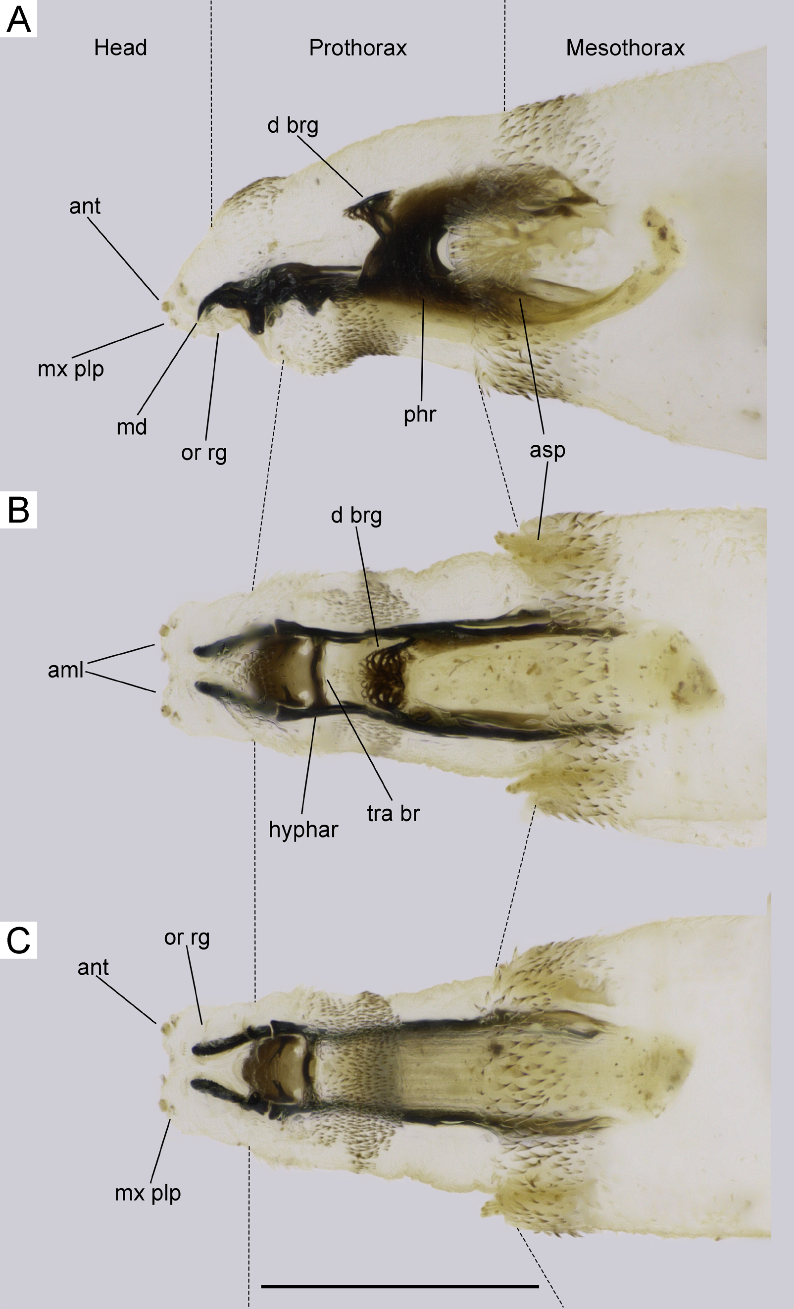
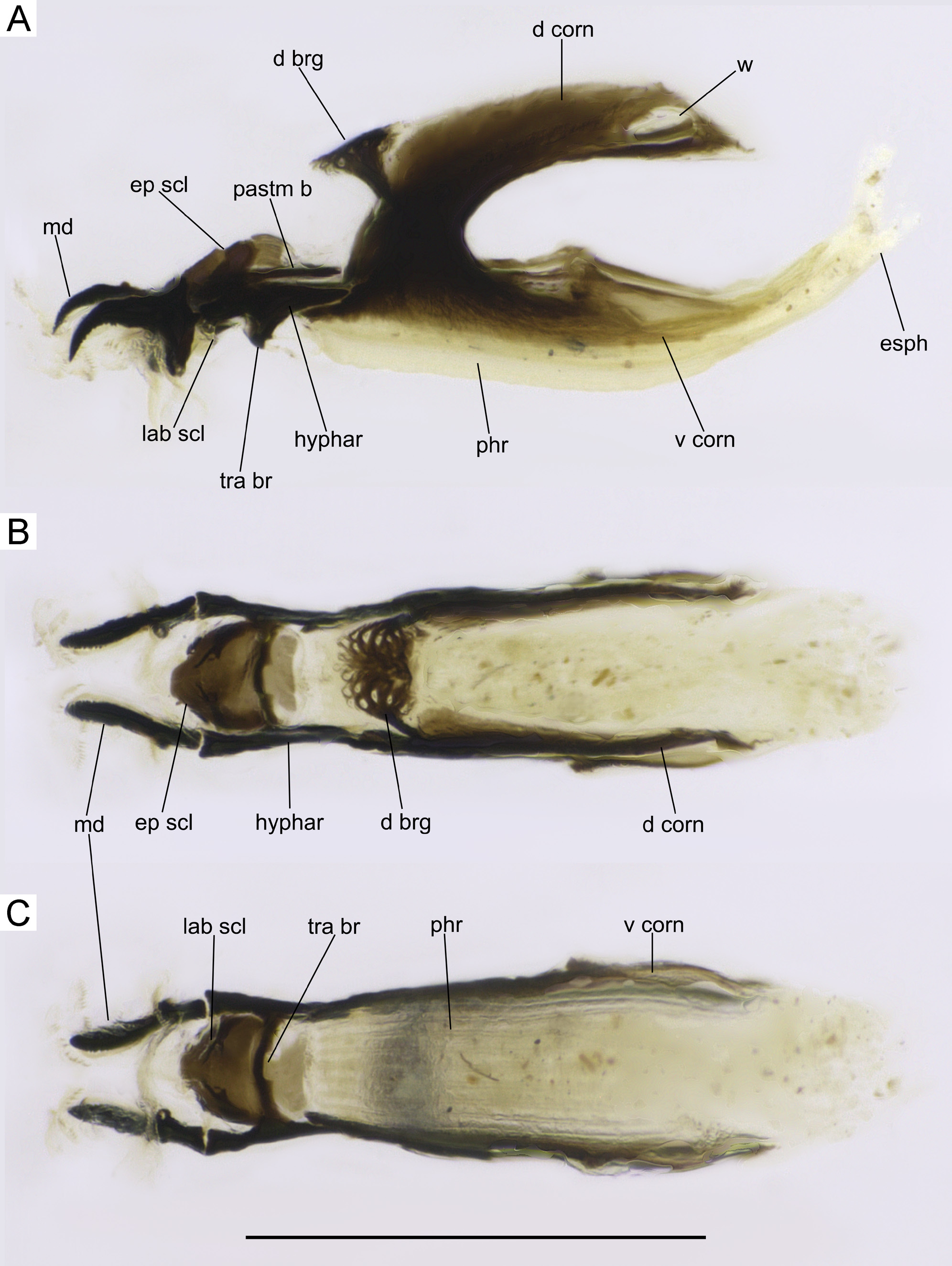
Cephalopharyngeal skeleton ( Figs. 6 View FIGURE 6 , 7 View FIGURE 7 ): mostly dark brown to black pigmented; length 0.90 mm (n=1); mandibles length 0.14mm (n=1) paired and not connected dorsally; narrow, curved downward anteriorly, ventral surface denticulate, broad posteriorly with ventral projection about third of mandible length ( Fig. 7A View FIGURE 7 ); mandible with 2/3 of its length on pseudocephalic segment and 1/3 on anterior part of prothoracic segment ( Fig. 6 View FIGURE 6 A–C); hypopharyngeal sclerite length 0.15 mm (n=1), slightly shorter than mandibles, not fused with mandibles and pharyngeal sclerite, connected ventrally by a narrow, slightly anteriorly arched posterior transverse bar ( Fig. 7C View FIGURE 7 ), restricted to prothoracic segment ( Fig. 6 View FIGURE 6 A–C); labial sclerite under hypopharyngeal sclerite not connected anteriorly with other sclerites, bifurcate, forming two branches (Y-shaped) with rounded apex, posterior branch shorter, half the length of the anterior branch ( Fig. 7C View FIGURE 7 ); epipharyngeal sclerite with mid-anterior margin truncate with a small recession anterolaterally, forming a curve laterally, without windows, laminate ( Fig. 7B View FIGURE 7 ); parastomal bar not connected to pharyngeal sclerite ( Fig. 7A View FIGURE 7 ); pharyngeal sclerite whitish ventrally, occupying posterior half of prothorax and anterior half of mesothorax ( Fig. 6 View FIGURE 6 A–C), dorsal cornu large, connected dorsally by a dorsal bridge, each dorsal cornu with a rounded window posteriorly ( Fig. 7A View FIGURE 7 ); dorsal bridge formed by a strong arm on each side and very broad medially with numerous windows with reticulate aspect ( Fig. 7B View FIGURE 7 ); ventral cornu as broad as dorsal cornu, without windows, with a slight projection mid-dorsally; floor of pharyngeal sclerite with 8–9 longitudinal striae ( Fig. 7C View FIGURE 7 ) (based on 1 dissected specimen).
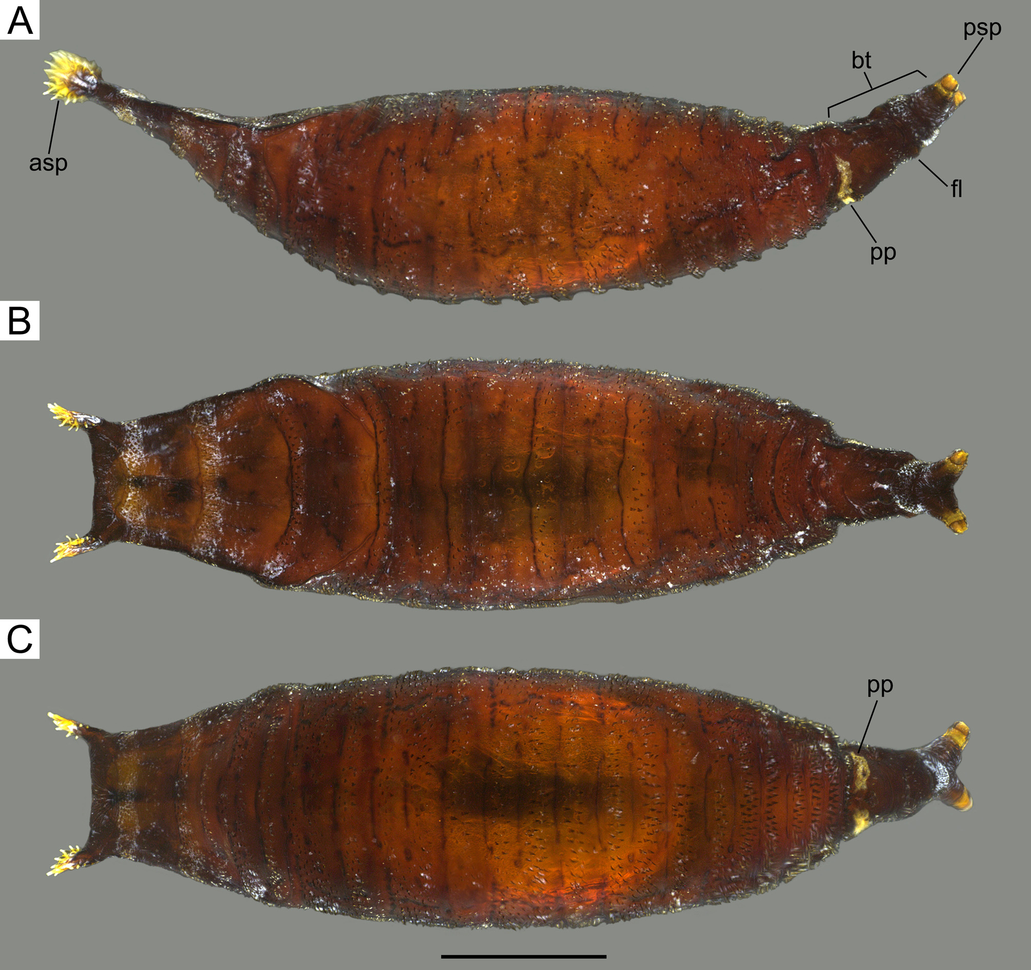
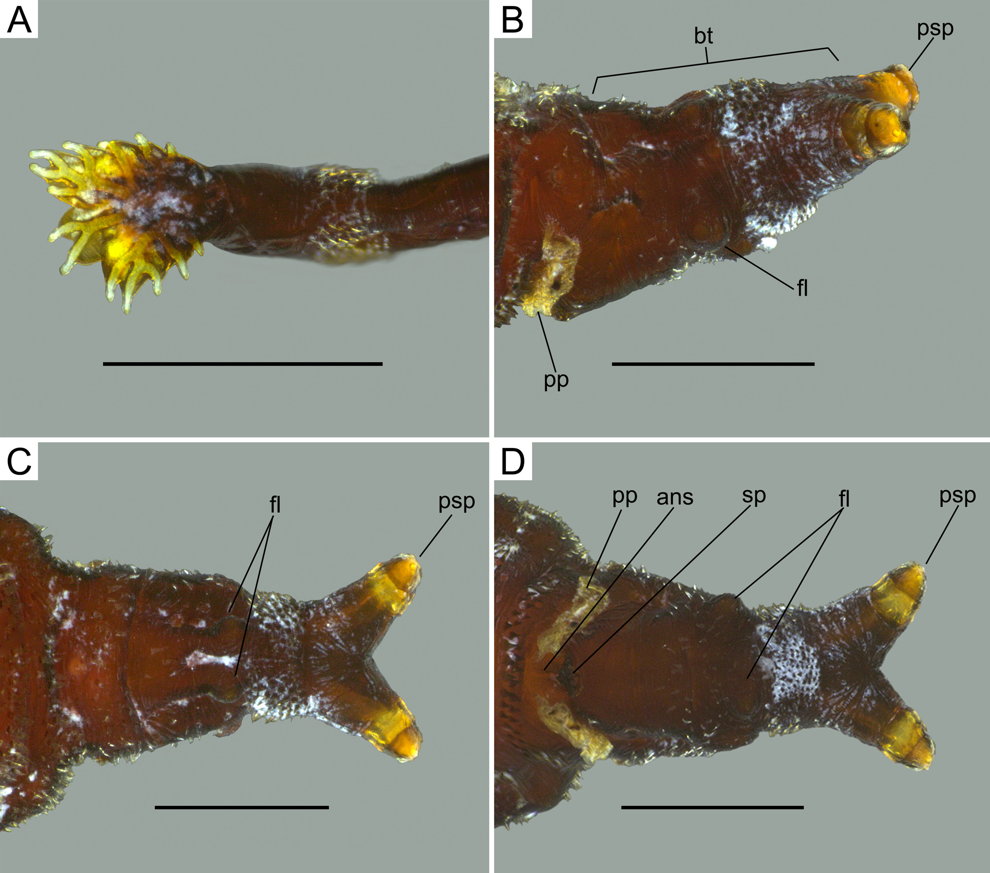
Puparium ( Figs. 8 View FIGURE 8 , 9 View FIGURE 9 ): main body length 5.10–5.85 mm (n=10) (including anterior spiracles and breathing tube), breathing tube length 0.63–0.82 mm (n=10); maximum width in dorsal view 1.36–1.82 mm (n=10); dark reddish brown; venter arcuate and dorsum only slightly arcuate medially in lateral view; anterior end (thoracic segments) flattened dorsoventrally, slightly curving upward in lateral view ( Fig. 8A View FIGURE 8 ); anterior margin of segment 2 tapered; posterior end conical and curved upward in lateral view ( Fig. 8 View FIGURE 8 A–C); anterior spiracles ( Fig. 9A View FIGURE 9 ) yellowish, arising from anterolateral corners of puparium with 13–15 finger-like projections interconnected by thin lamella resembling fan-shape; segment 1 and almost entire segment 2 invaginated; dorsal cephalic cap flattened, slightly depressed on segments 4 and 5, delineated by a weak line that extends laterally along segments 3–4 and curving inward along segment 5 reaching the posterior margin of segment 5 ( Fig. 8B View FIGURE 8 ); segments 6–10 with two annuli dorsally; segment 12 with perianal pad yellowish, wrinkled ( Fig. 8C View FIGURE 8 ); breathing tube tapering towards apex, bifurcate; posterior spiracle yellowish; arrangement of spinules similar to third instar larva ( Fig. 9 View FIGURE 9 B–D) (based on 10 specimens that pupariated in the laboratory).
No known copyright restrictions apply. See Agosti, D., Egloff, W., 2009. Taxonomic information exchange and copyright: the Plazi approach. BMC Research Notes 2009, 2:53 for further explanation.
|
Kingdom |
|
|
Phylum |
|
|
Class |
|
|
Order |
|
|
Family |
|
|
Genus |