
Hermanella angeli, Almeida, Everlin, Costa, Sabrina & Mariano, Rodolfo, 2016
|
publication ID |
https://doi.org/10.11646/zootaxa.4078.1.10 |
|
publication LSID |
lsid:zoobank.org:pub:5282C105-27A2-4056-A994-1F53C4FA8E7B |
|
DOI |
https://doi.org/10.5281/zenodo.6085099 |
|
persistent identifier |
https://treatment.plazi.org/id/2803FD03-FFD6-FFFB-7284-F968A6EC5780 |
|
treatment provided by |
Plazi |
|
scientific name |
Hermanella angeli |
| status |
sp. nov. |
Hermanella angeli sp. nov.
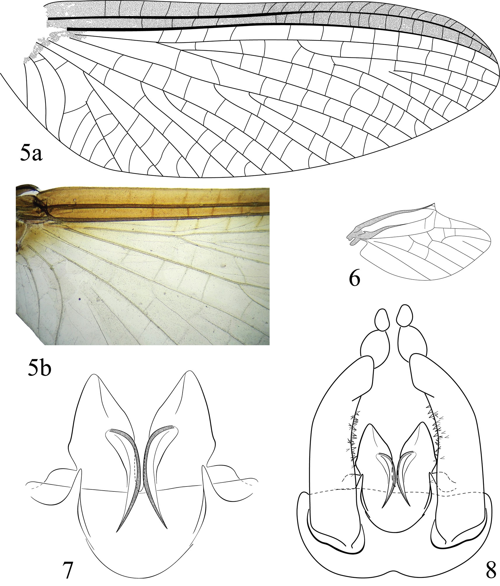
( Figures 1–19 View FIGURES 1 – 4 View FIGURES 5 – 8 View FIGURES 9 – 15 View FIGURES 16 – 19 )
Material examined. Holotype: Male imago, BRAZIL, Bahia State: Itacaré, Cachoeira Bom Sossego, 31.VII.2012, 14°20’05.2”S 39°01’27.4”W, Mariano, R; Almeida, E & Costa, S. leg. ( MZUSP). Paratypes: five females and three male imagos: same data as holotype but in LOA; Ten nymphs, four females, one male, same data as holotype but 24.IX.2012, Mariano, R, Costa, S. & Santos, D. leg. ( MZUSP); One male imago, Igrapiúna, Reserva Ecológica da Michelin, Córrego da Mata, Trilha do Guigó, 22.IX.2012, Calor, A.R. & Oliveria, R.C. leg. (LOA); Ten nymphs, same date but Córrego das Matas, 19.VIII.2014, Mariano, R., Almeida, E., Pires, R.S. & Sousa, M.M.L. leg. (LOA).
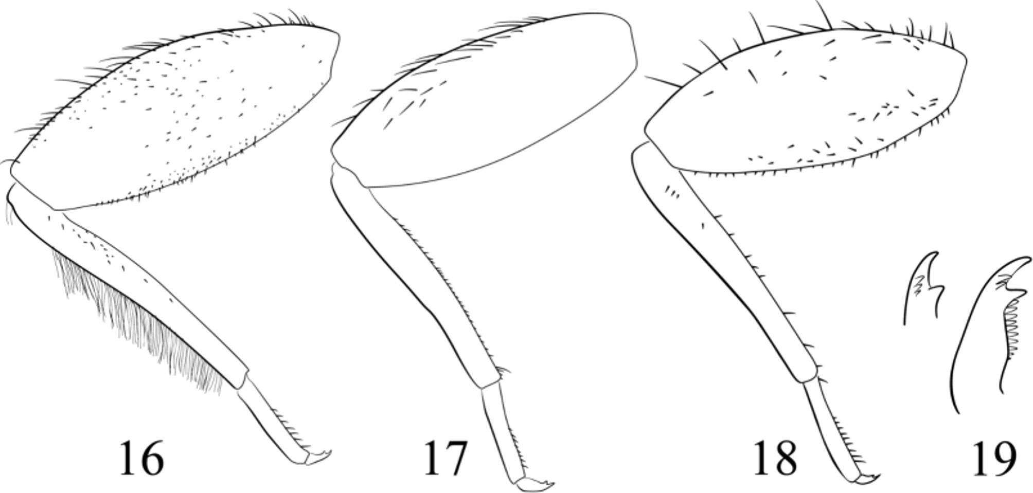
Diagnosis. Hermanella angeli sp. nov. is distinguished from the others congeners by the following combination of characteristics. In the male imago: (1) forewing with costal and subcostal area brown ( Fig. 5 View FIGURES 5 – 8 b); (2) thorax and abdomen terga predominantly brown as in Fig. 2–3 View FIGURES 1 – 4 ; (3) penis divided on apical 1/8 ( Fig. 8 View FIGURES 5 – 8 ); (4) penis lobe with a ventral, long, curved spine ( Fig. 7 View FIGURES 5 – 8 ); (5) prosternum narrow ( Fig. 1 View FIGURES 1 – 4 ). In the nymphs: (1) tarsal claws with 2–3 accessory denticles ( Fig. 19 View FIGURES 16 – 19 ); (2) mandible without setae near basal articulation ( Fig. 13 View FIGURES 9 – 15 a); (3) prostheca of left mandible with spine on inner margin ( Fig. 13 View FIGURES 9 – 15 b); (4) plate-like gills terminated in a finger like process ( Fig. 11 View FIGURES 9 – 15 ).
Male imago. Length: body 7.3–8.9 mm; forewing 8.9–9.1 mm; hind wing 2.8 mm.
Head: light brown with a black macula near antenna; upper portion of compound eye pale brown, lower portion grayish black. Antenna: scape brownish with a black ring on apex; pedicel brownish and flagellum pale. Thorax: pronotum chestnut, with posterolateral blackish band; mesonotum chestnut with some dark sutures, scutellum pale yellow and scuto–scutellar impression yellowish; metanotum chestnut with dark sutures; prosternum narrow ( Fig. 1 View FIGURES 1 – 4 ). Wings ( Figs 5, 6 View FIGURES 5 – 8 ): membrane of forewing hyaline, costal and subcostal area brown, base radial area light brown; C, Sc and R1 brown; cross veins of C and Sc area brown. Membrane of hind wing hyaline, brownish at base; C, Sc and R1 veins yellowish brown; longitudinal and cross veins translucents. Legs: coxae and trochanters light brown; femora chestnut with lateral region, basal ½ and apex tinged with black ( Fig. 2 View FIGURES 1 – 4 ); tarsi whitish tinged with brown. Leg I: tibia dark brown. Leg II: tibia pale yellow. Leg III: tibia pale yellow with apex tinged with black. Abdomen ( Figs 3–4 View FIGURES 1 – 4 ): terga I–V chestnut tinged with black; tergum VI gradually brown to chestnut; terga VII–IX chestnut with posterior region tinged with black; tergum X chestnut with black maculae. Genitalia ( Figs 7–8 View FIGURES 5 – 8 ): styliger plate light brown, forceps whitish; segment I of forceps 1/2 length of segment II, segment II 1 /8 length of segment I; penis divided on basal 1/8, each penis lobe whitish with a ventral, long, curved yellowish spine ( Fig. 7 View FIGURES 5 – 8 ). Caudal filaments white with every annulus marked with black.
Female imago. Length: body, 5.4–6.8 mm; forewing, 7.2–7.8 mm; hind wing, 1.4–1.5 mm. Color pattern similar to male imago except for abdomen uniformly dark brown.
Nymph. Length: body 8.1–10.3 mm, antenna 3.1 mm, terminal filament 13.7 mm. General coloration orange brown ( Figs 9, 10 View FIGURES 9 – 15 ).
Head: on female and male general coloration brown. Male head with a white macula on frons ( Fig. 10 View FIGURES 9 – 15 ); upper portion of male’s compound eye brown, lower portion black. Female eyes black. Antenna yellowish. Mouthparts ( Figs 12–15 View FIGURES 9 – 15 ): clypeus without a medial projection, maximum width of labrum 1.6 times maximum width of clypeus. Labrum yellowish brown, with dorsal proximal row of 32 long setae on each side; numerous ventral setae directed obliquely to sagittal plane ( Fig. 12 View FIGURES 9 – 15 ). Mandible yellowish brown, median region translucent, molars orangish brown; outer margin of mandible strongly curved forming a square angle ( Fig. 13 View FIGURES 9 – 15 a); without setae near basal articulation; incisors of left mandible with 3–5 spines on inner margin ( Fig. 13 View FIGURES 9 – 15 b). Maxilla light yellowish brown; galea–lacinia with prominent tusk on inner apical margin (nearly 1/5 the apical width of galea-lacinia); basal segment of palpus light brown ( Fig. 14 View FIGURES 9 – 15 ). Labium light yellowish brown ( Fig. 15 View FIGURES 9 – 15 ). Thorax: terga orangish brown, pleura and sterna yellowish. Anterolateral margin of pronotum with two spine-like setae. Legs yellowish brown, slightly washed with brown. Leg I ( Fig. 16 View FIGURES 16 – 19 ): tibia with basal and subapical brown bands, tarsus with a basal brown band. Legs II and III ( Figs 17, 18 View FIGURES 16 – 19 ): femora with an apical brown band, a long setae on posterior margin, dorsal surface covered with thick, short spines; tibiae with basal and subapical brown bands, tarsi with a basal brown band. Tarsi with spine-like setae along inner margin. Tarsal claws with subapical denticle much larger than others, 9–10 median denticles subequal in size and 2–3 accessory denticles ( Fig. 19 View FIGURES 16 – 19 ). Abdomen: terga orangish chestnut, segments II, III and VI–IX with a medial and submedial brown marks ( Fig. 9 View FIGURES 9 – 15 ); sterna yellowish white. Posterolateral projections on abdominal segments VIII–IX. Gills on abdominal segments I–VII, grayish black, trachea darker and apical filaments pale ( Fig. 11 View FIGURES 9 – 15 ). Caudal filaments yellowish chestnut.
Life cicle association. Reared.
Etymology. angeli (genitive singular), specific name in honor the Angelo B. M. Machado, on the occasion of his 80th birthday.
Remarks. Several works have been published in the last ten years on the Hermanella group, showing how complex is the study of this particular group ( Peters et al. 2005; Sartori 2005; Polegatto & Batista 2007; Kluge 2007; Mariano et al. 2010; Mariano 2011; Lima et al. 2012; Nascimento & Salles 2013). Hermanella angeli sp. nov. is another intermediary species in the Hermanella complex, as seen in the discussion on H. mazama by Nascimento & Salles (2013). The new species possess several characteristics between Hermanella (i.e. prosternum narrow on imago and by the absence of median projection on clypeus in nymphs) and Needhamella (i.e. presence of accessory denticles and gills with finger-like ends).
Male imagos of the H. angeli sp. nov. show the same color pattern on C and Sc area of forewings as in most of the Hermanella species, but differing basically on the shape of the styliger plate. The nymph is the only species in the Hermanella that possess accessory denticles on tarsal claws. Following the key proposed by Nascimento & Salles 2013, H. angeli sp. nov. would key out in the couplet that leads to H. grandis and H. froehlichi . The male imagos of H. grandis , however, are unknown and because of that we modified the key as follows:
8(7). Length of forewings 8.9–9.1 mm; color pattern of abdomen as in Fig. 3 View FIGURES 1 – 4 ...................... Hermanella angeli sp nov. 8. Length of forewings 6.8–8.7 mm; color pattern of abdomen as in Fig. 144Q of Domínguez et al. (2006)..................................................................................................... Hermanella froehlichi


| MZUSP |
Museu de Zoologia da Universidade de Sao Paulo |
No known copyright restrictions apply. See Agosti, D., Egloff, W., 2009. Taxonomic information exchange and copyright: the Plazi approach. BMC Research Notes 2009, 2:53 for further explanation.
|
Kingdom |
|
|
Phylum |
|
|
Class |
|
|
Order |
|
|
Family |
|
|
Genus |