
Sinobathyscia tianma, Wang & Perreau & Růžička & Song, 2017
|
publication ID |
https://doi.org/ 10.11646/zootaxa.4303.3.2 |
|
publication LSID |
lsid:zoobank.org:pub:260F1C3F-4FC8-4DA4-8AEE-0DD11A99185F |
|
DOI |
https://doi.org/10.5281/zenodo.6030427 |
|
persistent identifier |
https://treatment.plazi.org/id/291D87F1-FF9D-CA6A-FF0A-F8DAFB7DF853 |
|
treatment provided by |
Plazi |
|
scientific name |
Sinobathyscia tianma |
| status |
sp. nov. |
Sinobathyscia tianma View in CoL sp. nov. (天马ĢḆṁṂffl)
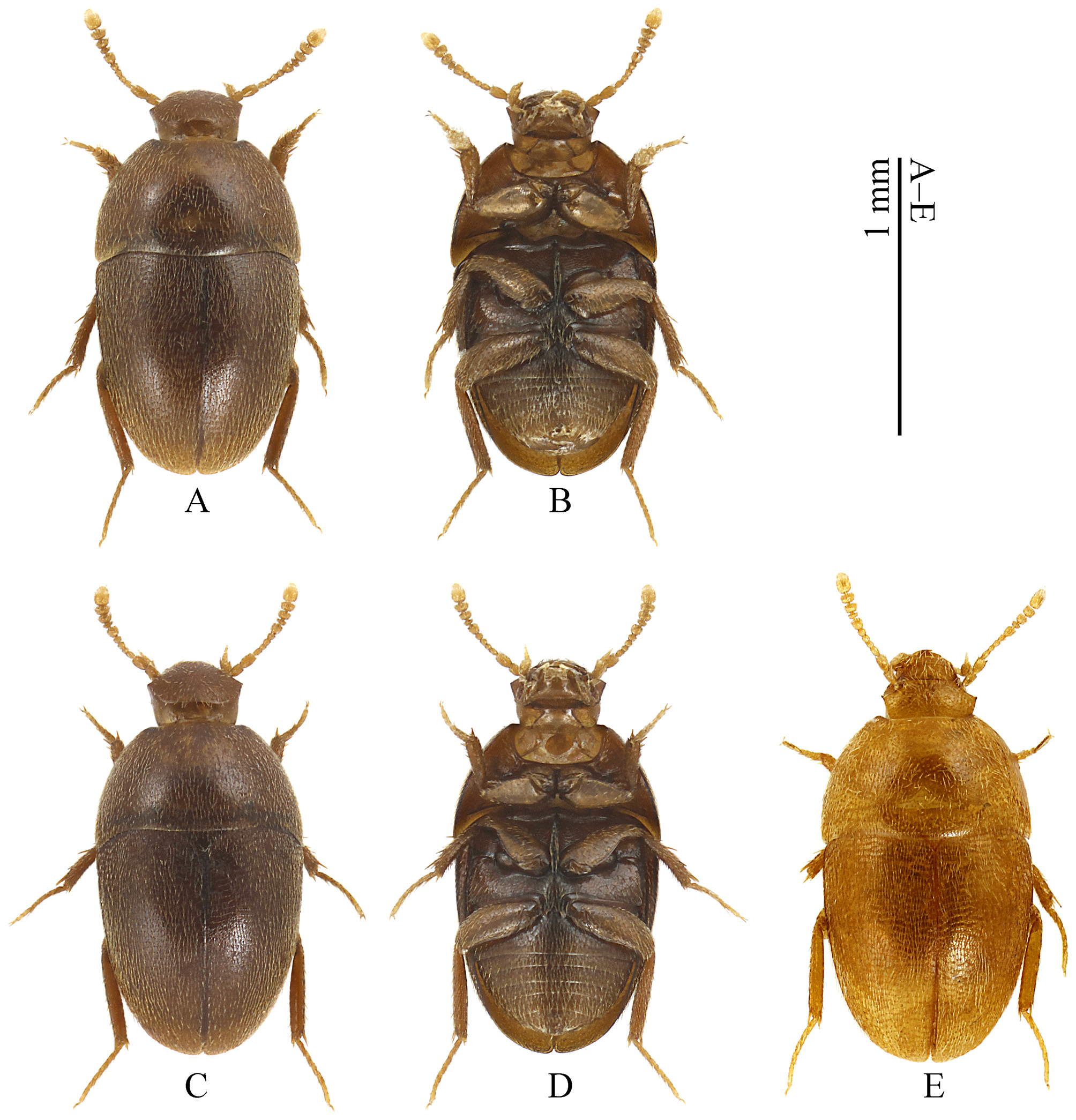
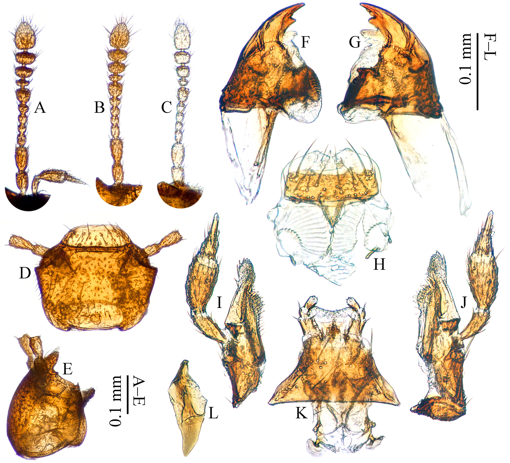
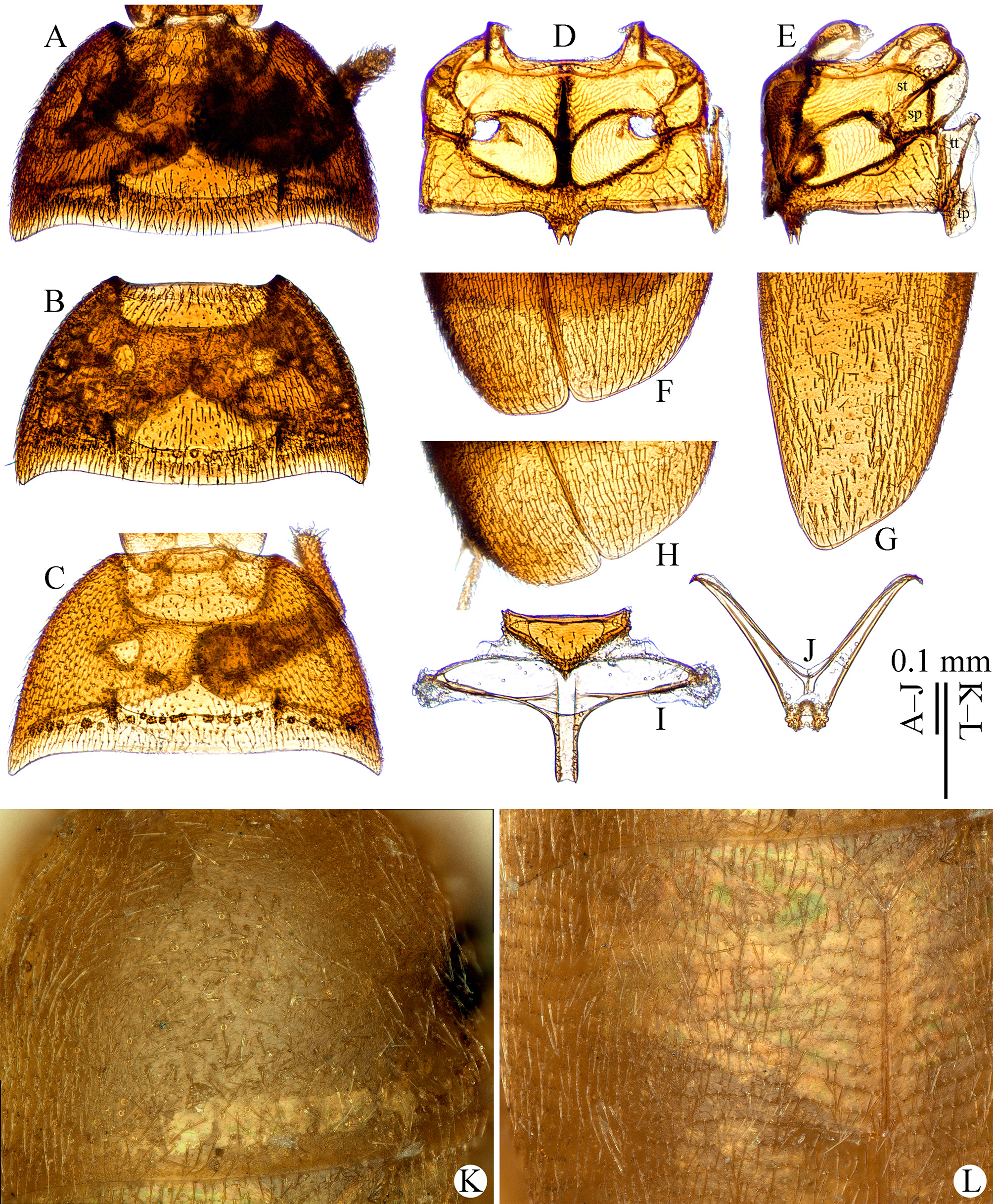
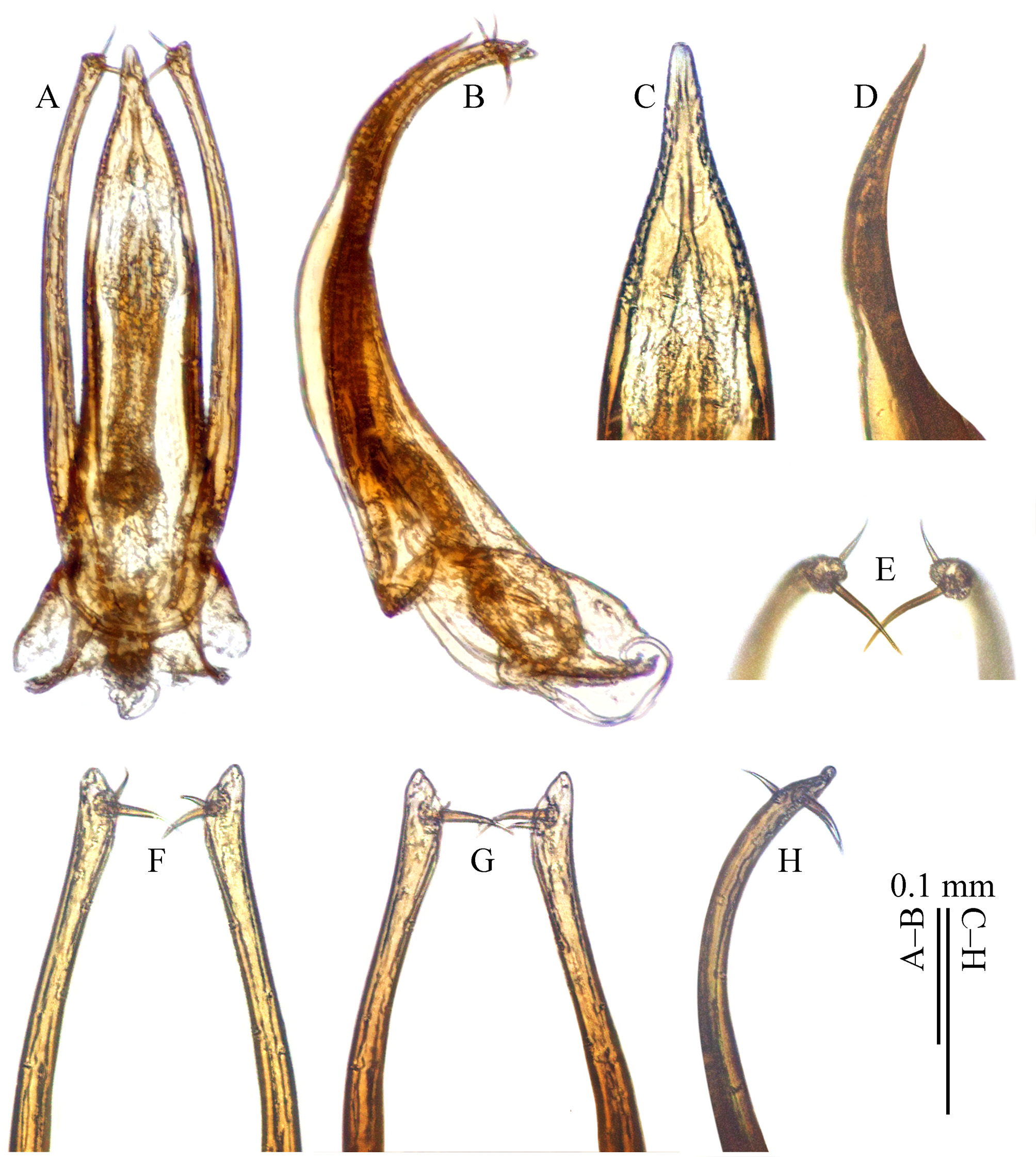
( Figs. 1 View FIGURE 1 A–D; 2A–B, D–L View FIGURE 2 ; 3A–B, D– G View FIGURE 3 , I–J; 4A–D, G–J, L View FIGURE 4 ; 5A–H View FIGURE 5 ; 6C–D, E View FIGURE 6 ; 7A–C View FIGURE 7 )
Type material. Holotype: ♂, CHINA, Shanghai: / Songjiang District, / Tianmashan (天 马山), / alt. 40 m, 31°04'35.95''N / 121°08'45.45''E, // sifted from rotten wood, / 19.XII.2016, / Xiao-Bin Song leg. ( SNUC). GoogleMaps
Paratypes: 1♂ 8♀♀, same data as holotype (1♂ 5♀♀ in SNUC, 1♀ in CJRZ, 1♀ in CMPR and 1♀ in BMNH). Description. Male holotype. Extended body length: 1.4 mm. Length (mm) of different body parts: head (0.2): antenna (0.5): pronotum (0.4): elytra (0.7); width (mm): head (0.3): pronotum (0.7): elytra (0.7). Proportion of antennomeres from base to tip in µm (length × width): 49 × 32, 58 × 34, 28 × 24, 20 × 22, 22 × 24, 22 × 27, 33 × 38, 15 × 35, 26 × 47, 30 × 54, 71 × 54. GoogleMaps
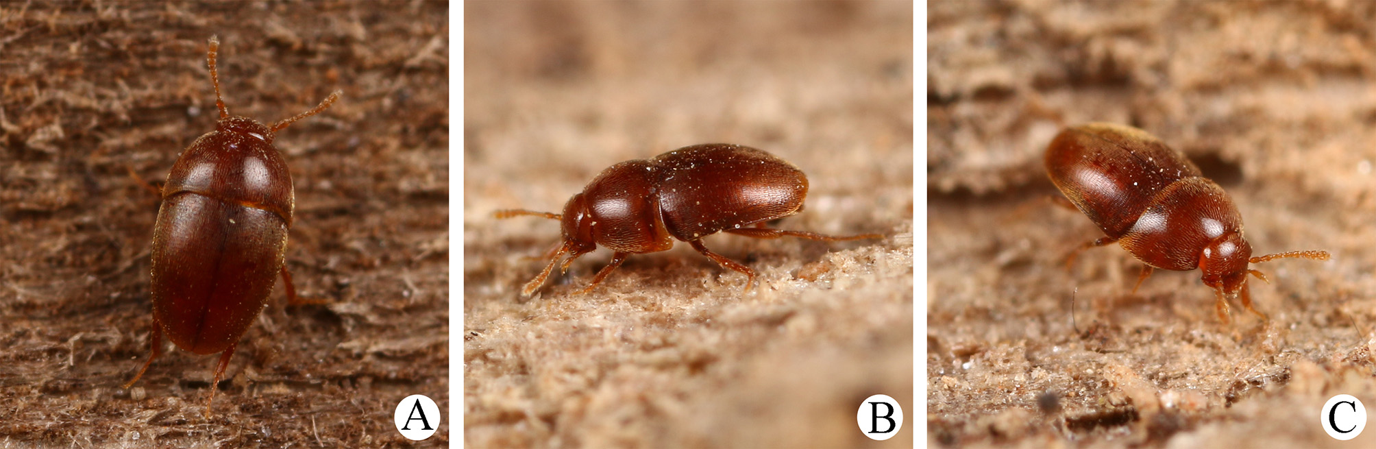
Habitus ( Figs. 1 View FIGURE 1 A–D) oval, much convex and sublustrous. Moderately pigmented: mostly brown; mouthparts, antennae and tarsi somewhat paler. Dorsum continuously clothed with fine, prostrate, yellowish pubescence. Insertions of pubescence on dorsal surfaces of elytra and femora aligned along superficial transverse striolations; interspace between two striolations microreticulate. Dorsal surfaces of head and pronotum finely and sparsely punctate, not transversely striolated; interspace microreticulate.
Head retractile, width/length = 1.4. Clypeus subtrapezoidal, front margin straight. Antennae ( Fig. 2 View FIGURE 2 A) slender and incompact, AL/HW = 1.4; antennomere I and II wide, about 1.4 times wider than antennomere III; antennomere VIII transverse, wider than twice of its length; antennomere IX and X nearly twice as wide as long; antennomere XI pear-like, length/width = 1.30.
Pronotum ( Fig. 3 View FIGURE 3 A) transverse, widest at base, width/length = 1.7. Sides gently arched, gradually narrowing from posterior to anterior; hind angles projected backwards and bluntly rounded. Posterior margin emarginate near hind angles.
Elytra oval, tightly combined with each other but can be separated using some force, as wide as long and widest at base. Sides evenly arched, gradually narrowing from bases to apices; apices ( Fig. 3 View FIGURE 3 F) almost obliquely truncated.
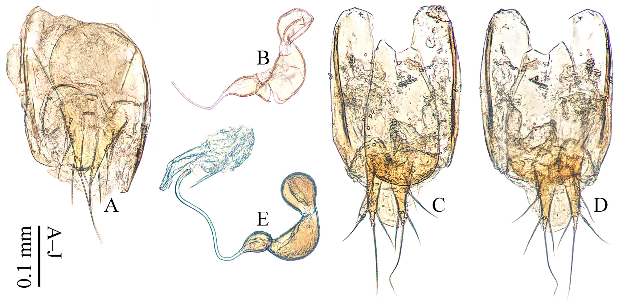
Abdominal ventrite VIII ( Fig. 4 View FIGURE 4 H) transverse, almost not emarginate on posterior margin, and a subrounded median patch strongly sclerotized posteriorly. Genital segment ( Fig. 4 View FIGURE 4 I) longer than wide, with spiculum gastrale poorly sclerotized, ill-defined; genital plate quite narrow; tergite IX rounded ventro-apically.
Prolegs ( Fig. 4 View FIGURE 4 A) short but strong, with protarsi pentamerous, basal three protarsomeres ( Fig. 4 View FIGURE 4 B) strongly expanded: (tibial width)/(basitarsal width) = 1.1. Protibiae strongly expanded towards apex. Profemora broad. Mesotibiae with inner side inconspicuously bisinuate, outer side with several big spines erecting from pubescence. Metatibiae straight, outer side with several small spines hidden in pubescence.
Aedeagus ( Fig. 5 View FIGURE 5 A) with median lobe slender, gradually narrowing apically and terminated to a lanceolate apex ( Fig. 5 View FIGURE 5 C) in dorsal view; parameres thick, gently convergent inwards, slightly expanded before apex and narrowly subrounded at apex, reaching the apex level of median lobe; two strong apical setae inserted on inner side, the posterior one distinctly longer and stronger than the anterior one, their arrangements as shown in Figs. 5 View FIGURE 5 E–H; basal lamella short. In lateral view ( Fig. 5 View FIGURE 5 B), median lobe regularly bent ventrad and acuminate at apex ( Fig. 5 View FIGURE 5 D); parameres with a rounded knob at apex ( Fig. 5 View FIGURE 5 H). Endophallus with two obvious bands of phanerae, a pair of small sclerites in the middle region and a U-shaped complex in the basal region.
Female. Similar to male in general appearance ( Figs. 1 View FIGURE 1 C–D), including elytral apices ( Fig. 3 View FIGURE 3 G), but distinct in the following characters: pronotum ( Fig. 3 View FIGURE 3 B) with hind angles narrower; protarsi ( Fig. 4 View FIGURE 4 D) tetramerous and simply linear; protibiae ( Fig. 4 View FIGURE 4 C) considerably narrower; ventrite VIII ( Figs. 4 View FIGURE 4 J, L) generally rounded at posterior edge, only slightly protruded in the median, spiculum ventrale quite narrow, nearly parallel-sided; tergite IX ( Fig. 6 View FIGURE 6 C) widely rounded at posterior edge, with four minute setae posteriorly; valvifer with one lateral seta; coxite ( Figs. 6 View FIGURE 6 C–D) with two subapical and two lateral setae; stylus minute ( Figs. 6 View FIGURE 6 C–D), cylindrical, with one long seta; spermatheca ( Fig. 6 View FIGURE 6 E) large, trilobed, and the middle one the largest.
Field observations. Specimens were found in an abandoned termite nest in rotten wood ( Fig. 7 View FIGURE 7 A–C), located near a regular latrine site of Meles meles (Linnaeus, 1758) (Mustelidae) or Nyctereutes procyonoides Gray, 1834 (Canidae) .
Distribution. China (Shanghai) ( Fig. 8 View FIGURE 8 ).
Etymology. The specific epithet is from the Chinese name (Pinyin) of the type locality “Tianmashan”, which means “flying horse”.
Diagnosis. This new species is similar to Sinobathyscia kurbatovi from Wuhan, Hubei, but with stable differences on the spiculum ventrale of female ventrite VIII, the most important character for identifying females of Cholevinae : in S. tianma sp. nov., the spiculum ventrale is narrow ( Figs. 4 View FIGURE 4 J, L); while in S. kurbatovi , the spiculum ventrale is much wider, about 1.4 times as wide as in the new species ( Figs. 4 View FIGURE 4 K, M).


| CMPR |
Centre for Medicinal Plants Research |
No known copyright restrictions apply. See Agosti, D., Egloff, W., 2009. Taxonomic information exchange and copyright: the Plazi approach. BMC Research Notes 2009, 2:53 for further explanation.
|
Kingdom |
|
|
Phylum |
|
|
Class |
|
|
Order |
|
|
Family |
|
|
Genus |