
Sinobathyscia Perreau, 1999, 2017
|
publication ID |
https://doi.org/ 10.11646/zootaxa.4303.3.2 |
|
publication LSID |
lsid:zoobank.org:pub:260F1C3F-4FC8-4DA4-8AEE-0DD11A99185F |
|
DOI |
https://doi.org/10.5281/zenodo.6030425 |
|
persistent identifier |
https://treatment.plazi.org/id/291D87F1-FF9F-CA6D-FF0A-FAA1FCDAF8E6 |
|
treatment provided by |
Plazi |
|
scientific name |
Sinobathyscia Perreau, 1999 |
| status |
|
Genus Sinobathyscia Perreau, 1999 View in CoL (ĢḆṁṂfflffi)
Perreau, 1999: 404 (SpecieS included: kurbatovi View in CoL ); Perreau, 2000: 170 (world catalogue; 1 SpecieS); Perreau, 2015: 210 (Palaearctic catalogue; 1 SpecieS).
Type species: Sinobathyscia kurbatovi Perreau, 1999 View in CoL , Fixed by original deSignation.
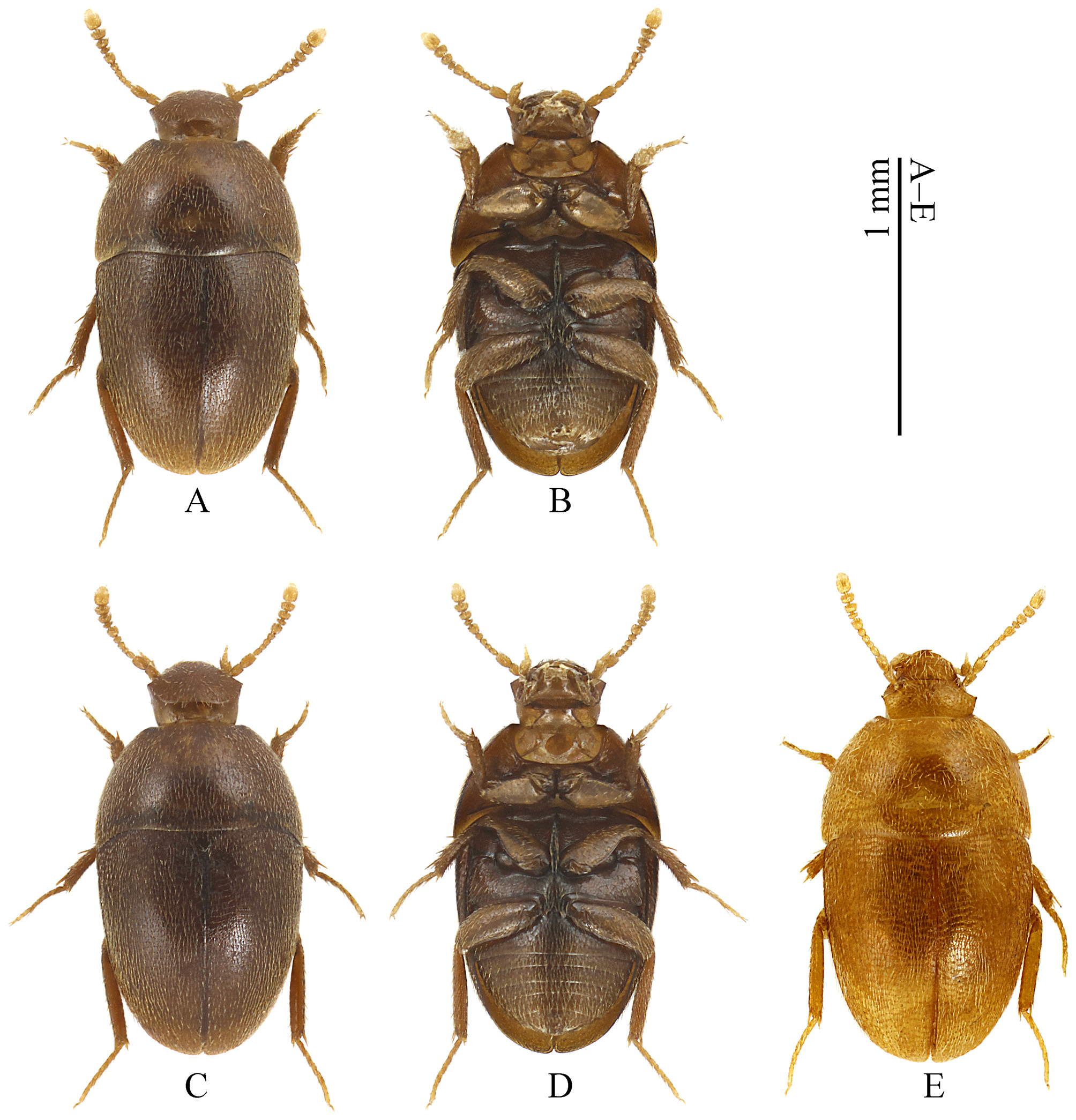
Redescription. Small size, less than 1.5 mm. Body ( Figs. 1 View FIGURE 1 A–E) Bathyscia- like, oval and rather convex. Dorsum consistently clothed with homogeneous fine, prostrate pubescence.
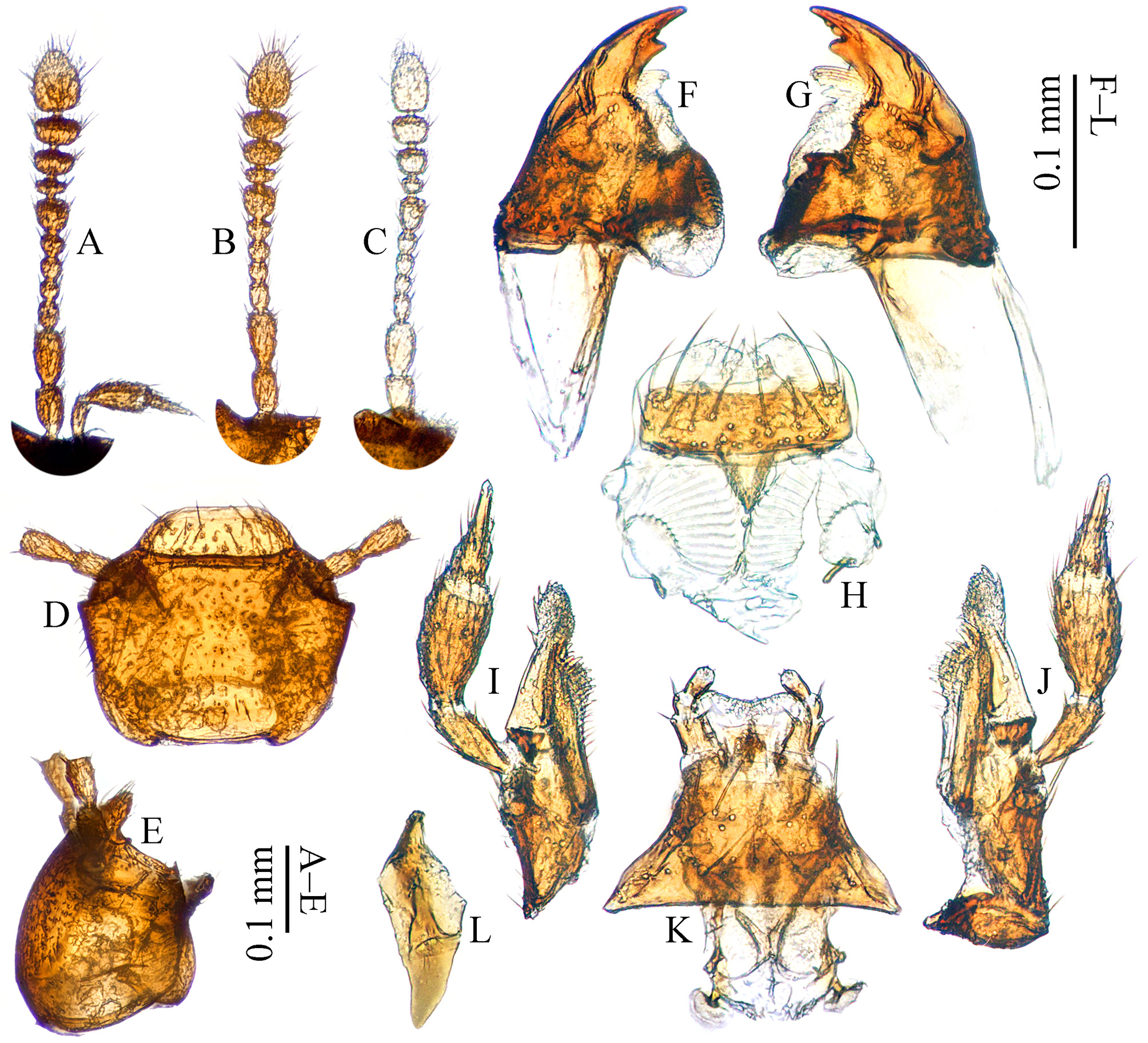
Head wider than long; clypeofrontal suture ( Fig. 2 View FIGURE 2 D) distinct. Anophthalmic, compound eyes ( Fig. 2 View FIGURE 2 E) completely absent. Occipital carina vestigial. Labrum ( Fig. 2 View FIGURE 2 H) transverse, rounded subrectangle; epipharyngeal area with torma. Left and right mandibles ( Figs. 2 View FIGURE 2 F–G) asymmetrical, each with a tooth on inner margin of subapex; subapical orthogonal penicillus made up of slender, fine setae; mola well-developed. Maxillary palp ( Figs. 2 View FIGURE 2 I–J) with penultimate palpomere slightly swollen, length/width = 1.7; ultimate palpomere quite slender, nearly as long as and about half as wide as penultimate palpomere. Labium as shown in Fig. 2 View FIGURE 2 K, premier labial palpomere only a little longer than ultimate palpomere, and ultimate palpomere nearly twice as long as penultimate palpomere. Antennae ( Figs. 2 View FIGURE 2 A–C) with basal two antennomeres much strong and nearly as long as each other. Cervical sclerite ( Fig. 2 View FIGURE 2 L) tooth-like, tapering posteriorly, apex subacute, length/width = 2.9.
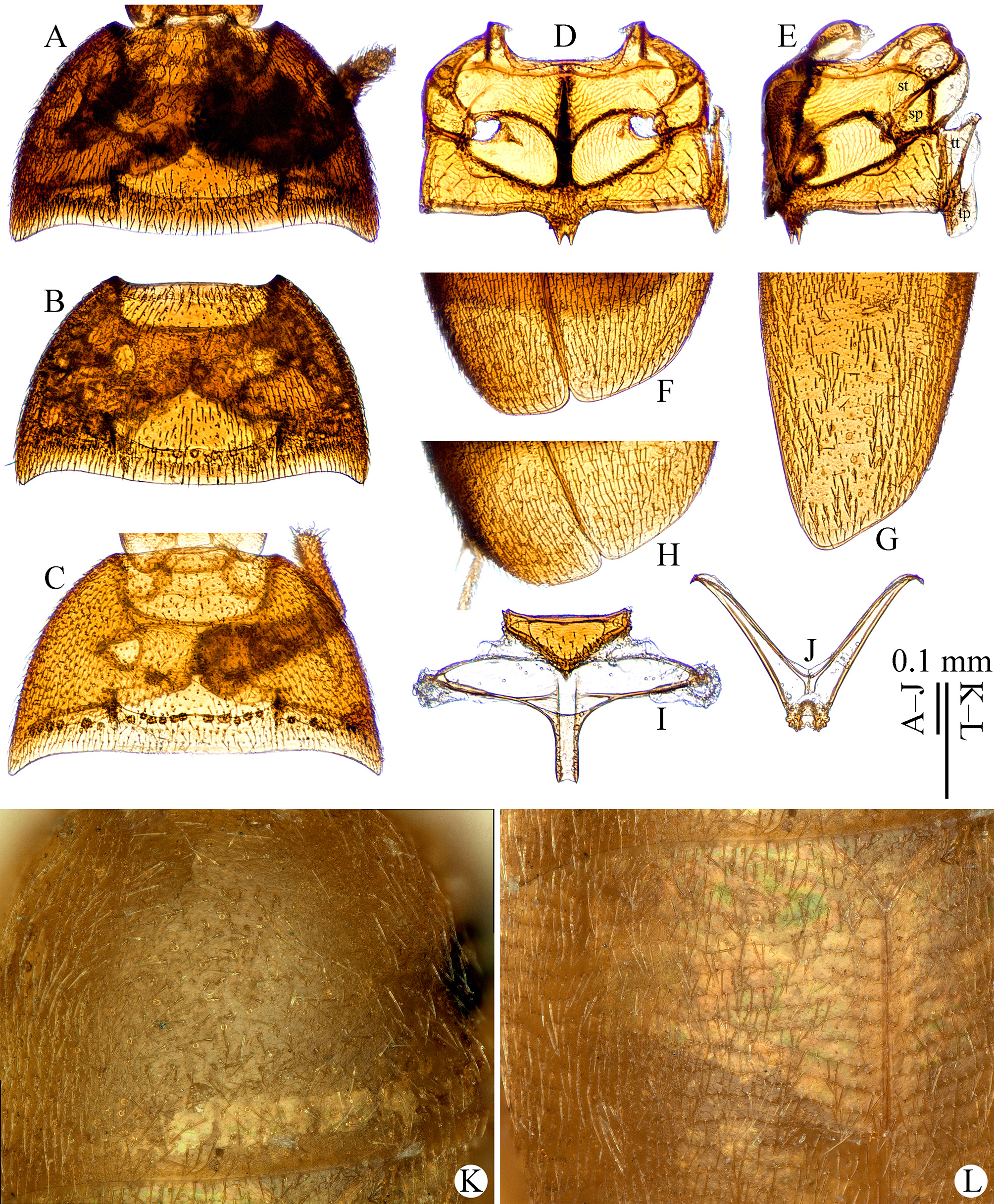
Pronotum ( Figs. 3 View FIGURE 3 A–C) widest at base, punctate and microreticulate but not transversely striolated ( Fig. 3 View FIGURE 3 K). Elytra not fused with each other, with fine transverse striolations ( Fig. 3 View FIGURE 3 L). Sutural striae absent. Epipleura commonly narrow. Metathoracic wings absent.
Scutellum much wide, tightly integrated with metanotum. Metanotum ( Fig. 3 View FIGURE 3 I) short but with moderately and narrowly elongated metatergal apophysis, reaching about the basal third of elytra. Metendosternite ( Fig. 3 View FIGURE 3 J) with stalk short, (stalk length)/(furcal arm width) = 0.3.
Meso- and metaventrite as shown in Figs. 3 View FIGURE 3 D–E, and shapes of mesepisternum, mesepimeron, metaepisternum and metepimeron can be fully seen in Fig. 3 View FIGURE 3 E; mesepimera wider than long, outer margin longer than inner margin. Mesoventral carina ( Fig. 3 View FIGURE 3 E) strongly developed, lamellate, high, somewhat thickened, and shortly extended under metaventrite. Metacoxal cavities distinctly separated.
Abdominal tergites ( Fig. 4 View FIGURE 4 G) only the last two sclerotized, the others membranous.
Protarsi pentamerous and expanded in male ( Fig. 4 View FIGURE 4 B), tetramerous and linear in female ( Figs. 4 View FIGURE 4 D, F). Protibiae with several small outer spines but not forming into a row ( Figs. 4 View FIGURE 4 A, C, E). Meso- and metatibiae without spine combs around apex. Outer spurs well-developed. Mesotarsi simply linear.
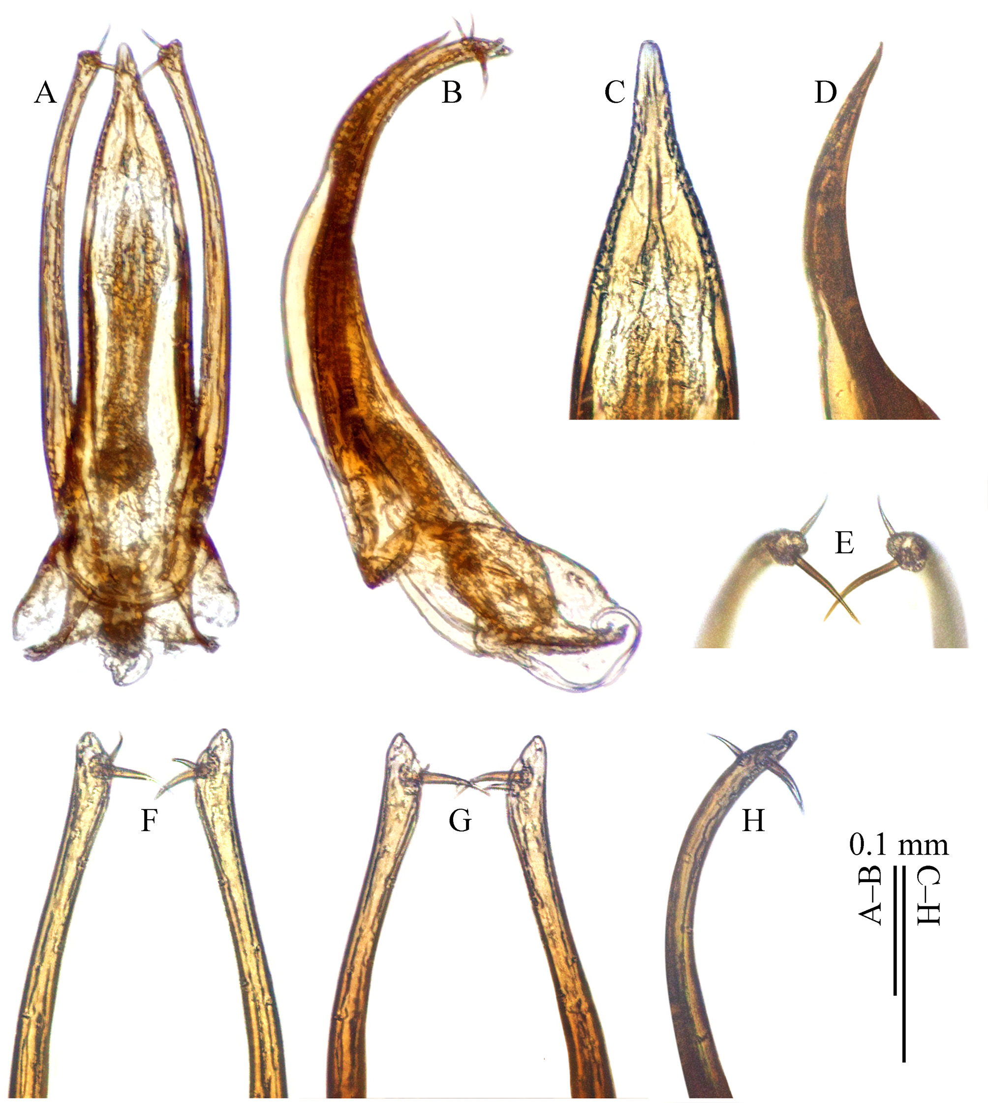
Male genital segment ( Fig. 4 View FIGURE 4 I) with spiculum gastrale poorly sclerotized, ill-defined. Aedeagus ( Fig. 5 View FIGURE 5 A) slender; parameres thick, reaching the apex level of median lobe; basal lama short; endophallus with two obvious bands of phanerae.
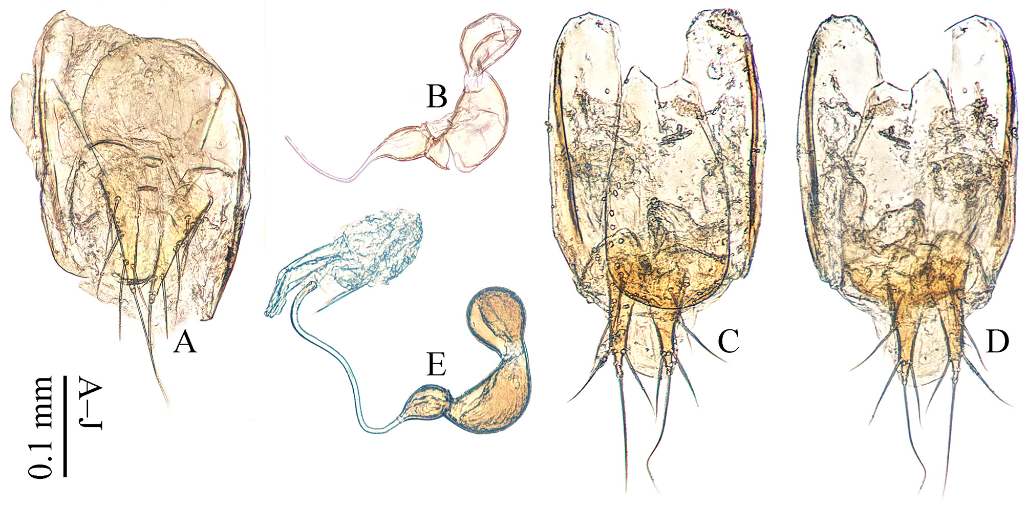
Spermatheca ( Figs. 6 View FIGURE 6 B, E) large, trilobed, the middle one the largest, and the two apical ones separated by a narrow unsclerotized region. Ovipositor with minute stylus.
Distribution. China (as in Fig. 8 View FIGURE 8 ).
Diagnosis. Sinobathyscia well resembles Coreobathyscia from South Korea, but the latter is larger than 2.0 mm in body length, antennae with basal two antennomeres distinctly more slender, parameres much exceeding the apex level of median lobe, endophallus without sclerites.


No known copyright restrictions apply. See Agosti, D., Egloff, W., 2009. Taxonomic information exchange and copyright: the Plazi approach. BMC Research Notes 2009, 2:53 for further explanation.
|
Kingdom |
|
|
Phylum |
|
|
Class |
|
|
Order |
|
|
Family |
Sinobathyscia Perreau, 1999
| Wang, Cheng-Bin, Perreau, Michel, Růžička, Jan & Song, Xiao-Bin 2017 |
kurbatovi
| Perreau 1999 |