
Deeveya bransoni Kornicker & Palmer 1987
|
publication ID |
https://doi.org/ 10.11646/zootaxa.1565.1.1 |
|
publication LSID |
lsid:zoobank.org:pub:A2CDD9CB-CA5E-418B-A471-9EEFDC5CCF16 |
|
persistent identifier |
https://treatment.plazi.org/id/2A5087FF-3E0F-FC44-3A91-FF0BFD3B6AE4 |
|
treatment provided by |
Felipe |
|
scientific name |
Deeveya bransoni Kornicker & Palmer 1987 |
| status |
|
Deeveya bransoni Kornicker & Palmer 1987
Figs. 27–39 View FIGURE 27 View FIGURE 28 View FIGURE 29 View FIGURE 30 View FIGURE 31 View FIGURE 32 View FIGURE 33 View FIGURE 34 View FIGURE 35 View FIGURE 36 View FIGURE 37 View FIGURE 38 View FIGURE 39
Deeveya bransoni Kornicker & Palmer 1987: 610 , figs. 1–5.— Kornicker et al. 1990: 37, figs. 22c, 23d,e, 29b, 30.—Kornicker et al. 2002: 12, figs. 7-12, 13a-k.
Holotype. USNM 193301 About USNM , A- 1 female on slide and in alcohol. Type locality. Evelyn Green’s Blue Hole, South Andros Island , Great Bahama Bank.
Material. Sanctuary Blue Hole, South Andros Island, Sta 99-061: USNM 1021408 About USNM , adult male on 4 slides and in alcohol; USNM 1021409–1021411 About USNM , 3 About USNM A-1 instars on 1 slide and in alcohol; USNM 1021413 About USNM , USNM 1022176 About USNM , 1 About USNM A-2 instar in alcohol; USNM 1021414 About USNM , 1 About USNM A-3 instar on 2 slides and in alcohol; USNM 1021415 About USNM , 1021416 About USNM , 2 About USNM A-3 instars in alcohol; USNM 1021417 About USNM , 1 About USNM A-5 instar in alcohol; USNM 1021418 About USNM , 1 About USNM A-5 instar on 1 slide and in alcohol; USNM 1021419 About USNM , 1 adult male in alcohol; USNM 1021420–1021424 About USNM , 5 About USNM A-1 instars in alcohol; USNM 1021425 About USNM , 1 About USNM A-4 right valve in alcohol; USNM 1021387 About USNM 1 adult male in alcohol (shell fragmented and not measured); USNM 1021427–1021429 About USNM , 3 adult females in alcohol; USNM 1022177 About USNM , 1 adult female in alcohol. Double Drop Blue Hole, South Andros Island: Sta 99-055 : USNM 1021430 About USNM , 1 adult female in alcohol; USNM 1021431 About USNM , 1 About USNM A-2 instar in alcohol (left valve broken); Sta 99-056 : USNM 1021432 About USNM , 2 About USNM A-2 instars in alcohol; USNM 1021433 About USNM , 2 About USNM A-1 instars in alcohol. Conch Sound Blue Hole, Andros Island: Sta 00- 023 , USNM 1021434 About USNM , 3 specimens in alcohol; USNM 1021435 About USNM , 1 specimen in alcohol. Sta 00-023, USNM 1021434 About USNM , 3 specimens in alcohol. Sta 00-024, USNM 1021435 About USNM , 1 specimen in alcohol. Sta 01-003: USNM 1021437 About USNM , 1 About USNM A-4 instar; USNM 10214438 About USNM , 1 About USNM A-5 instar in alcohol; USNM 1021439 About USNM , 27 specimens in alcohol. Sta 01-004, USNM 1021440 About USNM , 7 specimens in alcohol. Sta 01-012, USNM 1021441 About USNM , 8 specimens in alcohol.
Distribution. Great Bahama Bank: Andros Island (Evelyn Green’s Blue Hole, Stargate Blue Hole, Sanctuary Blue Hole, Double Drop Blue Hole, Conch Sound Blue Hole).
Remarks. Because many appendages of the adult male described in Kornicker et al. (2002: 15) were lost and thus incompletely described, a supplementary description of the adult male is presented herein.
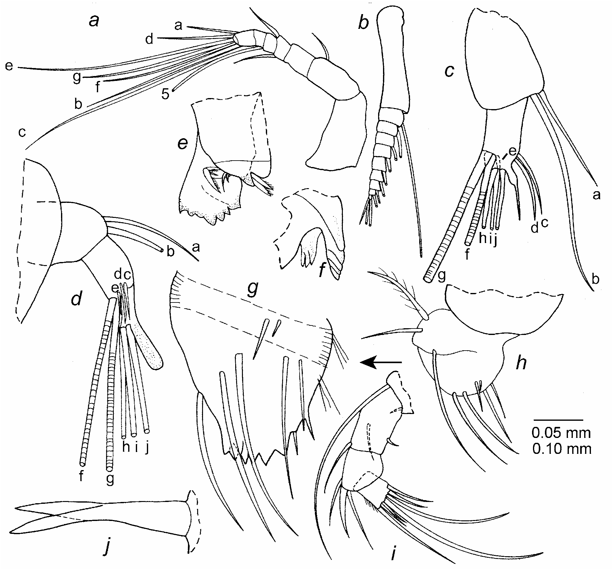
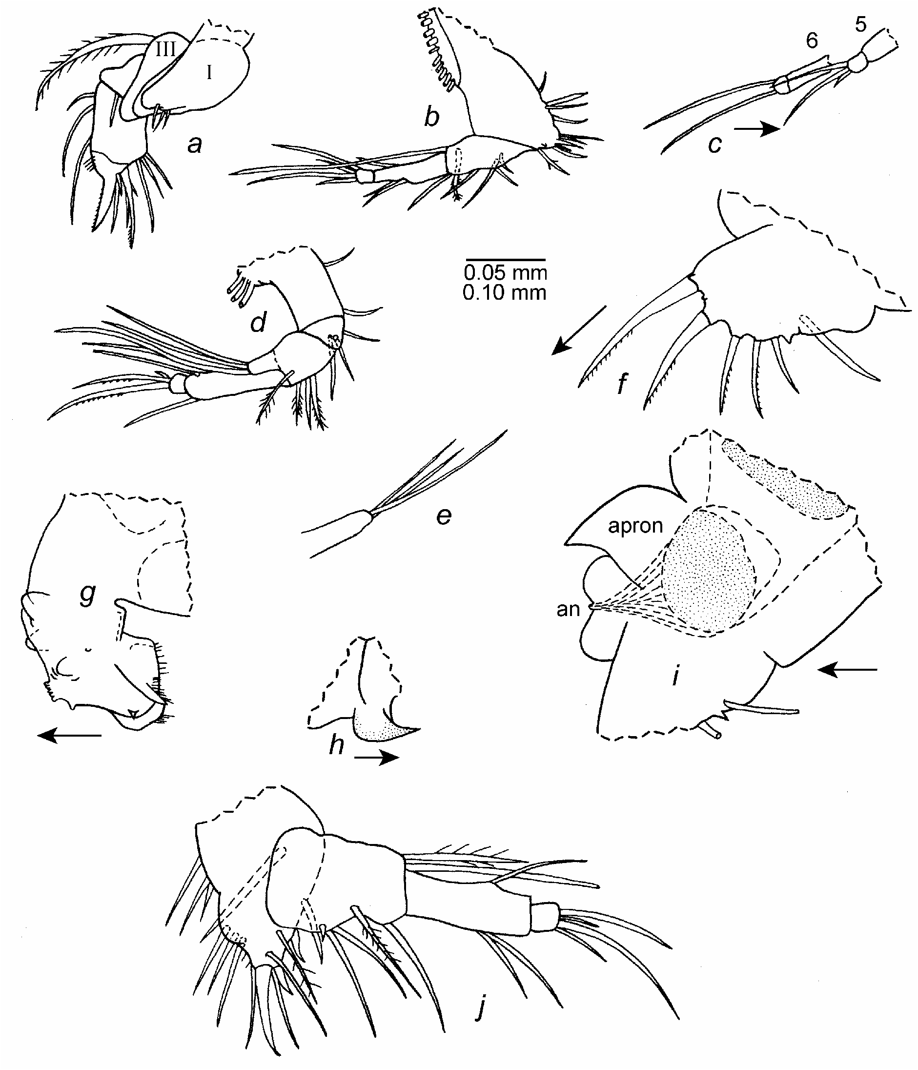
Supplementary description of adult male ( Figs. 27–32 View FIGURE 27 View FIGURE 28 View FIGURE 29 View FIGURE 30 View FIGURE 31 View FIGURE 32 ). Carapace similar in shape and ornamentation to that described in Kornicker et al. (2002:15: fig. 7 a–d) ( Fig. 27 a,b View FIGURE 27 ).
Central adductor muscle scars ( Fig. 27 a,b View FIGURE 27 ): With about 9 ovoid scars, 3 indistinctly divided.
Carapace size (length, height in mm): USNM 1021403, 2.00, 1.49; USNM 1021419, l.98, 1.38.
First antenna: As shown in Figs. 27 c,d View FIGURE 27 (appendage lost).
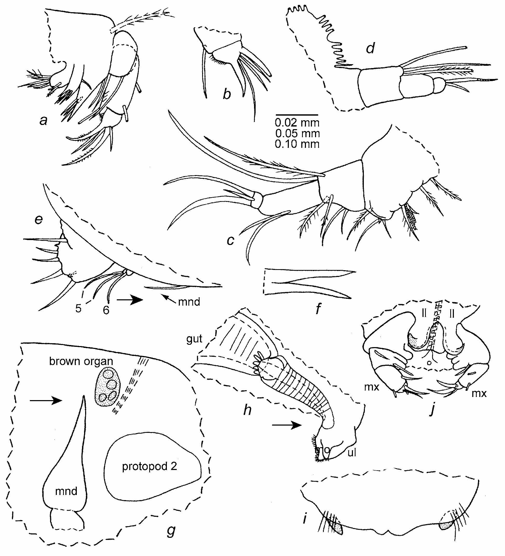
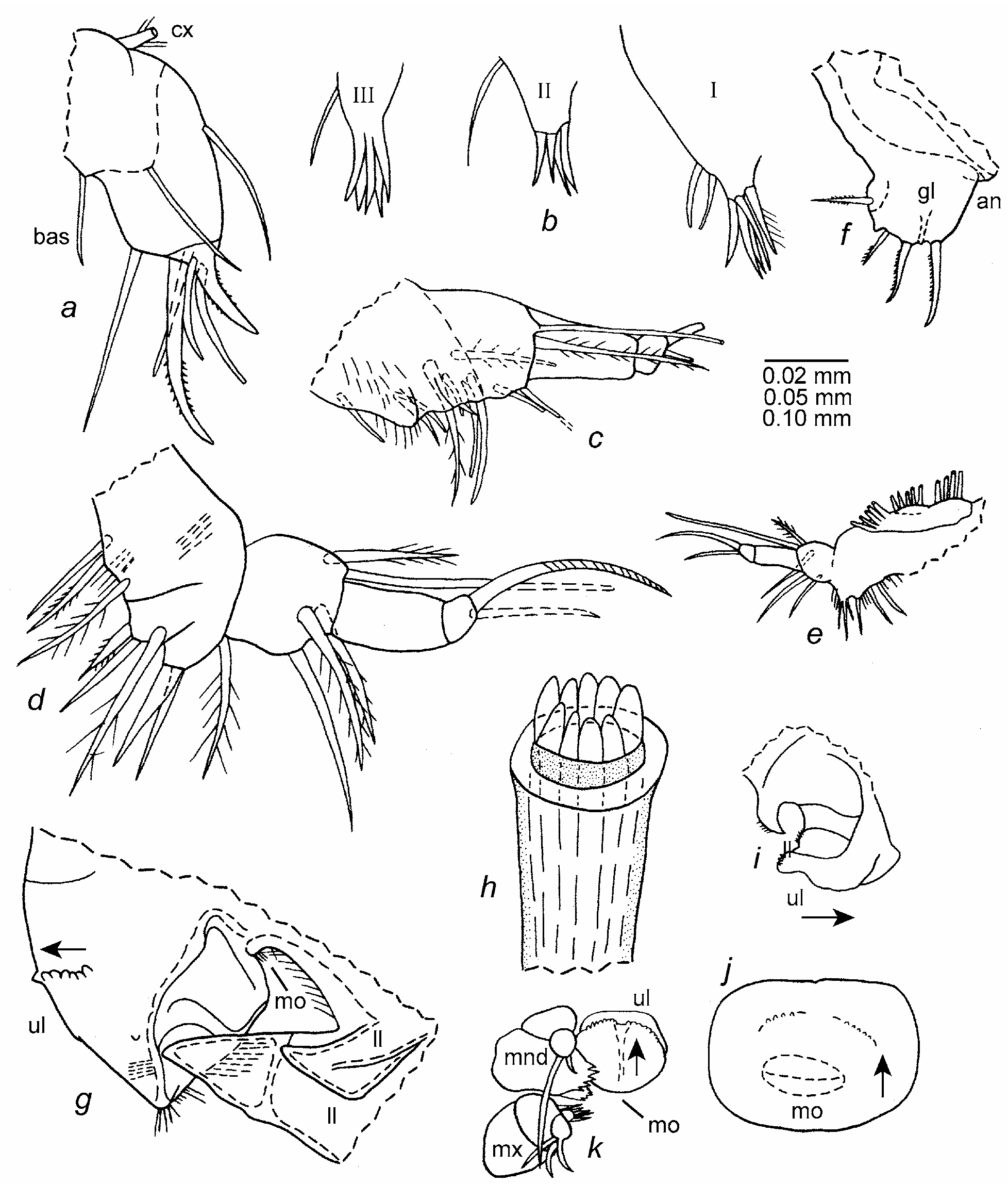
Second antenna ( Fig. 27 e–j View FIGURE 27 ): Protopod with distal lateral spines ( Fig. 27 g View FIGURE 27 ). Endopod 3-segmented ( Fig. 27 f,h,j View FIGURE 27 ): 1st and 2nd segments linear; 1st segment with 2 terminal spinous bristles (proximal a-bristle about one half length of distal b-bristle); 2nd segment with filament-like f-bristle, about one-half length of g-bristle; g-bristle with more-strongly developed rings than f-bristle; both f- and g-bristles with widely separated minute spines and with tapered tips with terminal papilla; 3rd segment small, separated from 2nd segment by suture, with filament-like h-, i-, and j-bristles (each less than one-half length of g-bristle and with tapering tip and terminal papilla) and with minute medial ventral peg and small medial bristle near midwidth ( Fig. 27 f,h,j View FIGURE 27 ). Exopod 9-segmented ( Fig. 27 i View FIGURE 27 ): 1st segment divided into long proximal and short distal parts; distal part with short ringed bristle with short marginal spines ( Fig. 27 i View FIGURE 27 ); bristle of 2nd segment long, with short ventral spines and dorsal natatory hairs; 9th segment with 4 bristles (longest bristle with dorsal spines and distal natatory hairs, next-to-longest bristle with only minute spines; shorter 2 bristles either bare or with minute spines). Fulcrum attached to posterior edge of protopod more strongly sclerotized along dorsal edge ( Fig. 27 e View FIGURE 27 ).
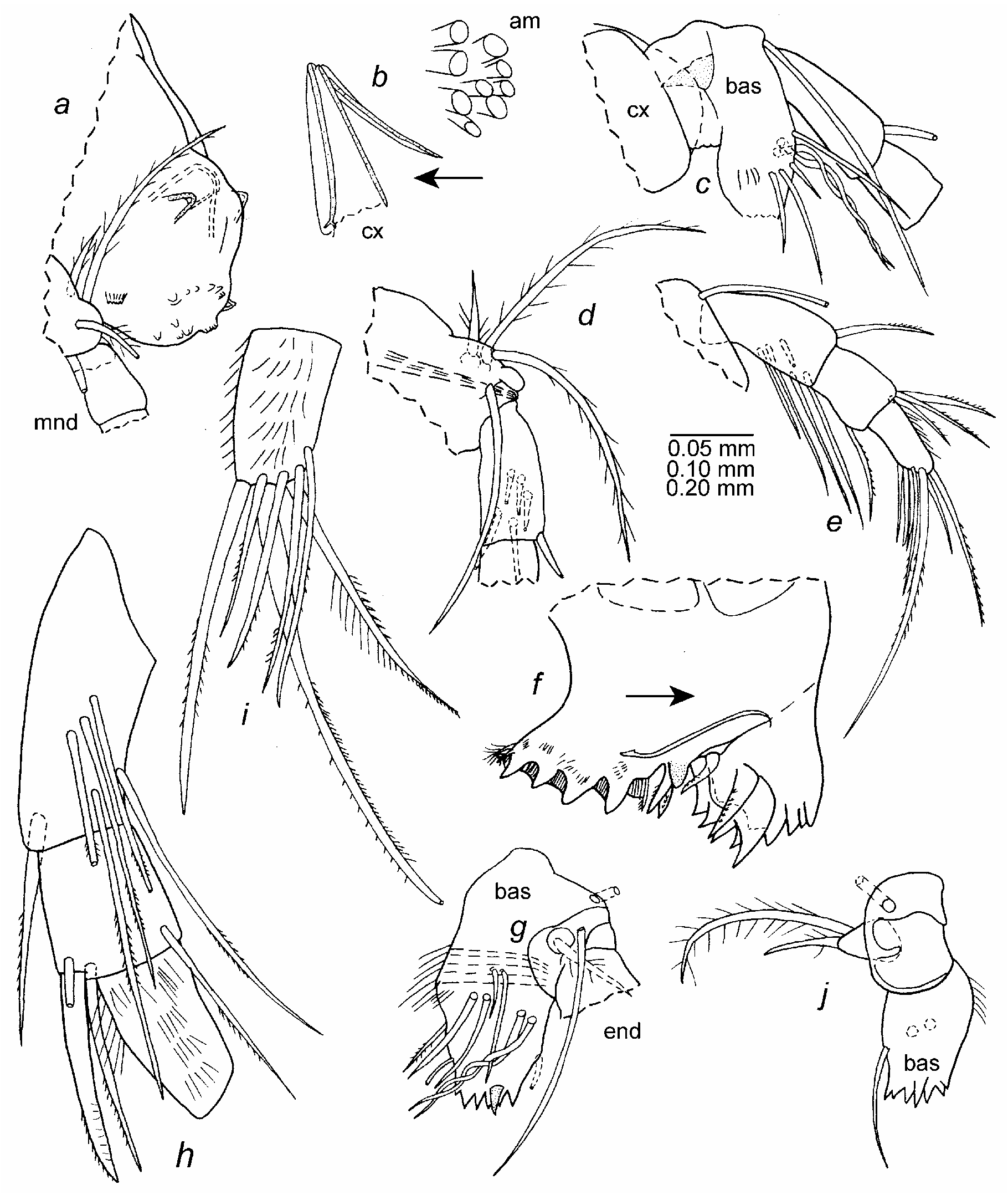
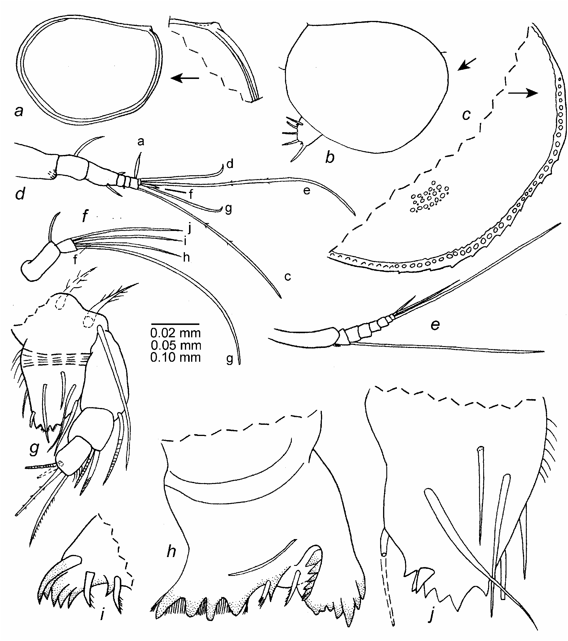
Mandible ( Fig. 28 View FIGURE 28 ): Coxa endite with proximal and distal sets of teeth separated by space ( Fig. 28 f View FIGURE 28 ); proximal set comprising 4 stout cusps, and with 2 short, spinous, distal bristles on anterior edge and 1 short, spinous, distal bristle on posterior edge; surface between cusps and anterior and posterior to cusps with abundant slender spines. Stout rounded tooth between proximal and distal sets of teeth, with 2 spinous bristles adjacent to tooth. Distal set of teeth comprising 2 flat teeth; proximal tooth with 5 cusps (posterior cusp stouter); distal tooth with 6 or 7 cusps (middle cusp stouter); 2 spinous bristles present lateral to distal set of teeth. Basis with 2 proximal bristles (1 stout plumose, 1 long, slender, either bare or with short spines), and 2 medial plumose bristles (1 long, 1 short) on short process ( Fig. 28 d, j View FIGURE 28 ). Basis endite ( Fig. 28 g View FIGURE 28 ): anterior margin with single bristle; posterior margin with proximal hairs, and 1 short bristle proximal to 1 short distal tubular bristle; medial side with few rows of long hairs near midlength and also just proximal to terminal cusps; lateral side with 6 slender distal bristles (2 longer than others and entwined with 5 crossings) and 1 short, stout tooth just proximal to distal edge of endite; ventral edge of endite with 6 terminal cusps (5 anterior cusps ser- rate proximally, 1 posterior cusp smaller than others). Endopod 3-segmented ( Fig. 28 e,h,i View FIGURE 28 ): 1st segment with 1 spinous, terminal, dorsal bristle, 1 spinous, distal, ventral bristle, and 4 spinous distal medial bristles (left limb of USNM 1021408 with additional lateral bristle); 2nd segment with 3 spinous, terminal, dorsal bristles (1 claw-like on edge of segment and with distal rings), and 1 spinous, terminal, ventral bristle; 3rd segment hirsute medially and along anterior margin, with 4 medial spinous bristles forming row, and 3 stout terminal bristles (middle bristle longest and with smooth, pointed, slightly recurved tip; posterior bristle with long spines at midlength).
Maxilla ( Figs. 29 View FIGURE 29 , 30 View FIGURE 30 ): Endite I with 2 proximal long bristles with long proximal hairs and 10 subterminal and terminal bristles (5 tubular, 5 stout, pointed, with either long hairs, minute spines, or minute teeth) ( Fig. 29 d View FIGURE 29 ); endite II with 2 long proximal bristles with short spines and 11 terminal and subterminal bristles (5 tubular, 6 claw-like with distal marginal teeth) ( Fig. 29 e View FIGURE 29 ); endite III with 1 long proximal bristle with base near basis and with short marginal spines, and 6 terminal bristles (2 tubular, 4 claw-like) ( Fig. 29 f View FIGURE 29 ). Coxa with stout plumose dorsal bristle. Basale with 2 bristles (1 tubular near ventral margin, 1 pointed near dorsal margin, both with minute indistinct marginal spines) ( Fig. 30 View FIGURE 30 ). Endopod ( Fig. 30 View FIGURE 30 ): 1st segment with 11 bristles (3 tubular); 2nd segment with hairs along anterior surface, 2 stout pectinate claws, and 6 tubular bristles (3 medial, 3 lateral).
Fifth limb ( Fig. 31 a–c View FIGURE 31 ): Epipod with bristles forming 3 groups: ventral group with 5 bristles; middle group with 6 bristles; dorsal group fragmented on specimen (with at least 3 bristles). Protopod with internal glandular field. Precoxa and coxa with total of 29 claws and bristles. Basis with 10 bristles. Exopod represented by 2 bristles (1 long bare, 1 short spinous). Endopod: 1st segment with 5 bristles (4 ventral, 1 dorsal); 2nd segment with 2 stout claw-like bristles and 2 slender bristles.
Sixth limb ( Fig. 31 d View FIGURE 31 ): Epipod with bristles forming 3 groups (ventral group with 5 long setose bristles; middle group with 6 long setose bristles; dorsal group with 7 setose bristles (6 long, 1 short dorsal). Precoxa and coxa with total of 8 bristles. Basis with 1 lateral and 6 ventral or medial bristles. Exopod well-developed, with 4 long bristles (1 bare, 3 with long marginal hairs). Endopod: 1st segment with 6 bristles on or near ventral margin; 2nd segment with 3 bristles (2 ventral, 1 dorsal); 3rd segment with 2 stout claw-like pectinate bristles and 2 slender bristles (1 long, ventral, 1 short, medial).
Seventh limb ( Fig. 31 e View FIGURE 31 ): Limb with 3 terminal bristles (1 long, 2 shorter).
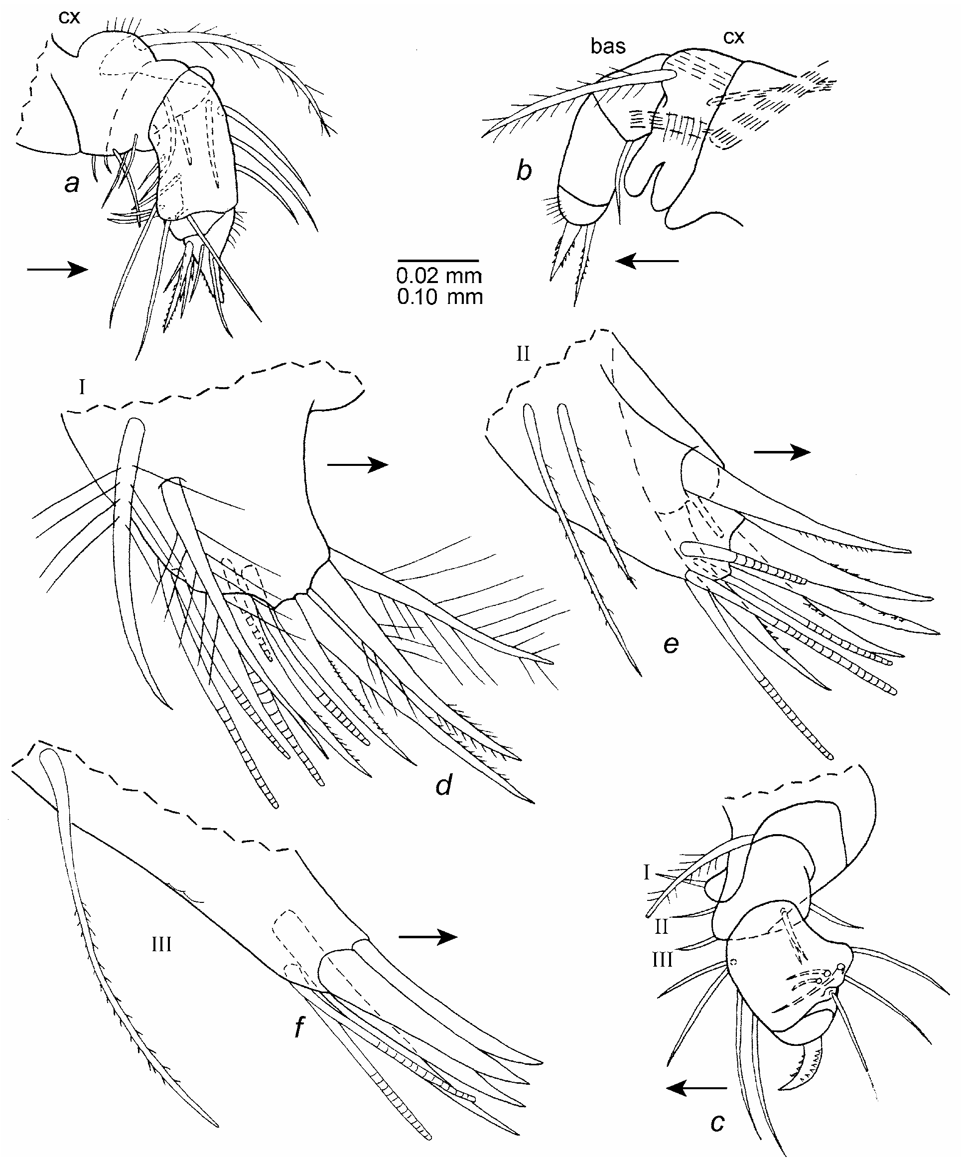
Furca ( Figs. 31 f View FIGURE 31 , 32 c View FIGURE 32 ): Each lamella with 7 claws (claw 4 slight smaller than claw 3); claws 1–4 with teeth along posterior margin; claws 5–7 with teeth along both margins. Small glandular peg present between claws 1 and 2. Apron bearing muscle present dorsal to anterior edge of lamellae; 2 lobes without internal muscles present between apron and lamellae ( Fig. 32 c View FIGURE 32 ): Lamellae followed by unpaired spinous bristle about same length as claw 3.
Bellonci Organ ( Fig. 27 c View FIGURE 27 ): Well developed, bifurcate at midlength, with each branch tapering to point.
Lips : Upper lip as shown in Figs. 28 a View FIGURE 28 , 32 a,b View FIGURE 32 . Lower lip a beak-shaped process on each side of mouth ( Fig. 32 b View FIGURE 32 ).
Copulatory organ ( Fig. 32 c–g View FIGURE 32 ): Similar to that illustrated in Kornicker et al. (2002: fig. 8g –j) in having 2 tubular processes at tip of anterior branch, but some variability observed in shape and distribution of minute teeth on large flat tooth ( Fig. 32 d,e,g View FIGURE 32 ).
Posterior of body ( Fig. 32 c View FIGURE 32 ): Unsegmented; posterodorsal edge of posterior of specimen with fringe and small spine, but both could be debris.
Remarks. The third segment of the first antenna of the adult male described herein ( Fig. 27 c View FIGURE 27 ) is more elongate than the third segment of the A- 1 female illustrated by Kornicker et al. (1990: fig. 22c).
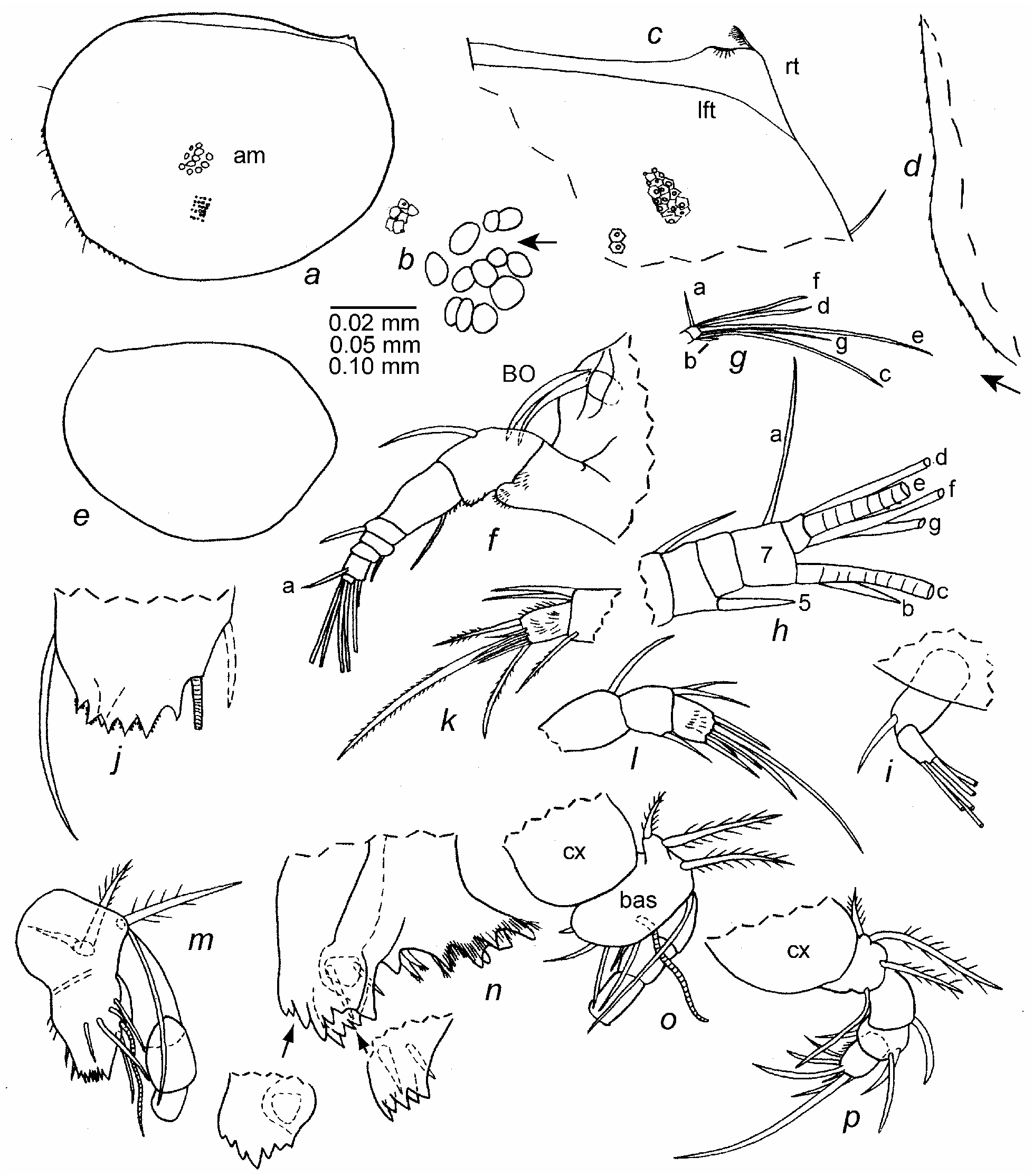
Supplementary description of adult female ( Figs. 33 a–d View FIGURE 33 , 39 f View FIGURE 39 ). Carapace shape similar to that of adult female illustrated in Kornicker et al. (2002: fig. 10a) ( Fig. 33 a–d View FIGURE 33 ). Ornamentation similar to that of adult male ( Fig. 39 f View FIGURE 39 ).
Central adductor muscle attachments ( Fig. 33 a,b View FIGURE 33 ): Consisting of about 12 ovoid scars.
Carapace size (length, height in mm): USNM 1021430, 2.04, 1.56; USNM 1022177, 2.07, 1.57; USNM 1021427, 1.95, 1.44; USNM 1021428, 2.07, 1.44; USNM 1021429, 2.01, 1.36. Furca: Each lamella with 7 claws. Lamellae followed by unpaired bristle. Supplementary description of A-1 instar (sex not determined unless noted). Carapace shape and ornamentation ( Fig. 39 e View FIGURE 39 ): Similar to that of adult male.
Carapace size (length, height in mm): USNM 1021409 About USNM , female, 1.59, 1.17; USNM 1021410 About USNM , female, 1.49, 1.16; USNM 1021411 About USNM , male, 1.59, 1.18; USNM 1021420 About USNM , male, 1.53, 1.20; USNM 1021421 About USNM , 1.58 About USNM , 1.16 About USNM ; USNM 1021422 About USNM , 1.61 About USNM , 1.22 About USNM ; USNM 1021423 About USNM , 1.65 About USNM , 1.19 About USNM ; USNM 1021424 About USNM , 1.57 About USNM , 1.12 About USNM ; USNM 1021433 About USNM , 2 specimens : 1.62, 1.22; 1.59, 1.19.
Furca. Each lamella with 7 claws. Lamellae followed by unpaired bristle.
Supplementary description of A-2 instar (sex not determined). Carapace similar in shape and ornamentation ( Fig. 39 d View FIGURE 39 ) to that of adult male.
Carapace size (length, height in mm): USNM 1021413 About USNM , 1.20 About USNM , 0.92; USNM 1021431 About USNM , 1.17 About USNM , 0.92; USNM 1022178 About USNM , 2 specimens : 1.21, 0.90; 1.17, 0.90.
Second antenna: 9th exopod segment with 4 bristles.
Maxilla: 2nd endopod segment with 2 claws and 4 bristles.
Sixth limb: Precoxa and Coxa with 5 bristles. Basis with 6 bristles.
Furca: Each lamella with 6 claws followed by small triangular process with pointed or bifid tip (incipient 7th claw). Lamellae followed by unpaired bristle.
Description of A-3 instar (sex unknown) ( Figs. 33 e–p View FIGURE 33 , 34 View FIGURE 34 , 39 c View FIGURE 39 ). Carapace similar in shape and ornamentation to that of adult male ( Figs. 33 e View FIGURE 33 , 39 c View FIGURE 39 ).
Carapace size (length, height in mm): USNM 1021414, 0.94, 0.72; USNM 1021415, 0.90, 0.72; USNM 1021416, 0.91, 0.71.
First antenna ( Fig. 33 f–h View FIGURE 33 ): Differs from A-2 instar mainly in having short ventral bristle on 5th segment (just reaching 8th segment), and short b-bristle on 7th segment (about same length as combined lengths of segments 6 and 7). Also, suture separating 3rd and 4th segments only weakly developed. c-bristle slightly shorter than e-bristle; e-bristle long, ringed, and with widely separated marginal spines; d-, f-, and g-bristles filament-like, about ½ length of e-bristle.
Second antenna ( Fig. 33 i View FIGURE 33 ): Endopod differs from A-2 instar in having only 1 dorsal bristle on 1st segment. 9th exopod segment with 3 bristles (1 long, 1 medium, 1 short).
Mandible: Coxa similar to that of A-2 instar ( Fig. 33 n View FIGURE 33 ). Basis ( Fig. 33 j,m,o,p View FIGURE 33 ) with only 1 of the 2 entwined lateral distal bristles present on the A-2 instar ( Fig. 33 m,o View FIGURE 33 ). Endopod ( Fig. 33 k–m,o,p View FIGURE 33 ): 1st segment with 1 dorsal bristle; 2nd segment with 1 ventral and 2 dorsal bristles; 3rd segment with 3 short medial bristles forming row and 3 stout terminal bristles (ventral of these bare or with short spines; middle bristle longest, with smooth, pointed, slightly recurved tip and marginal spines; dorsal bristle straight spinous, with short marginal spines).
Maxilla ( Fig. 34 a View FIGURE 34 ): Endite I with 2 proximal and about 7 terminal bristles (2 or 3 tubular); endite II with 1 proximal and about 7 terminal bristles (1 tubular); endite III with 1 proximal and about 5 terminal bristles (1 tubular). Coxa with stout spinous dorsal bristle. Basis with 2 terminal bristles. Endopod: 1st segment with 3 anterior bristles and 3 distal posterior bristles; 2nd segment with hairs along anterior margin, 2 stout, pectinate, terminal claws, and 4 slender tubular bristles.
Fifth limb ( Fig. 34 b,c,f,j View FIGURE 34 ): Epipod: dorsal group with 4 long bristles; middle group with 6 long bristles; ventral group with 4 long and 1 short bristle. Precoxa and coxa with 15 bristles. Basis with 5 ventral bristles. Exopod represented by 2 bristles (1 long, bare, 1 short, spinous). Endopod: 1st segment with 2 ventral and 1 dorsal bristle; 2nd segment with 2 long and 2 short bristles.
Sixth limb ( Fig. 34 c,d View FIGURE 34 ): Tip of limb extends well past tip of 5th limb ( Fig. 34 c View FIGURE 34 ). Epipod: dorsal group with 5 long bristles; middle group with 6 long bristles; ventral group with 1 short and 4 long bristles. Precoxa and coxa with 5 ventral bristles. Basis with 4 bristles. Exopod with 4 long bristles. Endopod: 1st and 2nd segments fused, with 2 bristles (1 dorsal, 1 ventral) on 2nd segment; 3rd segment with 2 stout claw-like pectinate bristles and 1 short slender bristle.
Seventh limb ( Fig. 34 e View FIGURE 34 ): With 3 terminal bristles (1 long, 2 shorter).
Furca ( Fig. 34 f,i View FIGURE 34 ): Each lamella with 5 claws followed by small triangular process with pointed tip (incipient 6th claw). Lamellae followed by unpaired bristle. Small lateral glandular process between claws 1 and 2. Apron similar to that of adult. (On specimen studied apron separated from furca by 2 lobes, 1 on each side of anus ( Fig. 34 i View FIGURE 34 )).
Bellonci Organ ( Fig. 33 f View FIGURE 33 ): Bifurcate distally, with branches tapering to pointed tip. Lips : Upper lip ( Fig. 34 g View FIGURE 34 ) and lower lip ( Fig. 34 h View FIGURE 34 ), similar to those of later instars and adults. Gut content: Gut filled with unidentified amber-colored particulate matter. Description of A-4 instar (sex unknown) ( Figs. 35 View FIGURE 35 , 36 View FIGURE 36 , 39 b View FIGURE 39 ). Carapace similar in shape to A-3 instar ( Fig. 35 a–c View FIGURE 35 ). Right valve with small projecting glandular process at posterior end of dorsal margin; process with small bristle.
Ornamentation ( Figs. 35 c View FIGURE 35 , 37 b View FIGURE 37 ): Surface in transmitted light with bright round spots inside very weakly developed reticulations ( Fig. 39 b View FIGURE 39 ). Smaller round bright spots present between larger bright spots, few bristles along valve margins. The spots form concentric circles on the valve surface.
Carapace size (length, height in mm): USNM 1021425, right valve, 0.73, 0.57; USNM 1021437, 0.74, 0.59.
First antenna ( Fig. 35 d View FIGURE 35 ): 1st segment with spines in distal ventral corner. 2nd segment with 1 dorsal bristle. 3rd and 4th segments fused but indentation on ventral margin indicates place of separation; 3rd segment with 1 ventral bristle; 4th segment with 1 short dorsal bristle. 5th segment bare or with minute ventral terminal nub. 6th segment bare. 7th segment with short a-bristle and long c-bristle with minute widely separated marginal spines. 8th segment with medium d-bristle, long e-bristle with widely separated minute spines and with terminal papilla, medium g-bristle with terminal papilla, and very short f-bristle. 1st and 2nd segments linear.
Second antenna ( Figs. 35 e,f View FIGURE 35 , 36 g View FIGURE 36 ): Exopod with 1st segment not divided into 2 parts, with small terminal ventral bristle. Segments 2 to 8 with long ventral bristle. Segment 9 with 2 bristles (1 medium, 1 small). Endopod: segment 1 with 1 dorsal bristle; segment 2 with short f-bristle and long g-bristle; segment 3 fused to segment 2, with long h-, i-, and j-bristles.
Mandible ( Figs. 35 g –j View FIGURE 35 , 36 g View FIGURE 36 ): Coxa endite with proximal and distal sets of teeth separated by space ( Fig. 35 h,i View FIGURE 35 ): proximal set comprising 4 stout cusps and with 2 short spinous bristles (1 on anterior edge, 1 on posterior edge); surface between cusps with abundant slender spines; stout pointed tooth between proximal and distal sets of teeth, with 1 spinous bristle lateral to tooth. Distal set of teeth comprising 2 flat teeth; proximal and distal teeth each with 6 cusps; stout lateral bristle proximal to proximal tooth. Basis with 2 plumose medial bristles near dorsal margin and 1 long slender lateral bristle near endopod ( Fig. 35 g View FIGURE 35 ). Basis endite: left limb ( Fig. 35 j View FIGURE 35 ): anterior margin with single bristle; posterior margin with proximal hairs and 1 short bristle proximal to short distal tubular bristle; lateral side with 3 bristles (1 long posterior to midwidth, 1 long near posterior margin, 1 short proximal and anterior to long bristles; none entwined); ventral edge of endite with 6 cusps; single lateral tooth near distal edge of endite. Right limb (probably aberrant) ( Fig. 35 g View FIGURE 35 ): differs from left limb in having basis endite with only 4 terminal teeth and no distal lateral tooth). Endopod with 3 segments ( Fig. 35 g View FIGURE 35 ): 1st segment with 1 dorsal terminal bristle; 2nd segment with 2 dorsal terminal bristles (1 claw-like); 3rd segment hirsute medially and along anterior margin, with 1 medial bristle and 3 terminal bristles.
Maxilla ( Fig. 36 a,b,j View FIGURE 36 ): Endite I with 2 proximal and 5 terminal bristles (1 tubular); endite II with 1 proximal and 4 or 5 terminal bristles (2 tubular); endite III with 1 proximal and 4 terminal bristles (1 tubular). Coxa with 1 plumose dorsal bristle. Basis with 1 ventral bristle and 1 bristle at midwidth. Endopod with 2 segments: 1st segment with 2 anterior and 1 posterior bristle. 2nd segment with 2 claw-like bristles and 3 slender bristles; ventral edge of segment bare or with spines.
Fifth limb ( Fig. 36 c,e View FIGURE 36 ): Epipod with plumose bristles forming 3 groups: 4 bristles in dorsal group and 6 in middle group; ventral group fragmented, with 2 bristles on remaining part. Precoxa and coxa with total of 11 bristles and claws (2 bristles tubular, 1 claw short tooth-like). Basis with 4 bristles. Exopod represented by 2 long bristles (1 bare, 1 plumose). Endopod with 2 segments: 1st segment with 2 distal ventral bristles; 2nd segment with 2 slender claw-like bristles and 1 short slender bristle.
Sixth limb ( Fig. 36 d,e View FIGURE 36 ): Epipod with plumose bristles forming 3 groups: 4 bristles in dorsal and middle groups, 5 in ventral group (ventral bristle short). Precoxa and coxa fragmented during dissection; no bristles observed prior to dissection [instar A-4 of D. jillae bears 1 bristle ( Kornicker & Iliffe 1985: Fig. 15h View FIGURE 15 )]. Basis without bristles. Exopod represented by small lobe with 3 long bristles (inner bristle plumose, others bare). Endopod with 2 segments: 1st segment bare; 2nd segment with 2 terminal bristles. Terminal segment not extending past distal segment of 5th limb ( Fig. 36 e View FIGURE 36 ). Sutures between segments indistinct.
Seventh limb: Absent.
Furca ( Fig. 36 e View FIGURE 36 ): Each lamella with 4 claws followed by small triangular process fused to lamella. Claws separated from lamella by suture. Small projecting lateral glandular process present midway between claws 1 and 2. Unpaired lateral bristle present posterior to lamellae.
Bellonci Organ ( Fig. 36 f View FIGURE 36 ): Well developed, bifurcate distally, branches taper to point.
Lips : Upper lip ( Fig. 36 h,i View FIGURE 36 ) and lower lip ( Fig. 36 j View FIGURE 36 ), in general, similar to those of A-3 instar. Dorsal end of esophagus with transparent flap-like structures ( Fig. 36 h View FIGURE 36 ).
Brown Organ ( Fig. 36 g View FIGURE 36 ): With 4 ovoid cells.
Gut content: Unidentified matter, somewhat stringy.
Description of A-5 instar (sex unknown) ( Figs. 37–39 a View FIGURE 37 View FIGURE 38 View FIGURE 39 ). Carapace similar in shape to that of A-3 instar ( Fig. 37 a,c View FIGURE 37 ).
Ornamentation ( Figs. 37 a,b View FIGURE 37 , 39 a View FIGURE 39 ): Valve viewed in transmitted light with closely spaced large round bright spots appearing inside very weakly developed reticulations ( Fig. 39 a View FIGURE 39 ). Few single bristles along valve edge.
Infold ( Fig. 37 c View FIGURE 37 ): Broad transparent infold along anterior, ventral, and posterior margins.
Carapace size (length, height in mm): USNM 1021418, 0.57, 0.44; USNM 1021417, 0.59, 0.49; USNM 1021438, 0.56, 0.46.
First antenna ( Fig. 37 d View FIGURE 37 ): Differs from A-3 instar in having 3rd and 4th segments fused (without suture separating them), and in lacking a dorsal bristle on the 2nd segment, a ventral bristle on the 3rd segment, a dorsal bristle on the 4th segment, a ventral bristle on the 5th segment, a- and b-bristles on the 7th segment, and an f-bristle on the 8th segment. Also, the c-bristle of the 7th segment is much shorter.
Second antenna ( Fig. 37 e–g View FIGURE 37 ): Endopod differs from A-3 instar in lacking an f-bristle on the 2nd segment ( Fig. 37 g View FIGURE 37 ). Exopod: 1st segment not divided into two parts, and without short terminal bristle present on A-4 instar ( Fig. 37 e View FIGURE 37 ). 9th segment with 2 bristles (1 medium, 1 short). Posterior edge of protopod with slender sclerite ( Fig. 37 f View FIGURE 37 ).
Mandible ( Fig. 37 h–l View FIGURE 37 ): Coxa endite ( Fig. 37i, j View FIGURE 37 ): proximal set of teeth similar to that of A-3 instar. Stout tooth between proximal and distal sets of teeth and 1 or 2 bristles adjacent to tooth. Distal set of teeth comprising 2 flat teeth (proximal set with 7 cusps; distal set with fewer cusps (exact number of teeth present obscured); 1 bristle present lateral to proximal set of teeth. Basis with long slender bristle near insertion of endopod and 2 plumose bristles near dorsal margin ( Fig. 37 h,k View FIGURE 37 ). Basis endite ( Fig. 37 k View FIGURE 37 ): anterior margin with single bristle; posterior margin with proximal hairs, 1 short proximal bristle, and 1 short distal tubular bristle; lateral side with 2 slender bristles; bristles entwined on adult absent; ventral edge of endite with 5 terminal cusps, and without short stout lateral tooth proximal to distal edge. Endopod 3-segmented ( Fig. 37 l View FIGURE 37 ): 1st segment with 1 terminal dorsal bristle and without medial bristles; 2nd segment with 2 terminal dorsal bristles (1 claw-like); 3rd segment hirsute medially and along anterior margin, with 1 medial bristle and 3 terminal bristles.
Maxilla ( Fig. 38 a,b,k View FIGURE 38 ): Endite I with 2 proximal bristles and about 5 terminal bristles; endite II with 1 proximal and 4 or 5 terminal bristles; endite III with 1 long proximal bristle and about 5 terminal bristles (some endite bristles tubular) ( Fig. 38 b View FIGURE 38 ). Coxa with stout plumose dorsal bristle. Basis with 2 terminal bristles. Endopod ( Fig. 38 a View FIGURE 38 ): 1st segment with 2 bristles (1 anterior, 1 tubular); 2nd segment with 2 stout pectinate claws and 3 tubular bristles; anterior edge of segment bare.
Fifth limb ( Fig. 38 c–e View FIGURE 38 ): Epipod: dorsal group with 4 long bristles; middle group with 5 or 6 long bristles; ventral group with 5 bristles (ventral of these shorter). Precoxa and coxa with long hairs and 9 bristles. Basis with 3 bristles. Exopod represented by 2 bristles (1 long bare, 1 shorter plumose). Endopod: 1st segment elongate, bare; 2nd segment with 2 terminal bristles (1 long and clawlike, 1 short). Sutures separating segments indistinct.
Sixth limb and Seventh limb s: Absent.
Furca ( Fig. 38 f View FIGURE 38 ): Differs from A-1 instar in having 3 instead of 5 claws. Anus visible just dorsal to anterior edge of lamellae.
Bellonci Organ ( Fig. 37 d View FIGURE 37 ): Similar to that of A-1 instar.
Lips and mouth ( Fig. 38 g –k View FIGURE 38 ): In general, upper lip similar to that of A-3 instar. Lower lip with a large and a small triangular process. Dorsal end of esophagus with transparent elongate structures ( Fig. 38 h View FIGURE 38 ) (similar structures in A-4 instar).
Gut content: Filled with fine-grained particulate matter containing numerous minute unidentified ovoids.
Remarks. The species is reported for the first time in Conch Sound Blue Hole, Sanctuary Blue Hole, and Double Drop Blue Hole.
Ontogenetic development ( Table 9). The present collection includes A-3, A-4, and A-5 instars. An A-4 instar of D. jillae was described by Kornicker & Iliffe (1989a: 19). In that publication the specimen had been identified erroneously as an A-3 instar (Kornicker et al. 2002: Table 5). The A-3 and A-5 instars of Deeveya have not been described previously.
Carapace: The carapaces of adult and later instars of species of Deeveya are ornamented with walled polygons, and disks that appear bright in transmitted light are present at the intersections of the polygonal walls. In the present collection walled polygons are developed on the carapaces of instars A-1, A-2, and A-3, but only weakly on carapaces of instars A-4 and A-5. The bright disks on the carapace of the A-4 and A-5 instars are relatively larger than those on the carapaces of the later instars as well as that of the adult ( Fig. 39 View FIGURE 39 ). The carapace of the A-4 instar has smaller round bright disks between the larger disks. The large bright disks on instars A-4 and A-5 are interpreted to be forerunners of reticulations, and are no longer present when the reticulations become well developed in later instars and adults. Thus, the large disks on instars A-4 and A-5 are equivalent to reticulations in valves of later instars and adults, and not to the disks on valves of those stages. The smaller disks on valves of the A-4 instar are equivalent to the disks of the adults. The disks form concentric ovals on the valves (this is more apparent on the A-4 instar described herein than on the other stages).
First antenna: Segments 3 and 4 are fused on instars A-5 and A-4 instars and are separated by a suture in later stages. Segment 2 of instar A-5 is without a dorsal bristle but bears 1 dorsal bristle on later stages. Segment 3 of instar A-5 is without a ventral bristle but bears 1 bristle on later stages. Segment 4 is without a dor- sal bristle on instar A-5, but bears either a short node or small bristle on instar A-4, and 1 bristle on later instars. Segment 5 is without a ventral bristle on instars A-5 and A-4 but bears a bristle on later instars; the bristle becomes longer on later instars. The 7th segment of instar A-5 lacks b- and c-bristles, and the 8th segment lacks a g-bristle. The 7th segment on the A-4 instar lacks only the b-bristles, and the 8th segment bears a small g-bristle. Instar A-3 and later stages bear on segments all bristles present on the adult; both the b-bristle of the 7th segment and the g-bristle of the 8th segment become longer on later instars.
Second antenna: Exopod: The 1st segment is not divided into two parts on instar A-5, is partly divided on A-4, and is divided on later stages. The 1st segment is without a bristle on instar A-5 and bears 1 short bristle on later stages. The 9th segment bears 2 bristles on instars A-5 and A-4, 3 on the A-3 instar, and 4 on later stages. Endopod: 1st segment with 1 dorsal bristle on instars A-5, A-4, and A-3, and 2 dorsal bristles on later stages. 2nd segment without f-bristle on instar A-5 and with bristle on later instars (f-bristle on instar A-4 very small). All stages with g-, h-, i-, and j-bristles.
Mandible: Basis: The distributions of bristles on the mandible of known stages are listed in Table 9. Of particular interest is the distribution of the two distal lateral bristles of the basis that are entwined on later stages. Both bristles are absent on the A-5 instar and one is absent on instar A-4; both bristles are present on the A-3 instar but are not entwined; the bristles cross each other 2 or 3 times on the A-2 instar, and 4 or 5 times on later stages. A distal lateral tooth present on the basis endite of the A-4 instar and later stages is absent on instar A-5.
Maxilla and fifth limb: See Table 9 for distribution of bristles.
Sixth limb: The 1st and 2nd endopod segments are fused in instars A-5, A-4, and A-3 and separated by a suture in later stages. The limb is absent on instar A-5, is weakly developed and does not extend past the 5th limb on instar A-4, and is well developed and extends past the 5th limb in later stages.
Seventh limb: The limb with bristles is absent on instars A-5 and A-4 and bears 3 bristles on later stages.
Furca: Each furcal lamella bears 3 claws on instar A-5, 4 claws on instar A-4, 5 claws on instar A-3, 6 claws on instar A-2, and 7 claws on instar A-1 and the adult. Except for the A-1 instar and adult, each lamella of earlier instars bears a small triangular process (incipient claw) following the last claw.
Bellonci Organ: Similar on all known stages.


No known copyright restrictions apply. See Agosti, D., Egloff, W., 2009. Taxonomic information exchange and copyright: the Plazi approach. BMC Research Notes 2009, 2:53 for further explanation.
|
Kingdom |
|
|
Phylum |
|
|
Class |
|
|
Order |
|
|
Family |
|
|
Genus |
Deeveya bransoni Kornicker & Palmer 1987
| Kornicker, Louis S., Iliffe, Thomas M. & Harrison-Nelson, Elizabeth 2007 |
Deeveya bransoni
| Kornicker, L. & Yager, J. & Williams, D. 1990: 37 |
| Kornicker, L. S. & Palmer, R. 1987: 610 |