
Spelaeoecia parkeri Kornicker et al. 2002
|
publication ID |
https://doi.org/ 10.11646/zootaxa.1565.1.1 |
|
publication LSID |
lsid:zoobank.org:pub:A2CDD9CB-CA5E-418B-A471-9EEFDC5CCF16 |
|
persistent identifier |
https://treatment.plazi.org/id/2A5087FF-3E15-FC24-3A91-F95EFED06FD4 |
|
treatment provided by |
Felipe |
|
scientific name |
Spelaeoecia parkeri Kornicker et al. 2002 |
| status |
|
Spelaeoecia parkeri Kornicker et al. 2002
Figs. 14–17 View FIGURE 14 View FIGURE 15 View FIGURE 16 View FIGURE 17
Spelaeoecia parkeri Kornicker et al. 2002: 26 , figs. 16–21.
Holotype. USNM 194600 About USNM , female.
Type locality. Mermaid’s Lair, Grand Bahama Island, Little Bahama Bank.
Material. Type locality: Sta. 98-038: USNM 1021391 About USNM , one adult male on slide and in alcohol; USNM
1021392, one adult female in alcohol. Sta 98-039: USNM 1021393 About USNM , one adult female in alcohol. Sta 01-018, USNM 1021394 About USNM , one specimen. Lucy’s Cave, Sweeting's Cay , Grand Bahama Island, Little Bahama Bank, Sta 01-017: USNM 1021395 About USNM , one adult male, one juvenile, both in alcohol.
Distribution. Little Bahama Bank, Grand Bahama Island, Sweeting's Cay: Mermaid’s Lair, depth 17–22 m, and Lucy’s Cave, depth 18– 25 m.
Discussion. The collection described by Kornicker et al. (2002: 26) contained one female and one early instar. The early instar was referred to S. parkeri . Additional specimens of Spelaeoecia collected at the type locality reveal that two species of the genus live in the cave. The early instar has a divided Bellonci Organ with pointed tips indicating that it is indeed S. parkeri , and not the second species, which is described herein as Spelaeoecia hox , and has a divided Bellonci Organ with rounded tips. The female holotype in the original description was questionably identified as an adult. Comparison with adult females in the present collection shows that the holotype is an adult.
In describing the holotype Kornicker et al. (2002: 26) stated, "rostrum of right valve of holotype twisted and appearing falsely as having a pointed tip ( Fig. 16a,f View FIGURE 16 )." The additional specimens in the present collection show that the pointed tip of the rostrum of the right valve is not false. A second species having a pointed tip on the rostrum of the right valve is Spelaeoecia saturna Kornicker & Yager 2002 .
Supplementary description of adult female ( Fig. 14 a–g View FIGURE 14 ). Glandular masses of minute cells visible from outside of valve just within free margins ( Fig. 14 c View FIGURE 14 ). Posterodorsal corner of right valve with minute bristle in area of projecting glandular process.
Ornamentation ( Fig. 14 e–g View FIGURE 14 ): Surface of valves with vertical lineations with some short connecting lineations.
Carapace size (length, height in mm): USNM 1021393, 1.66, 0.89; USNM 1021392, 1.58, 0.93.
Second antenna: With exopod withdrawn inside carapace, distal part of endopod lies lateral to exopod.
Bellonci Organ: Elongate, bifurcate with pointed tips on branches. (Similar to that of adult male illustrated herein in Fig. 15 j View FIGURE 15 ).
Genitalia ( Fig. 14 a,b View FIGURE 14 ): Small lobe with terminal bristle anterior to minute bristle.
Description of adult male ( Fig. 14 h–o View FIGURE 14 , 15–17 View FIGURE 15 View FIGURE 16 View FIGURE 17 ). Carapace ( Fig. 14 h–o View FIGURE 14 ) similar to that of adult female ( Fig. 14 a–g View FIGURE 14 ).
Central adductor muscle attachments ( Fig. 14 o View FIGURE 14 ): Consisting of about 8 ovoid attachments posterior to large mandibular attachment scar.
Shell glands: Posterodorsal corner of each valve with glandular openings (that of left valve minute) ( Fig. 14 j, l View FIGURE 14 ). Free margin with small glandular processes along inner edge of valves ( Fig. 14 m View FIGURE 14 ).
Carapace size (length, height in mm): USNM 1021391, 1.74, 0.93.
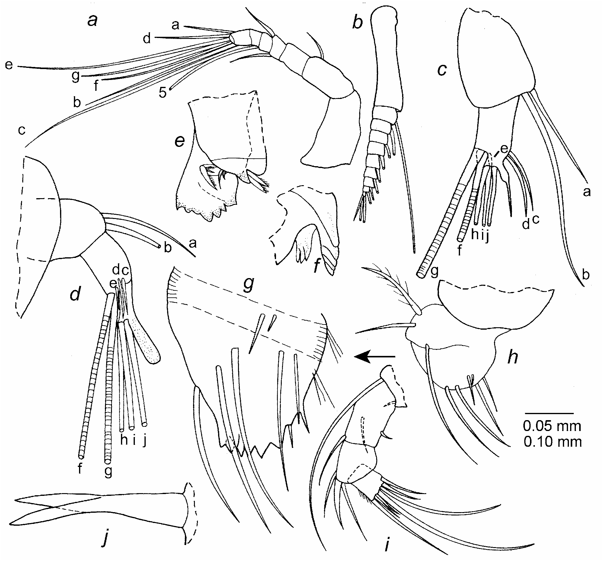
First antenna ( Fig. 15 a View FIGURE 15 ): Similar to that of adult female except spines not observed on segments or bristles.
Second antenna: Protopod and exopod ( Fig. 15 b View FIGURE 15 ) similar to those of adult female. Endopod with 3 segments: 1st segment with dorsal a- and b-bristles; 2nd segment with 2 terminal f- and g-bristles (g-bristle medial and longer and stouter than f-bristle), 2 slender lateral c- and d-bristles, and 1 smaller lateral e-bristle near base of f-bristle; 3rd segment with equilength h-, i-, and j-bristles; clasper of right limb stout with blunt tip ( Fig. 15 d View FIGURE 15 ); clasper of left limb shorter with long terminal bristle-like spine ( Fig. 15 c View FIGURE 15 ). With exopod withdrawn, distal part of endopod lies lateral to exopod.
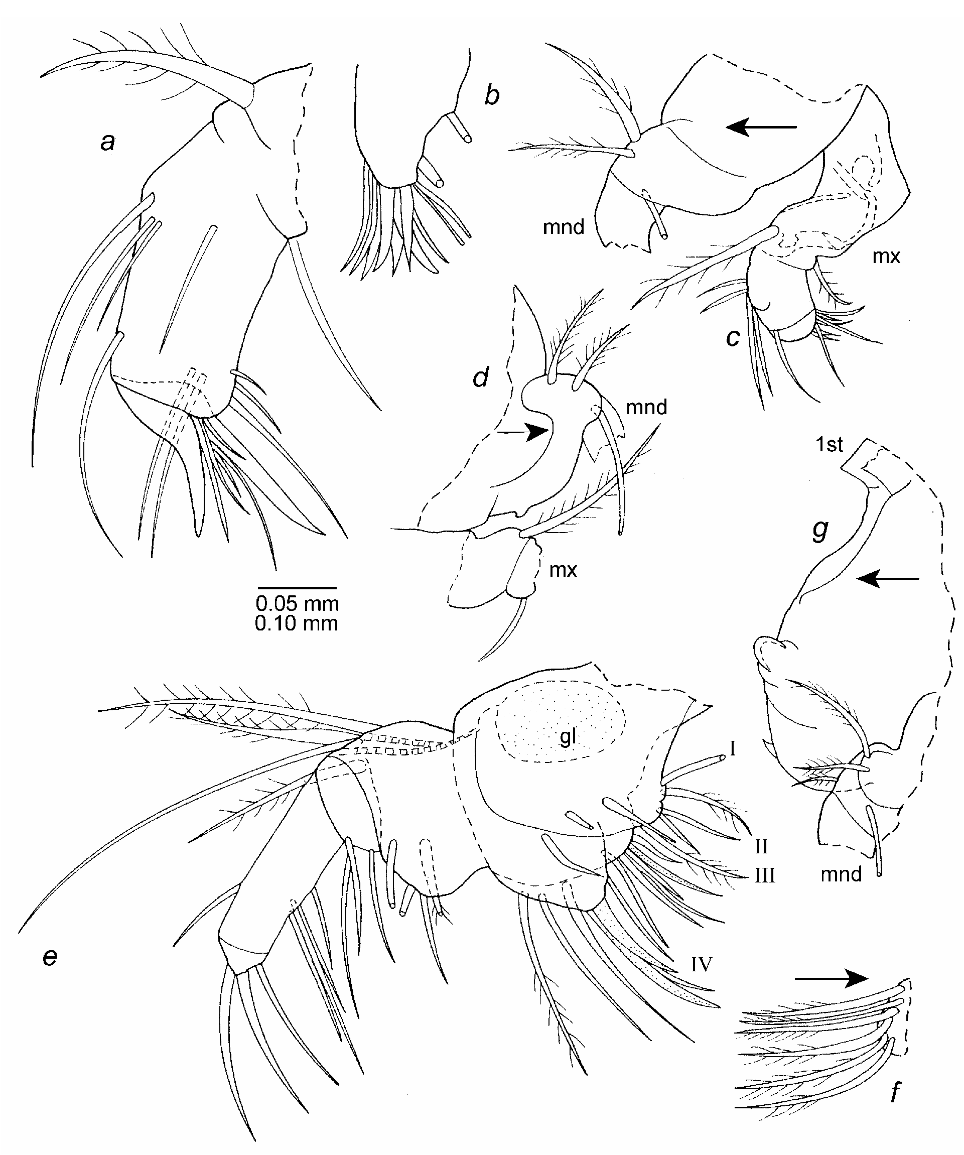
Mandible ( Figs. 15 e–i View FIGURE 15 , 16 c,d,g View FIGURE 16 ): Only 2 instead of 3 spinous dorsal bristles observed on basis. Limb otherwise similar to that of adult female. Plumose bristles near dorsal margin of basis project anteriorly when mandible in natural position on body ( Fig. 16 c,d,g View FIGURE 16 ).
Maxilla ( Fig. 16 a–d View FIGURE 16 ): Similar to that of adult female. Plumose bristles on dorsal margin of basis projects anteriorly when maxilla in natural position on body ( Fig. 16 c,d View FIGURE 16 ).
Fifth limb ( Fig. 16 e,f View FIGURE 16 ): Epipod with plumose bristles forming 3 groups (ventral and dorsal groups each with 5 bristles, middle group with 6 bristles ( Fig. 16 f View FIGURE 16 ). Protopod interpreted to have 4 endites (because endite sutures are poorly defined, number of bristles on a particular endite approximate); endite I with 2 ventral bristles; endite II with 4 bristles (2 ventral, 2 farther from ventral margin); endite III with 6 bristles (1 claw-like); endite IV with 9 bristles (2 claw-like); 21 total endite bristles; protopod with oval internal mass of cells questionably interpreted as gland. Basis: ventral margin divided into broad proximal part and narrow distal part; broad proximal part with 3 slender bristles on or near ventral margin and 1 plumose lateral bristle set back from ventral margin; narrow distal part with 3 slender ventral bristles and 1 lateral plumose bristle near dorsal margin. Exopod represented by 3 distal dorsal bristles (longest bare, others plumose). Endopod: 1st segment with 1 distal dorsal bristle and 4 ventral bristle near midlength; 2nd segment with 2 stout claw-like bristles and 1 slender ringed ventral bristle.
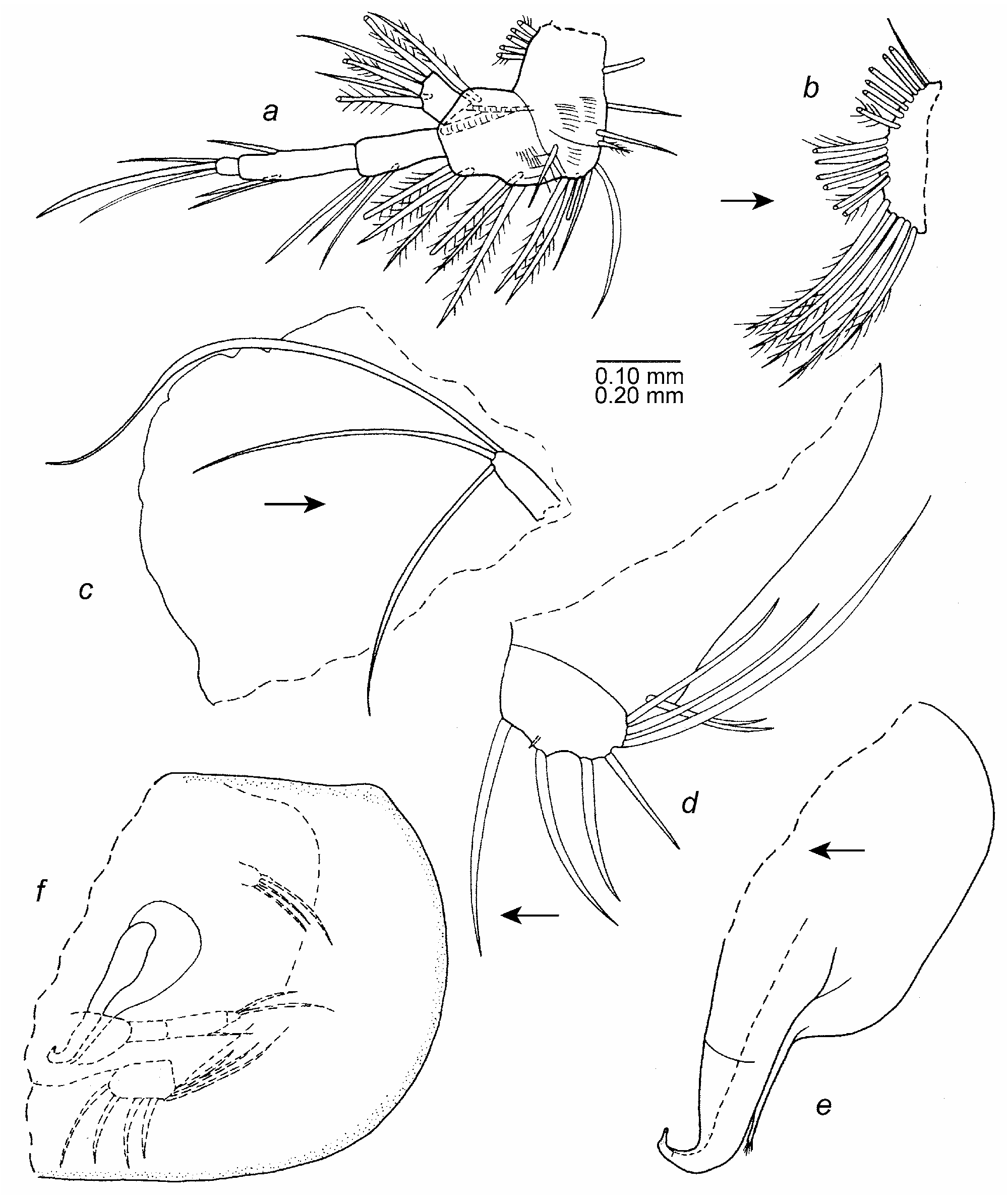
Sixth limb ( Fig. 17 a,b,f View FIGURE 17 ): Epipod with plumose bristles forming 3 groups (ventral group with 5 bristles, middle group with 6 bristles, dorsal group with 7 bristles including 1 short dorsal). Proximal protopod separated from basis by suture and divided by suture into 2 parts interpreted to be precoxa and coxa, both with long medial hairs: precoxa with 4 bristles; coxa with 5 or 6 bristles. Basis with long proximal medial hairs near ventral margin and 7 plumose bristles (6 on or near ventral margin, 1 distal lateral near dorsal margin). Exopod well developed, lateral to endopod, with 5 bristles (3 long plumose, 2 shorter bare). Endopod 3-segmented: 1st segment with 3 ventral bristles; 2nd segment with 3 bristles (2 ventral, 1 dorsal); 3rd segment with 3 long bare terminal bristles (middle bristle longer than others and claw-like), other bristles slender, ringed).
Seventh limb ( Fig. 17 c,f View FIGURE 17 ): Elongate with 3 terminal bristles (1 longer than others).
Furca ( Fig. 17 d, f View FIGURE 17 ): Each lamella with 4 claws followed by 3 bristles (anterior of these very long); minute teeth observed along posterior edges of claws 1 and 2. Minute glandular process present between claws 1 and 2 (closer to claw 2). Long bifurcate unpaired bristle following lamellae.
Bellonci Organ ( Fig. 15 j View FIGURE 15 ): Elongate, bifurcate with pointed tips extending past 1st segment of 1st antennae.
Lips: Similar to those of adult female. Profile of anterior part of upper lip shown in Fig. 16 g View FIGURE 16 .
Genitalia ( Fig. 17 e,f View FIGURE 17 ): Copulatory organ on left side of body medial to left sixth limb and lateral to furca. Tip of anterior branch with tubular upturned tip. Posterior branch with narrow tip bearing hairs.
Behavior. Each exopod of the second antennae of two females and one male in the collection is withdrawn inside the carapace, and the distal part of each endopod is located lateral to the exopod. This suggests that the endopod flairs outward when the exopod is withdrawn inside the carapace.
Remarks. This species was reported in Mermaid’s Lair in 1997 (Kornicker et al. 2002: 26). The present collection shows that it continued to inhabit the cave in 1998. The species is reported for the first time in Lucy’s Cave.

| USNM |
Smithsonian Institution, National Museum of Natural History |
No known copyright restrictions apply. See Agosti, D., Egloff, W., 2009. Taxonomic information exchange and copyright: the Plazi approach. BMC Research Notes 2009, 2:53 for further explanation.
|
Kingdom |
|
|
Phylum |
|
|
Class |
|
|
Order |
|
|
Family |
|
|
Genus |
Spelaeoecia parkeri Kornicker et al. 2002
| Kornicker, Louis S., Iliffe, Thomas M. & Harrison-Nelson, Elizabeth 2007 |
Spelaeoecia parkeri
| Kornicker 2002: 26 |