
Danielopolina (H.) palmeri, Kornicker & Iliffe & Harrison-Nelson, 2007
|
publication ID |
https://doi.org/10.11646/zootaxa.1565.1.1 |
|
publication LSID |
lsid:zoobank.org:pub:A2CDD9CB-CA5E-418B-A471-9EEFDC5CCF16 |
|
persistent identifier |
https://treatment.plazi.org/id/2A5087FF-3E2F-FC12-3A91-F95AFB896ACC |
|
treatment provided by |
Felipe |
|
scientific name |
Danielopolina (H.) palmeri |
| status |
sp. nov. |
Danielopolina (H.) palmeri View in CoL n.sp.
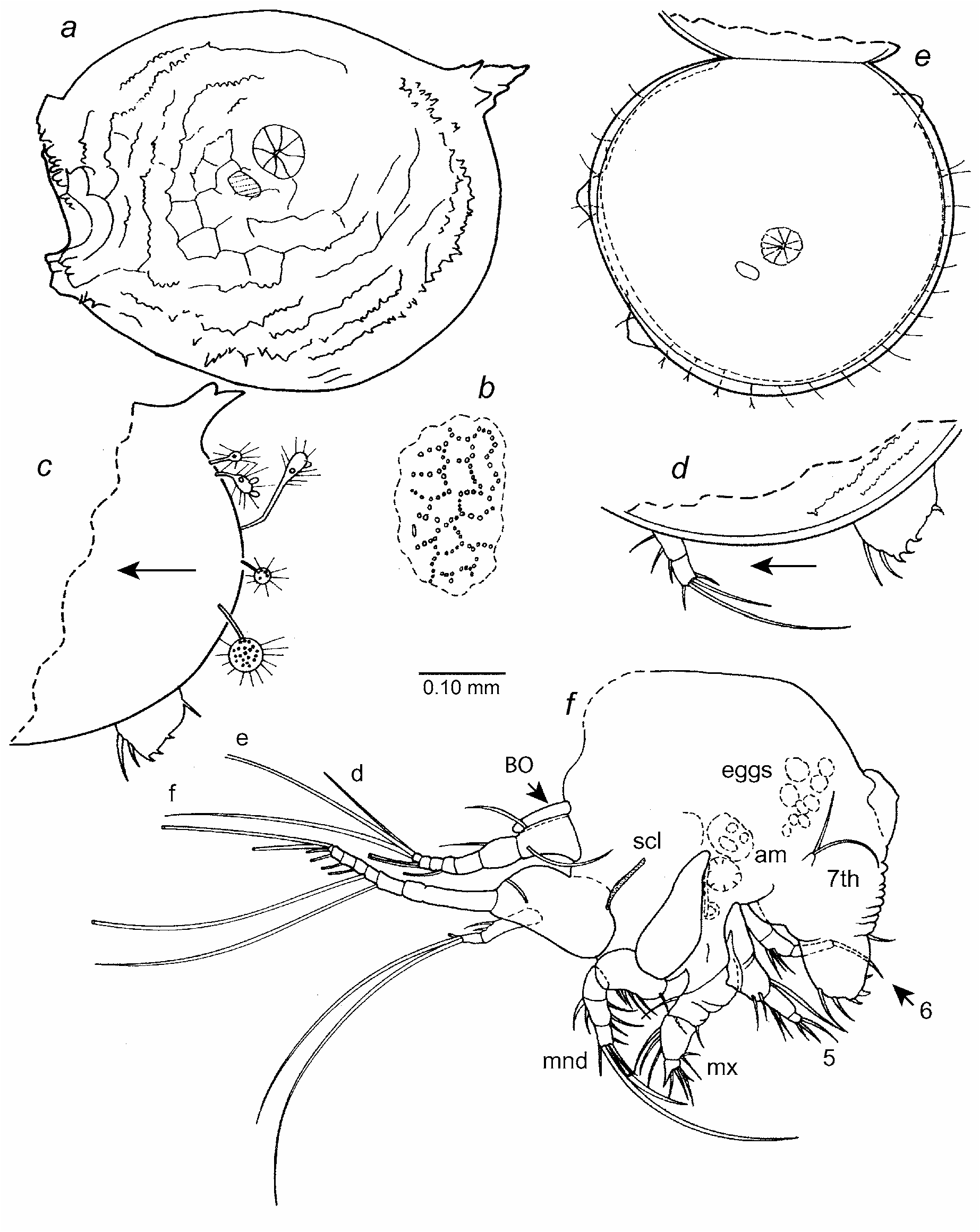
Figs. 10–13 a–i View FIGURE 10 View FIGURE 11 View FIGURE 12 View FIGURE 13
Etymology. Named posthumously in honor of British cave diver Rob Palmer, a pioneering explorer of the blue holes of the Bahamas and author of two books ( Palmer 1985, 1997) and numerous articles. Rob never returned from a deep dive in the Red Sea in 1997. The type locality for this species was first explored by Rob Palmer.
Holotype. USNM 1021375 About USNM , adult female on two slides and in alcohol.
Type locality. Sanctuary Blue Hole, South Andros Island, Great Bahama Bank, Sta 99-061.
Paratypes. Type locality, Sta 99-061: USNM 1021376 About USNM , adult female. USNM 1021377 About USNM , two early instars (probably second instars, length and height without processes (mm): 0.38, 0.27; 0.28, 0.23) .
Distribution. Type locality.
Description of adult female ( Figs. 10–13 a–i View FIGURE 10 View FIGURE 11 View FIGURE 12 View FIGURE 13 ). Carapace subround in lateral view with fairly straight margin between anterior and anteroventral processes ( Fig. 10 View FIGURE 10 ); hinge line straight ( Fig. 11 e View FIGURE 11 ). Short anterior and anteroventral processes with bases just lateral to valve edge; fragile spines on processes mostly broken off on holotype ( Fig.11 a View FIGURE 11 ); spines on processes completely broken off by dissecting needle leaving small firm triangular protuberance ( Fig. 11 a View FIGURE 11 ); a similar posterodorsal process in same place on each valve. Bristles present along edges of valve, those along anteroventral edge divided distally ( Fig. 11 e View FIGURE 11 ).
Ornamentation: Much of surface ornamentation missing on surface of carapace of holotype. Approximate location of some reticulations and spines of holotype shown in Fig. 11 a View FIGURE 11 , and minute subelliptical papillae forming walls of reticulations shown in Fig. 11 b View FIGURE 11 ; papillae at intersections of reticulate walls generally with small spines; minute spines forming rows near valve edges. Surface ornamentation well preserved on paratype ( Fig. 10 View FIGURE 10 ): minute papillae forming walls of reticulations, with spines at intersections; spines forming row just within valve edge and along dorsal edge of valve.
Selvage ( Fig. 11 d,e View FIGURE 11 ): Broad lamellar prolongation with smooth outer edge present along anterior, ventral, and posterior edges of valves.
Infold ( Fig. 11 e View FIGURE 11 ): Broad infold present along anterior, ventral, and posterior margins of valves.
Central adductor muscle attachments ( Figs. 10 View FIGURE 10 , 11 a,e View FIGURE 11 ): Comprising about 8 radially arranged attachments.
Carapace size (mm): USNM 1021375, length without processes 0.53, height without processes 0.47. USNM 1021376, length with anterior and posterodorsal processes 0.61, length without processes 0.49, height with anteroventral and posterodorsal processes 0.45, height without processes 0.42.
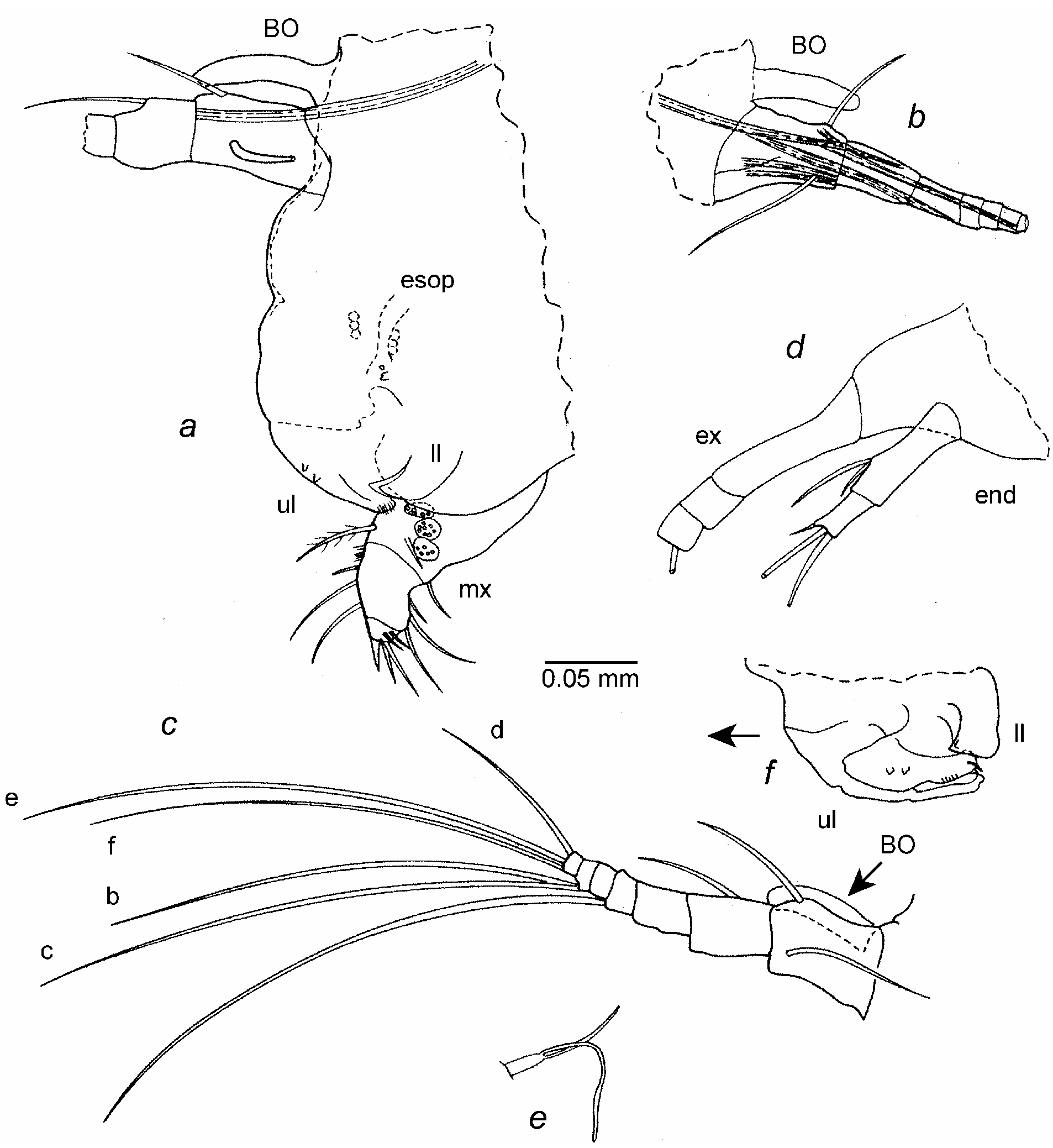
First antenna ( Figs. 11 f View FIGURE 11 , 12 a–c View FIGURE 12 ): 1st and 2nd segments linear (not forming right angle). 1st segment with 1 dorsal bristle and 1 lateral bristle oriented posteriorly. 2nd segment with 1 dorsal bristle. 3rd and 4th segments fused but place of boundary indicated by slight indentation in ventral margin. 5th segment with long terminal ventral filament. 6th segment bare. 7th segment with 2 long ventral bristles (b-bristle shorter than lateral c-bristle). 8th segment with 3 bristles (d-bristle shorter, f-bristle about one-half length of e-bristle).
Second antenna ( Fig. 12 d View FIGURE 12 ): Protopod bare. Endopod 3-segmented but with 2nd and 3rd segments fused. 1st segment with dorsal a- and b-bristles. 2nd segment with 2 long terminal bristles and indistinct, minute, distal, lateral spine near dorsal margin. 3rd segment narrow with short terminal bristle. Exopod 8 segmented: 1st segment divided weakly into long proximal and short distal parts; bristles of segments 2–7 long with indistinct natatory hairs; 8th segment with 2 bristles.
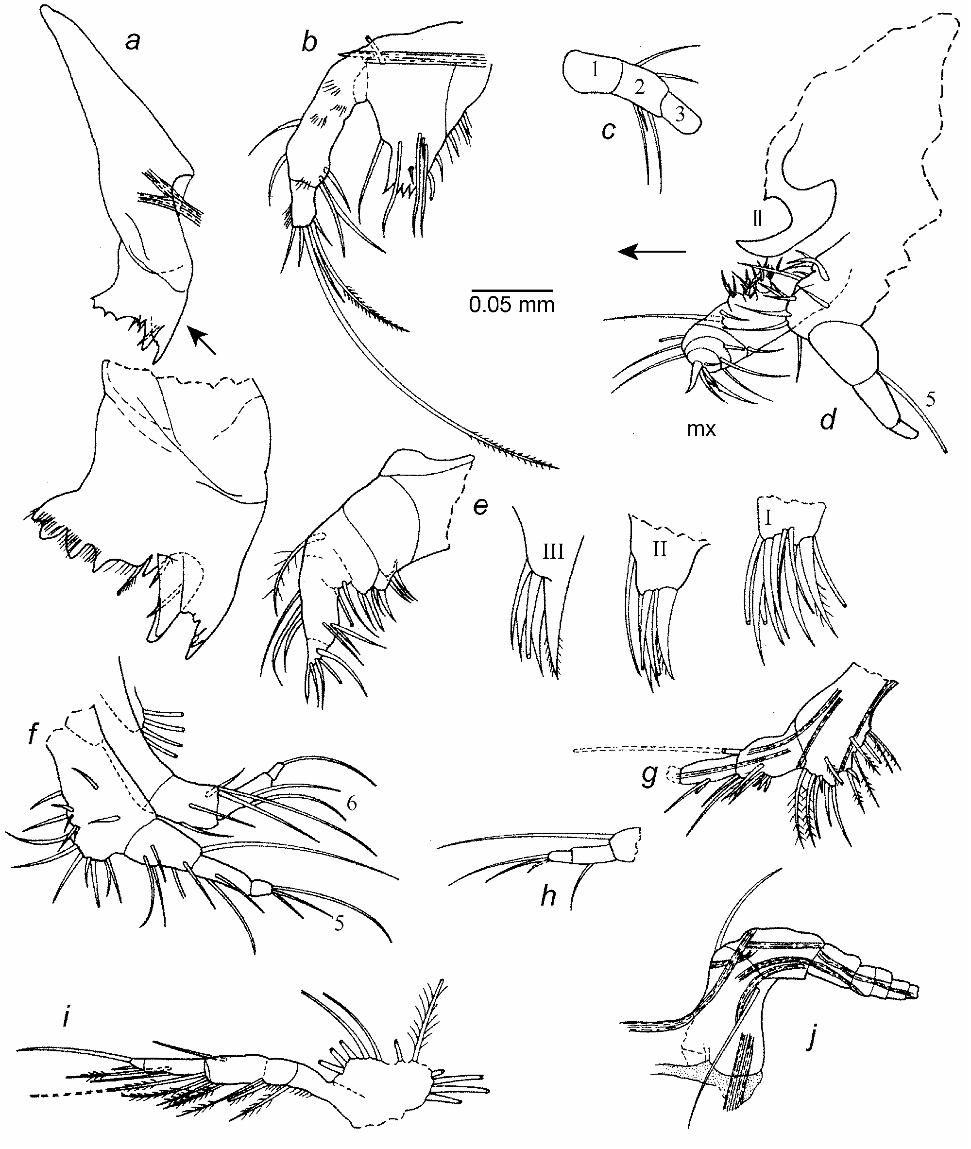
Mandible ( Fig. 13 a–c View FIGURE 13 ): Coxa endite with proximal and distal sets of teeth separated by space; proximal set comprising 4 broad cusps plus triangular tooth close to distal set of teeth ( Fig. 13 a View FIGURE 13 ); surface between cusps and surfaces just proximal to cusps with slender spines; 1 spinous bristle with base just posterior and another with base just anterior to triangular tooth; distal set of teeth consisting of 2 flat teeth, each with cusps; 1 slen- der bristle with base medial to distal set of teeth. Basis ( Fig. 13 b View FIGURE 13 ): tooth of endite with 5 triangular teeth, each with minute marginal cusp on each side (cusps on anterior 4 teeth better defined); posterior edge of endite spinous, with 2 short distal bristles (distal of these tubular with blunt tip); anterior margin of endite with long ringed bristle near midlength; lateral side of endite with 4 bristles (3 long, 1 short) near midlength and 1 short distal bristle; medial side of endite with 2 proximal bristles and long spines near midlength (spines not shown). Endopod 3 segmented (1st and 2nd segments of left limb fused, but interpreted to be an aberrancy ( Fig. 13 b View FIGURE 13 ); right limb with suture between 1st and 2nd segments ( Fig. 13 c View FIGURE 13 )): 1st segment with lateral spines; 2nd segment with distal lateral spines, 1 ringed distal ventral bristle, 2 ringed distal medial bristles with bases near ventral margin, and 2 ringed dorsal bristles; 3rd segment with dorsal and medial spines (medial spines not shown), 2 terminal lateral bristles (1 at midwidth about twice length of endopod and with distal marginal spines), 1 at ventral edge almost one-half length of bristle at midwidth and with distal marginal spines (longest spines at midlength), 3 shorter ringed terminal medial bristles, and 1 short ringed subterminal bristle on ventral margin.
Maxilla ( Fig. 13 d,e View FIGURE 13 ): Endite I with about 10 bristles (3 tubular); endite II with about 6 bristles (2 tubular); endite III with about 5 bristles (1 tubular). Coxa with long spinous dorsal bristle. Basis with 2 ventral bristles (1 spinous bristle near base of endite III, 1 terminal tubular bristle). Endopod 2 segmented: 1st segment with 3 dorsal bristles and 3 distal bristles on or near ventral margin; 2nd segment with stout, straight, unringed, nonarticulated, terminal claw and 4 ringed articulated bristles (1 medial tubular, 3 lateral (longest somewhat clawlike, spinous)).
Fifth limb ( Fig. 13 d,f–h View FIGURE 13 ): Epipod bristles not counted. Boundary of precoxa and coxa marked by muscle terminating at ventral edge of boundary ( Fig. 13 g View FIGURE 13 ). Precoxa with 6 ventral bristles (1 tubular) and 1 medial bristle set back from ventral margin. Coxa with 6 ventral bristles (2 claw-like bristles, 2 slender tubular bristles, 2 stout bristles with long marginal spines) and 1 long spinous bristle set back from ventral margin ( Fig. 13 g View FIGURE 13 ). Basis with 6 bristles on or near ventral margin and 1 spinous medial bristle set back from ventral margin. Exopod bristle long bare. Endopod 2 segmented: 1st segment with 2 ventral bristles near midlength; 2nd segment with 1 long and 2 short bristles.
Sixth limb ( Fig. 13 f,i View FIGURE 13 ): Epipod with plumose bristles forming 3 groups: proximal group with 5 bristles; bristles of second group fragmented during dissection; distal group with 4 or 5 bristles. Precoxa spinous, with 1 spinous ventral bristle. Coxa spinous, with 2 spinous ventral bristles. Basis with 3 spinous bristles (2 ventral and 1 dorsal). Small exopod with 2 unequal spinous bristles. Exopod of left limb medial and close to ventral margin of basis ( Fig. 13 i View FIGURE 13 ); exopod of right limb also medial but close to dorsal margin ( Fig. 13 f View FIGURE 13 ). Endopod 2 segmented: 1st segment with 2 ventral bristles near midlength; 2nd segment with 2 bristles (1 long terminal and 1 short, subterminal, ventral).
Seventh limb ( Fig. 12 e View FIGURE 12 ). Elongate with 2 long terminal bristles.
Furca ( Fig. 11 c,d,f View FIGURE 11 ): Each lamella with 2 long articulated anterior claws and 3 short nonarticulated ventral claws; all claws with indistinct spines along posterior margin (spines not shown); claw 1 indistinctly ringed. Stout unpaired process on posterior of body just proximal to furca.
Bellonci Organ ( Figs. 11 f View FIGURE 11 , 12 a–c View FIGURE 12 ): Well defined, elongate, with rounded tip.
Lips ( Fig. 12 a, f View FIGURE 12 ): Anterior face of lip with 2 small processes; tip of upper lip with spines and small process. Lower lip with a triangular process at each side of mouth.
Posterior of body ( Fig. 11 f View FIGURE 11 ): With 9 or 10 short ridges along posterior edge.
Genitalia. None observed.
Eggs ( Fig. 11 f View FIGURE 11 ): With several unextruded eggs.
Gut content: Gut viewed through body filled with brown unidentified particles.
Protistan: Carapace with several elongate and round stemmed protistans along posterior margin ( Fig. 11 c View FIGURE 11 ). Similar protistans also present on dorsal margin of 1st segment of left 1st antenna.
Feeding: Endite I of maxilla anterior and projecting farther medially than endites II and III ( Fig. 13 d View FIGURE 13 ).
Remarks. The holotype is interpreted to be an adult female because of the presence of fairly large unextruded eggs. However, genitalia were not observed, so it could be an A- 1 female. Paratype USNM 1021376 was not dissected in order not to fragment the ornamentation of the carapace. It is slightly smaller than the holotype, and no unextruded eggs were visible when the body was viewed through the valve, but the valve somewhat obscured the body. The furca has the same number of the claws as the holotype, and the endopod of the second antenna is similar to that of the holotype. The specimen is assumed to be an adult female, but it could be an A- 1 female.
Comparisons. The ornamentation of the carapace of D. (H.) palmeri differs from that of D. (H.) exuma and D. (H.) orghidani in having spines at most intersections of reticulate walls and, in general, being more spinous. The carapace of D. (H.) palmeri differs from that of D. (H.) bahamensis and D. (H.) styx in having a posterodorsal elongate process on each valve. The seventh segment of the first antenna of D. (H.) palmeri is without the a-bristle, which is present on D. (H.) kakuki , D. (H.) styx , D. (H.) wilkensi , and D. (H.) bahamensis . The first endopod segment of the mandible of D. (H.) palmeri is without a dorsal bristle, which is present on D. (H.) bahamensis , D. (H.) orghidani , and D. (H.) kakuki . The furca of D. (H.) palmeri differs from that of D. (H.) elizabethae in having two articulated claws rather than one on each lamella.

No known copyright restrictions apply. See Agosti, D., Egloff, W., 2009. Taxonomic information exchange and copyright: the Plazi approach. BMC Research Notes 2009, 2:53 for further explanation.