
Eusarsiella paniculata Kornicker 1986
|
publication ID |
https://doi.org/ 10.11646/zootaxa.1565.1.1 |
|
publication LSID |
lsid:zoobank.org:pub:A2CDD9CB-CA5E-418B-A471-9EEFDC5CCF16 |
|
DOI |
https://doi.org/10.5281/zenodo.5095936 |
|
persistent identifier |
https://treatment.plazi.org/id/2A5087FF-3E43-FC74-3A91-F881FA6B699C |
|
treatment provided by |
Felipe |
|
scientific name |
Eusarsiella paniculata Kornicker 1986 |
| status |
|
Eusarsiella paniculata Kornicker 1986 View in CoL
Figs. 66–68 View FIGURE 66 View FIGURE 67 View FIGURE 68
Eusarsiella paniculata Kornicker 1986 View in CoL , 58: figs. 28d–g, 29, 30.
Holotype. USNM 157973 About USNM , ovigerous female on slide and in alcohol.
Type locality. West Florida continental shelf, depth 58.5 m .
Material. Sta 00-025, Conch Sound Blue Hole, Andros Island, Great Bahama Bank: USNM 1021471 About USNM , one instar A- 1 female on slide and in alcohol ; USNM 1021472 About USNM , one A- 1 male in alcohol ; USNM 1021473 About USNM , one A-2 instar (sex unknown) in alcohol .
Distribution. West Florida continental shelf, depth 22.5–58.5 m. Conch Sound Blue Hole (about 365 m inside cave), Andros Island, Great Bahama Bank, depth 25 m.
Description of A- 1 female ( Figs. 66–68 a,b View FIGURE 66 View FIGURE 67 View FIGURE 68 ). Carapace oval in lateral view with pointed caudal process triangular in lateral view ( Fig. 66a View FIGURE 66 ). Surface with gel-like substance coating valves and short bristles.
Ornamentation ( Fig. 66 a,c View FIGURE 66 ): Surface with numerous processes bearing short tapered bristles, 1 very long bristle, and 1 shorter more slender bristle ( Fig. 66 a View FIGURE 66 ); surface with shallow round fossae with bare bottoms; surface between fossae with small spine-like pointed bristles of various lengths; spines also abundant along valve edge including caudal process.
Infold: Anterior infold with minute bristle near midheight. Infold of caudal process with 4 or 5 bare bristles forming vertical row and 1 similar bristle closer to the inner edge of infold ( Fig. 66 d,e View FIGURE 66 ).
Selvage: Broad lamellar prolongation with smooth outer edge present along entire margin and extending well past tip of caudal process.
Central adductor muscle attachments ( Fig. 66 b View FIGURE 66 ): consisting of about 14 ovoid scars.
Carapace size (length, height in mm): USNM 1021472, length including caudal process 1.26, length excluding caudal process 1.14, height 0.96.
First antenna ( Fig. 66 f View FIGURE 66 ): 1st segment bare. 2nd with dorsal bristle. 3rd and 4th segments fused; 3rd with 2 terminal bristles (1 ventral, 1 dorsal); 4th with 3 bristles (2 ventral, 1 dorsal). 5th segment with long ventral bristle with minute proximal filament. 6th segment with short medial bristle. 7th segment: a-bristle longer than bristle of sixth segment; b-bristle slightly shorter than a-bristle; c-bristle long, with 2 minute proximal filaments. 8th segment: d- and e-bristle long, bare; f-bristle long; g-bristle long with 2 minute proximal filaments.
Second antenna ( Fig. 67 a,b View FIGURE 67 ): Protopodite bare ( Fig. 67 a View FIGURE 67 ). Endopod with 2 segments: segment 1 broad with 2 ringed anterior bristles; segment 2 short with ringed terminal bristle about same length as bristles of 1st segment ( Fig. 67 b View FIGURE 67 ). Exopod: 1st segment with short recurved medial spine; bristles of segments 2–8 with stout proximal ventral spines and distal natatory hairs; 9th segment with 2 bristles (1 long with proximal ventral spines and distal natatory hairs, 1 short bare).
Mandible ( Fig. 67 c View FIGURE 67 ): Coxa endite consisting of short stout spine; ventral margin of coxa with short hairs. Basis with 6 bristle with 3 medial, 3 smaller on ventral margin, and 2 spine-like small dorsal subterminal bristles. Exopod absent. Endopod: 1st segment with distal medial spines, row of medial spines along distal margin, 3 stouter terminal dorsal spines, and minute spine-like medial bristle at base of stout ventral claw. 2nd segment with minute terminal dorsal bristle and stout ventral claw. 3rd segment with 2 minute bristles (1 ventral, 1 dorsal) and stout terminal claw.
Maxilla ( Fig. 67 d–f View FIGURE 67 ): Endite I with 6 terminal bristles; endite II with 4 bristles; endite III with 6 bristles. Coxa with short dorsal bristle. Basis with 1 bristle near exopod. Exopod with 2 bristles. Endopod: 1st segment with spinous alpha- and beta-bristles. 2nd segment with 2 lateral a-bristles, 1 medial c-bristle, and 5 pectinate terminal bristles.
Fifth limb ( Fig. 67 g View FIGURE 67 ): Epipod with 35 plumose bristles. Coxa with small bristle. Basis with 2 endites: endite I with 2 bristles; endite II with 3 bristles. Endopod with 2 fused segments: segment 1 with 3 bristles; segment 2 with 2 bristles. Exopod represented by short bristle. Limb hirsute.
Sixth limb ( Fig. 67 h View FIGURE 67 ): Single endite with 3 bristles (1 longer than others). End segment with 11 bristles plus 2 plumose posterior bristles.
Seventh limb ( Fig. 67 i View FIGURE 67 ): Each limb with 6 tapered bristle (2 proximal with 3 or 4 bells; 4 terminal with 4 or 5 bells). Terminus with minute terminal teeth. Each limb with 69–72 rings.
Furca ( Fig. 67 j View FIGURE 67 ): Each lamella with 5 claws with claw 1 fused to lamella. Claw 1 with 5 stout teeth, many small teeth, and long spines forming medial row near base; claws 2 and 3 with small teeth; claw 5 very small. Left lamella with ventral spines following claws and posterior to right lamella by width of claw 1 at base. Both lamellae with small anterior spine at about midheight.
Bellonci Organ: Lost during dissection.
Eyes: Lateral eye with 5 divided, amber-colored ommatidia ( Fig. 67 a View FIGURE 67 ). Medial eye larger than lateral eye ( Fig. 67 k View FIGURE 67 ).
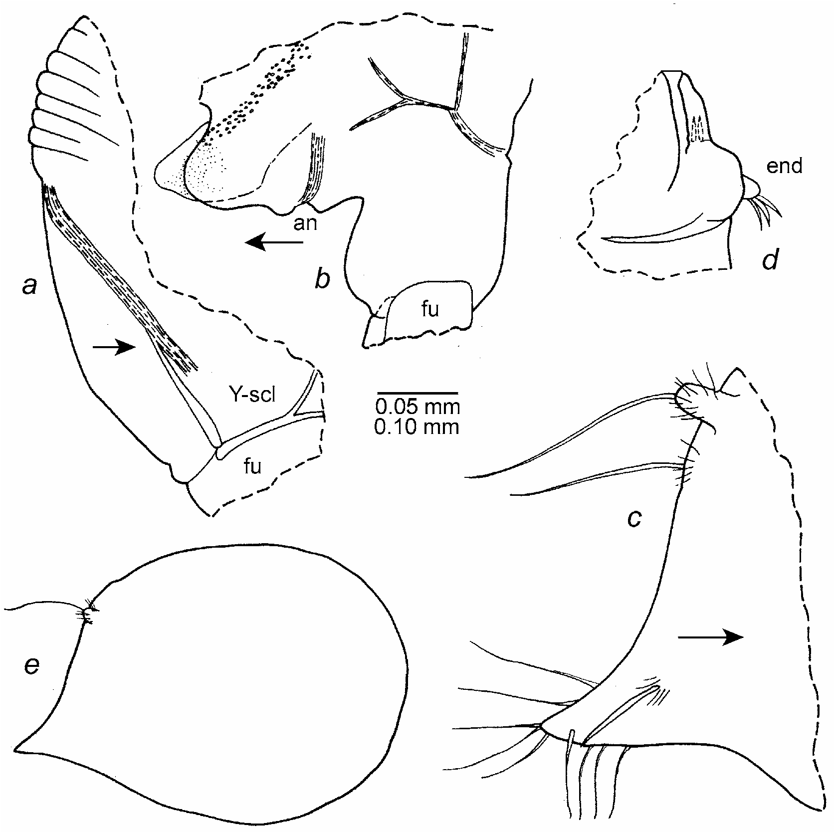
Genitalia? ( Fig. 68 b View FIGURE 68 ): Broad rounded lobe on each side of longer tapered lobe, all bare. Each rounded lobe with duct (containing minute globules) terminating anterior to Y-sclerite.
Y-Sclerite ( Fig. 68 a,b View FIGURE 68 ): With ventral branch.
Posterior of body ( Fig. 68 a View FIGURE 68 ): Posterior of body smooth, bare. Posterodorsal part of body dorsal to girdle scalloped.
Gut content: Copepod and unidentified crustacean.
Remarks Concerning Genitalia. The process just anterior to the anus that is questionably designated the genitalia herein is a puzzle. The ducts on each side show it to be part of the specimen and not foreign. Except for not being paired and being bare, it resembles the male copulatory limb more than it does female genitalia.
Description of A- 1 male ( Fig. 68 c,d View FIGURE 68 ). Carapace shape, ornamentation, infold, and selvage similar to those of A- 1 female ( Fig. 68 c View FIGURE 68 ). Gel-like coating present.
Carapace size (length, height in mm): USNM 1021472, length with caudal process 1.17, length without caudal process 1.04, height 0.88.
First antenna: Distribution of bristles similar to those of A- 1 female.
Second antenna: Protopod and exopod similar to those of A- 1 female. Endopod with 2 segments ( Fig. 68d View FIGURE 68 ): segment 1 with 2 ringed anterior bristles; segment 2 with 3 ringed bristles slightly longer than those of segment 1.
Mandible: Details not studied, but limb in general similar to that of A- 1 female.
Maxilla: Endite I, coxa, basis, exopod, and endopod similar to those of A- 1 female. Endites II and III obscured .
Fifth limb: Epipod with 37 bristles. Remainder of limb similar to that of A- 1 female.
Sixth limb: Obscured, but in general, similar to that of A- 1 female.
Seventh limb: Number of bristles similar to those of A- 1 female. Terminal segment without teeth.
Furca: Similar to that of A- 1 female.
Bellonci Organ: Obscured.
Eyes: Lateral eye with 5 divided amber-colored ommatidia. Medial eye obscured.
Posterior of body: Similar to that of A- 1 female.
Genitalia: Obscured.
Y-Sclerite: With ventral branch.
Gut content: Unidentified fragments.
Description of A-2 instar (sex unknown). Carapace shape similar to that of A- 1 female ( Fig. 68 e View FIGURE 68 ).
Ornamentation: Nodes less well developed than on A- 1 female (only posterodorsal node shown in Fig. 68 e View FIGURE 68 ); others mostly represented by long and short bristles. Gel-like coating present.
Carapace size (length, height in mm): USNM 1021473, length including caudal process 0.93, length without caudal process 0.80, height 0.66.
Furca: Similar to that of A- 1 female.
Comparisons. The female carapace of E. paniculata differs from that of E. syrinx in having a rounded rather than a pointed tip on the caudal process and in having more processes projecting outward from the valve edge. The bristles on the carapace processes of E. capillaris are broader and have blunter tips than those on E. paniculata . The carapace of Eusarsiella cornuta Poulsen 1965 from the West Indies is without processes along the shell edge.
Discussion. Kornicker (1986: 58) described an adult female. An A- 1 female and male and an A-2 instar are described herein. The right valve of the A- 1 male was removed, and the description of appendages is based on views of the specimen within the left valve. The A-2 instar was not opened. The identification of the sexes of the A- 1 male and female is based on the A- 1 male having three bristles on the second segment of the endopod of the second antenna and not having teeth at the tip of the seventh limb. The A- 1 female bears one terminal bristle on the second segment of the second antenna and minute terminal teeth on the seventh limb.


No known copyright restrictions apply. See Agosti, D., Egloff, W., 2009. Taxonomic information exchange and copyright: the Plazi approach. BMC Research Notes 2009, 2:53 for further explanation.
|
Kingdom |
|
|
Phylum |
|
|
Class |
|
|
Order |
|
|
Family |
|
|
Genus |
Eusarsiella paniculata Kornicker 1986
| Kornicker, Louis S., Iliffe, Thomas M. & Harrison-Nelson, Elizabeth 2007 |
Eusarsiella paniculata
| Kornicker 1986 |