
Synasterope matrix, Kornicker & Iliffe & Harrison-Nelson, 2007
|
publication ID |
https://doi.org/ 10.11646/zootaxa.1565.1.1 |
|
publication LSID |
lsid:zoobank.org:pub:A2CDD9CB-CA5E-418B-A471-9EEFDC5CCF16 |
|
persistent identifier |
https://treatment.plazi.org/id/2A5087FF-3EBE-FC80-3A91-F915FE386F5C |
|
treatment provided by |
Felipe |
|
scientific name |
Synasterope matrix |
| status |
sp. nov. |
Synasterope matrix View in CoL , new species
Figs. 79–81 View FIGURE 79 View FIGURE 80 View FIGURE 81
Etymology. From the Latin matrix (mother, womb, source).
Holotype. USNM 1021493 About USNM , adult male on slide and in alcohol.
Type locality. Sta 99-065, Bottomly’s Blue Hole, Stocking Island , Great Exuma, Exuma Cays , Great Bahama Bank , depth 27–29 m.
Paratypes. None.
Distribution. Type locality.
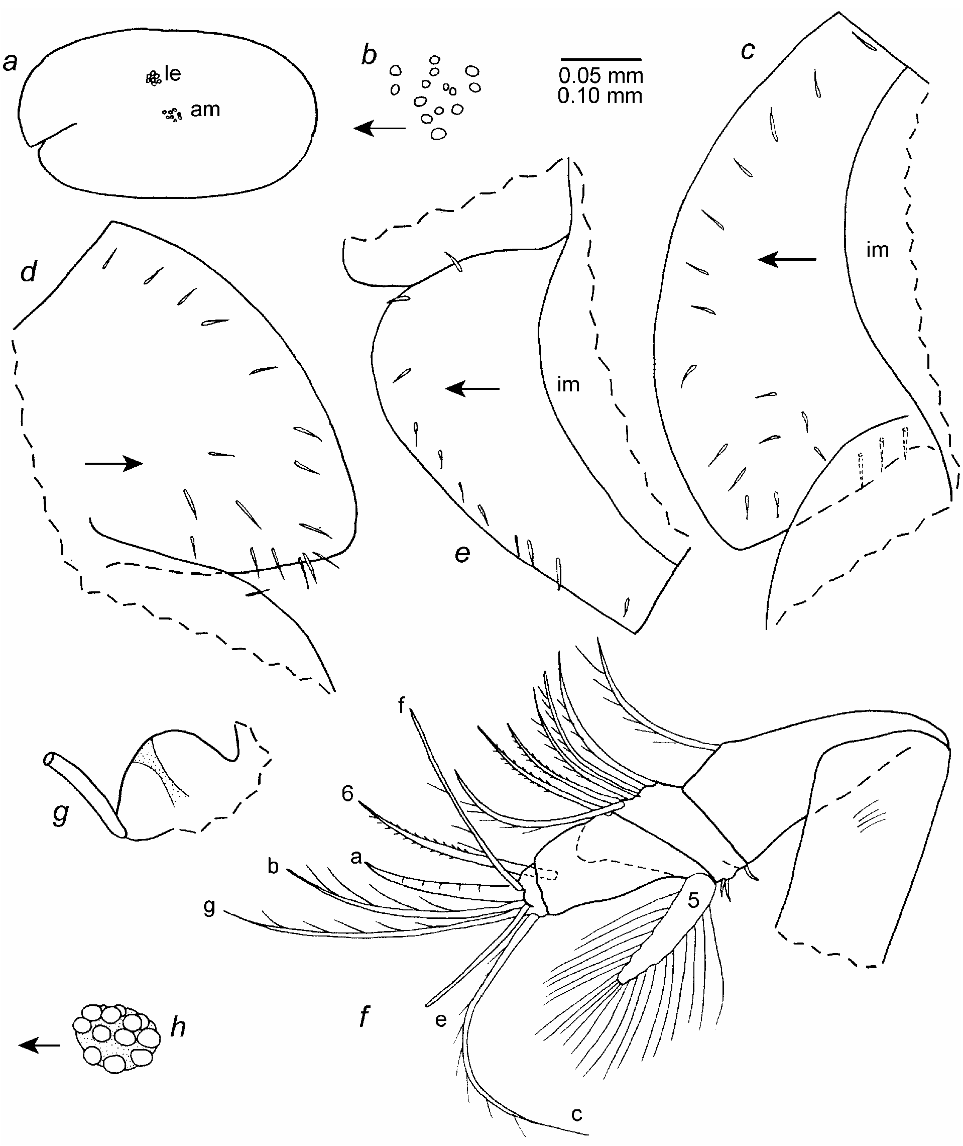
Description of adult male ( Figs. 79–81 View FIGURE 79 View FIGURE 80 View FIGURE 81 ). Carapace elongate with evenly rounded anterior and posterior margins ( Fig. 79 a View FIGURE 79 ).
Ornamentation: Vertical row of few long hairs near posterior end of valve.
Infold: Obscure on slide, especially posterior end of valves. Infold of rostrum with about 19 bristles ( Fig. 79 c,d View FIGURE 79 ). Anteroventral infold with about 11 bristles ( Fig. 79 e View FIGURE 79 ). Ventral infold with about 7 bristles. Flap-like
posterior bristles present but number obscured. Few small bristles present between flap-like bristles and posterior margin of valve. No processes observed between posteroventral list and valve margin.
Central adductor muscle attachments ( Fig. 79 a,b View FIGURE 79 ): Consisting of about 14 ovoid attachments ( Fig. 79 b View FIGURE 79 ).
Carapace size (length, height in mm): USNM 1021493, 0.87, 0.48.
First antenna ( Fig.79 f View FIGURE 79 ): 1st segment with indistinct lateral hairs. 2nd segment with spinous dorsal bristle. 3rd and 4th segments fused; 3rd segment with small bristle on short ventral margin and 5 spinous dorsal bristles (3 single with long spines, 2 paired (lateral with long spines, medial with short spines)) on long dorsal margin. 4th segment with long terminal dorsal bristle and 2 small ventral bristles. 5th segment fused to 6th, with stout ventral bristle with about 40 (not all shown) long filaments (terminal 3 filaments stouter than others; bristle of right limb broken off near base). 6th segment long, with long medial bristle with short spines. 7th segment: a-bristle claw-like, bare; b-bristle with 4 long dorsal filaments; long c-bristle with about 8 filaments. 8th segment: d-bristle absent; bare e-bristle about ½ length of c-bristle; f-bristle bent dorsally, with 3 marginal filaments; g-bristle long with about 5 filaments.
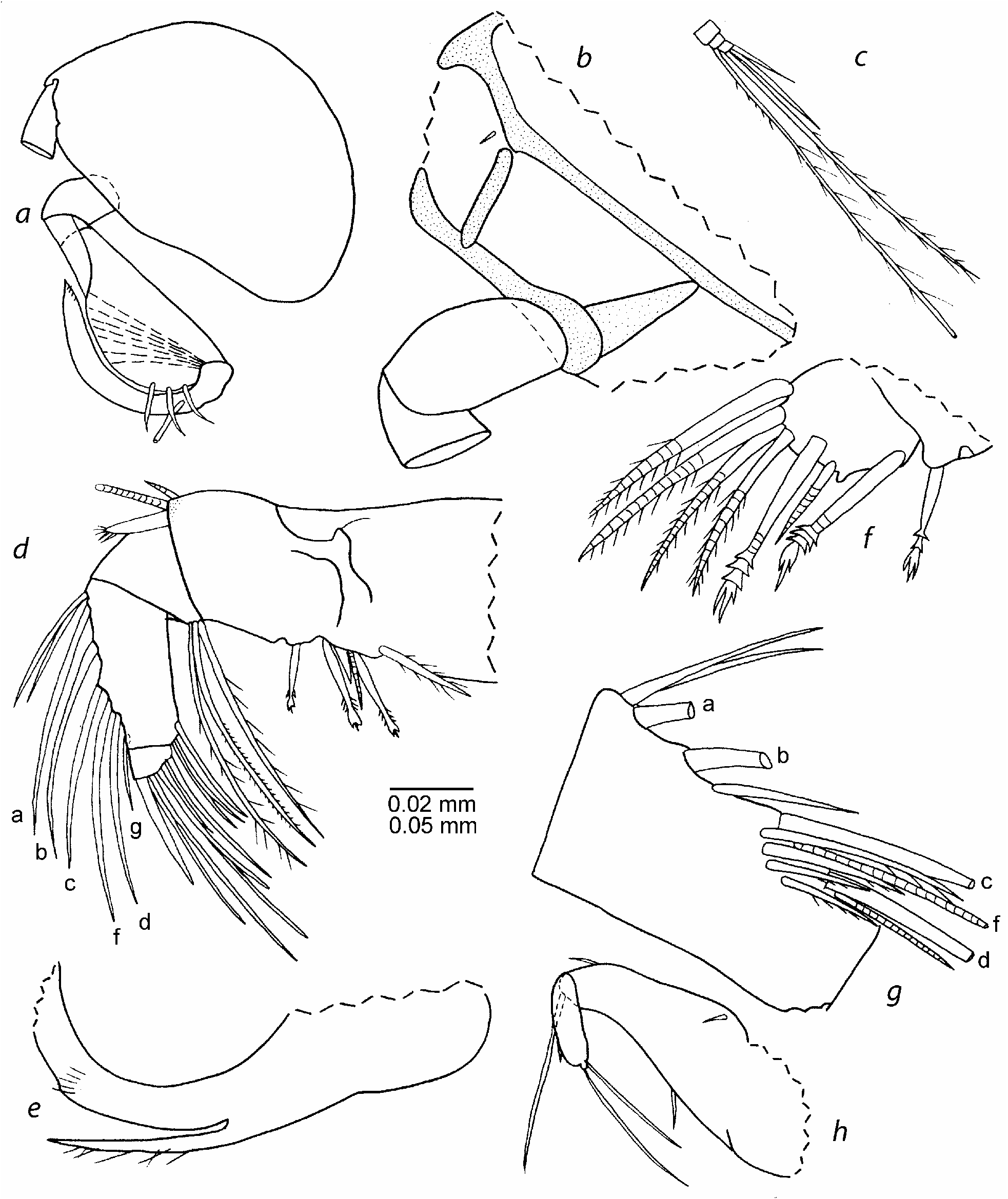
Second antenna: Protopod with small distal medial bristle ( Fig. 80b View FIGURE 80 ). Endopod with 3 segments ( Fig. 80 a View FIGURE 80 ): segment 1 short bare; segment 2 broad distally, with 3 short bare dorsal bristles; segment 3 reflexed on 2nd segment, long and narrow, with bare proximal bristle and serrations at tip. Exopod: 2nd segment only slightly longer than 3rd; bristle of 2nd segment reaching 9th segment, bare; bristles of segments 3–6 with slen- der ventral spines and distal natatory hairs; bristles of segments 7 and 8 with natatory hairs; 9th segment with 3 bristles (ventral bristle about ½ length of bristle of 8th segment; 2 dorsal bristles short bare) ( Fig. 80 c View FIGURE 80 ). Segments 2–8 with indistinct minute spines along distal dorsal edge; stout basal spines absent.
Mandible ( Fig. 80 d–g View FIGURE 80 ): Details of coxa endite obscured ( Fig. 80 e View FIGURE 80 ). Basis endite with 4 spinous end bristles, glandular peg, 1 fairly long dwarf bristle, and 2 triaenid bristles with 3 pairs of spines proximal to terminal pair ( Fig. 80 f View FIGURE 80 ). Basis: ventral margin with proximal U-shaped boss and trianid bristle (with 2 pairs of marginal spines proximal to terminal pair) proximal to boss; dorsal margin with 2 terminal bristles (1 long, 1 short). Exopod longer than one-half dorsal margin of 1st segment of endopod, with hirsute tip and 2 small subterminal bristles ( Fig. 80 d View FIGURE 80 ). Endopod: 1st segment with 3 long ventral bristles (2 with long spines to tip, 1 with short spines). 2nd segment: ventral margin with 3 terminal bristles with short spines; dorsal margin with a-, b-, c-, d-, f- and g-bristles (c-bristle only slightly broader than bases of b- and d-bristles), and 2 short bristles proximal to a-bristle; medial surface of segment with 1 short bare bristle between b- and c-bristles (bristle very close to ventral margin), and 4 spinous cleaning bristles near base of c-bristle. 3rd segment with straight claw and 6 bristles.
Maxilla ( Fig. 80 h View FIGURE 80 ): Epipod and endites fragmented during dissection. Basis: medial surface with 1 proximal bristle near dorsal margin; dorsal margin with short distal bristle; ventral margin with 1 backward-pointing bristle near midlength; ventral margin with long terminal bristle. Endopod: 1st segment with short alphabristle and long beta-bristle. 2nd segment with terminal bristle reaching past beta-bristle.
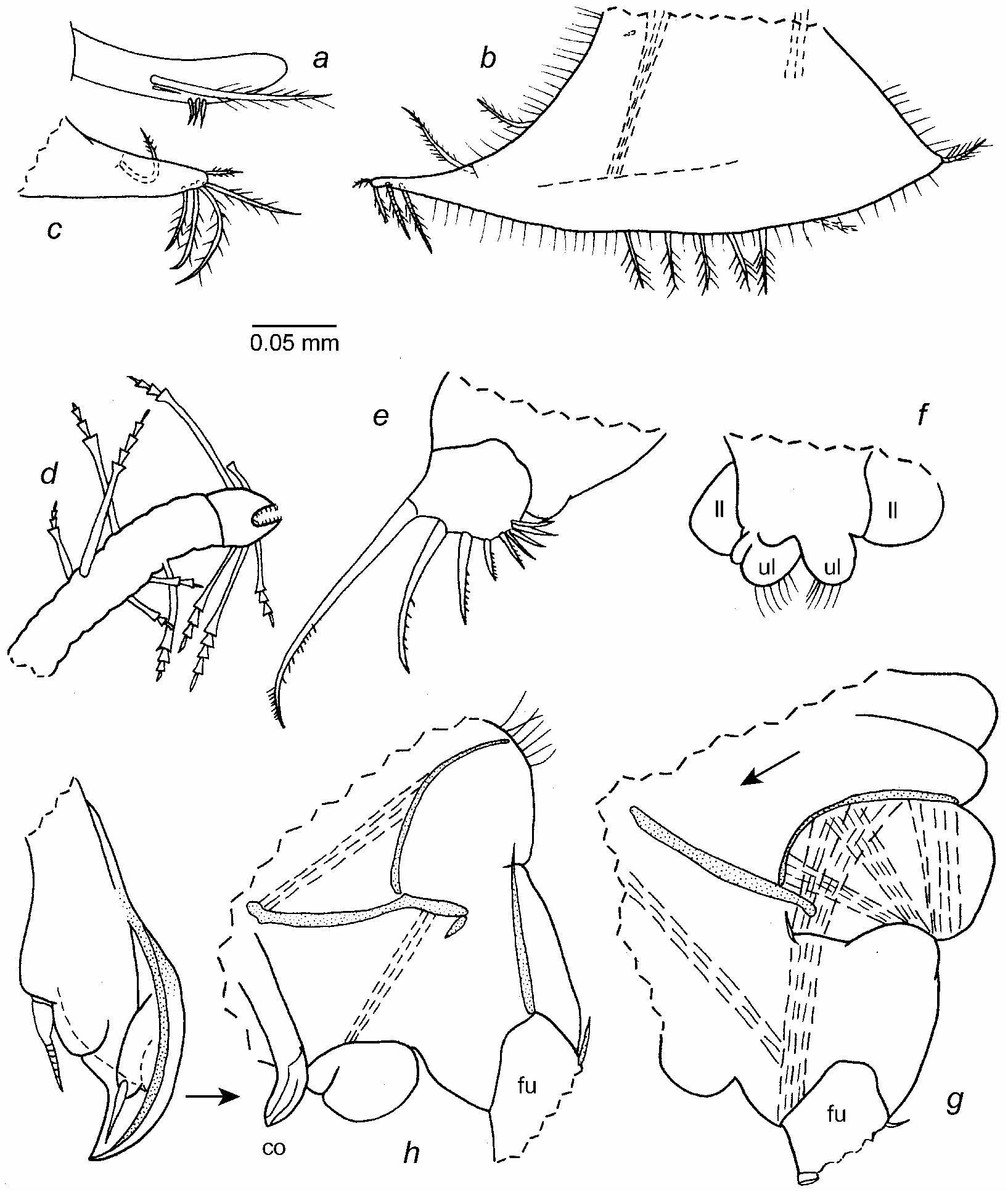
Fifth limb ( Fig. 81 a View FIGURE 81 ): Comb: lateral side with stout spinous exopodial bristle; 1 short slender spine just ventral to base of exopodial bristle and 3 short bristles closer to ventral margin.
Sixth limb ( Fig. 81 b,c View FIGURE 81 ): Small medial bristle near proximal anterior corner. Anterior margin with slender spinous bristle at upper and lower endites. Skirt: narrow lateral flap with slender hirsute anterior bristle; anterior end with 3 or 4 ventral bristles; ventral margin with 5 or 6 spinous bristles near midlength; posteroventral corner with short spinous bristle; margin of skirt hirsute.
Seventh limb ( Fig. 81 d View FIGURE 81 ): Proximal group with 6 bristles (3 on each side), each with 2 to 4 bells; distal group with 5 or 6 bristles (2 or 3 on each side), each with 3 or 4 bells. Terminus misshapen, with opposing combs. Illustrated left limb with 3 short proximal bristles; these bristles long on right limb.
Furca ( Fig. 81 e View FIGURE 81 ): Each lamella with 9 claws; posterior 4 claws bristle-like with rings (3 bent backward) Claws 1–5 with teeth along posterior edge, some longer than others; claw 1 with distal anterior spines; right lamella anterior to left by width of base of claw 1.
Bellonci Organ: Elongate ( Fig. 79 g View FIGURE 79 ) (tip missing on illustrated organ).
Eyes: Lateral eye with 12 ommatidia and light brown pigment between them ( Fig. 79 a,h View FIGURE 79 ). Medial eye slightly larger than lateral eye ( Fig. 79 g View FIGURE 79 ).
Lips : Lower and upper lips hirsute, oval in anterior view ( Fig. 81 f View FIGURE 81 ).
Genitalia ( Fig. 81 h View FIGURE 81 ): Copulatory organ drawn back on body, with sclerotized hook-like process and 2 bristles.
Posterior of body ( Fig. 81 h View FIGURE 81 ): Posterodorsal corner with hair-like spines.
Gills ( Fig. 81 g View FIGURE 81 ): With 7 or 8 gills on each side (2 shown).
Y-Sclerite ( Fig. 81 g,h View FIGURE 81 ): Without ventral branch.
Gut content: Unrecognized particulate matter.
Comparisons. The new species S. matrix was collected in the same sample as Synasterope browni . The sensory bristle of the first antenna of the adult male of S. matrix bears about 40 filaments compared to over 100 for S. browni . The dorsal margin of the basis of the mandible of the adult male of S. matrix bears one long and one short terminal bristles, compared to two long bristles on S. browni . The sixth limb of S. matrix has more bristles along the ventral margin than on S. setisparsa ( Kornicker 1958) and fewer bristles than the eight species in the “Key to Selected Species of S ynasterope” from the Gulf of Mexico and west Atlantic presented in Kornicker (1986:68, 69).
No known copyright restrictions apply. See Agosti, D., Egloff, W., 2009. Taxonomic information exchange and copyright: the Plazi approach. BMC Research Notes 2009, 2:53 for further explanation.