
Litarachna cf. amnicola Cook, 1986
|
publication ID |
https://doi.org/ 10.5281/zenodo.187078 |
|
DOI |
https://doi.org/10.5281/zenodo.6213092 |
|
persistent identifier |
https://treatment.plazi.org/id/2B075B36-120C-FFEA-21FF-6B0BFE6B1C12 |
|
treatment provided by |
Plazi |
|
scientific name |
Litarachna cf. amnicola Cook, 1986 |
| status |
|
Litarachna cf. amnicola Cook, 1986
( Figs. 12–18 View FIGURES 12 – 16 View FIGURES 17 – 18 )
Material. Tasmania, Tamar valley, Tamar river, North of Launceston, marsh around lagoon, muddy bottom, temperature 11.8 °C, 41°22.863΄S 147°03.993΄E, 23m asl., 14 September 2007, leg. Karanović, one male, six females, one deutonymph (one male, one female and one deutonymph of them dissected and slide mounted in Hoyer's fluid).
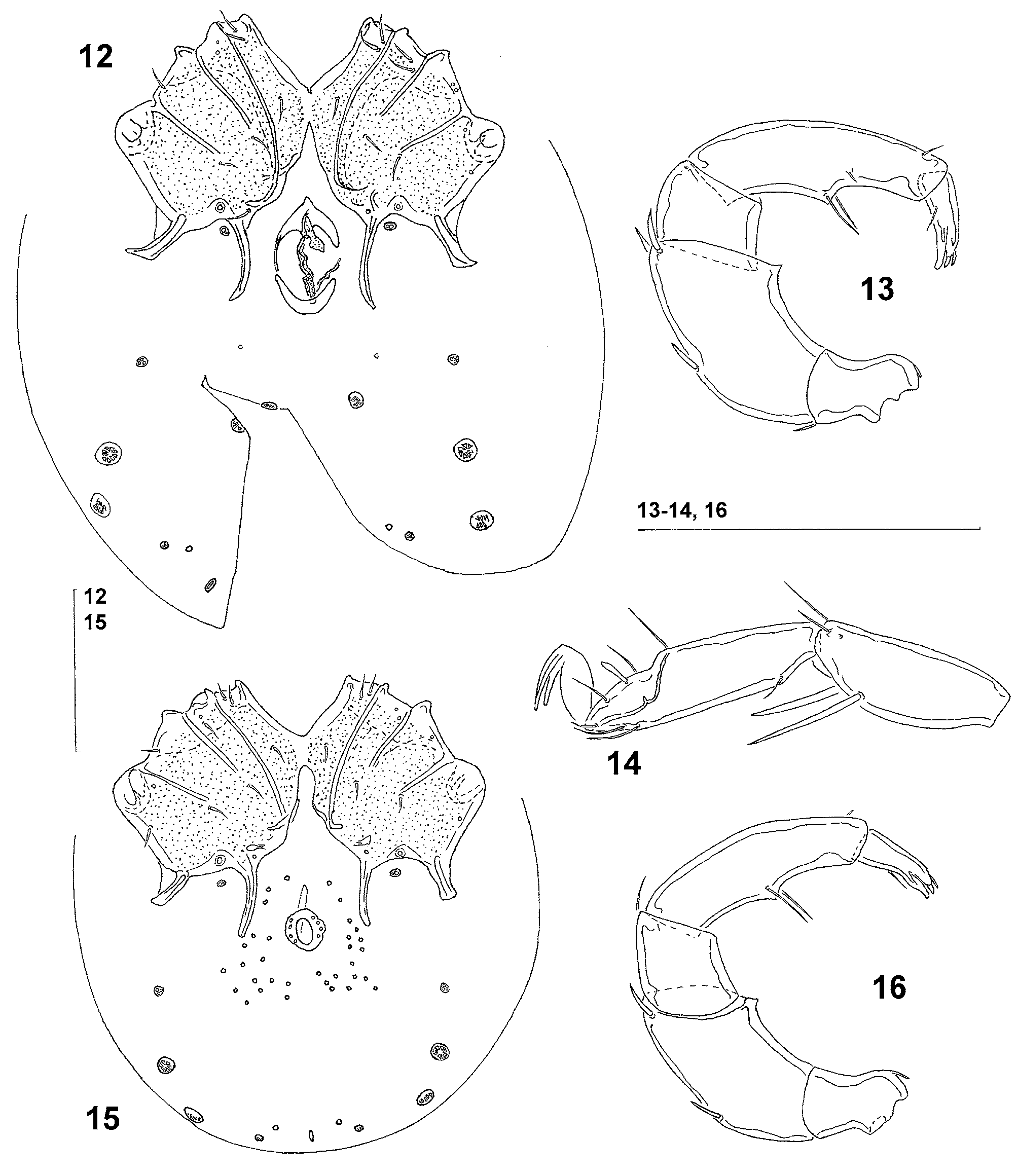
Morphology. Male: Idiosoma L/W 306/231. First coxal plates fused medially. Suture lines of coxal plates II/III and III/IV medially incomplete. Lateral apodemes of fourth coxal plates shorter than medial apodemes, the latter extending beyond anterior margin of genital field ( Fig. 15 View FIGURES 12 – 16 ). Genital field L/W 26/24, consisting of a sclerotized ring with four pairs of setae, 16–17 pairs of perigenital setae free in integument around genital field. A pair of small platelets with coxoglandularia 4 and associated setae placed between the posterior lateral and medial apodemes of the fourth coxal plates; the large glandularia-like structure fused with the fourth coxal plates. Posterior to the genital field a pair of platelets with three pores and three pairs of wheel-like acetabula (sensu Cook, 1996) or specialized glandularia (sensu Tuzovskij, 1978), with many radiating spokes Two of these wheel-like structures large, the most posterior one small with relatively few radiating spokes. Excretory pore unsclerotized, near the posterior idiosoma margin. Palp ( Fig. 16 View FIGURES 12 – 16 ) total L 202, dorsal L and %L (in parentheses): P-1 17 (8.4), P-2 62 (30.7), P-3 26 (12.9), P-4 70 (34.7), P-5 27 (13.4); L P-2/P-4 ratio 0.89; palp as in female. Lengths of I-Leg-4–6: 39, 55, 70; IV-Leg-4–6: 76, 86, 105; III-Leg-5, IV-Leg-4 and IV-Leg-5 with one swimming seta.
Female: Idiosoma L/W 343/263. First coxal plates fused medially. Suture lines of first and second coxal plates complete, suture lines of second and third coxal plates and suture lines of third and fourth coxal plates incomplete. Lateral apodemes of fourth coxal plates shorter than medial apodemes, the latter extending to posterior margin of genital field ( Fig. 12 View FIGURES 12 – 16 ). Genital field L/W 77/39. Pregenital and postgenital sclerite strongly bowed. A pair of small platelets with (according to Wiles et al. 2002) coxoglandularia 4 and associated setae placed between the posterior lateral and medial apodemes of the fourth coxal plates; the large glandularia-like structure fused with the fourth coxal plates. Posterior to the genital field a pair of platelets with three pores and four pairs of wheel-like acetabula (sensu Cook, 1996) or specialized glandularia (sensu Tuzovskij, 1978). Three of these wheel-like structures large with many radiating spokes, the most posterior one small with relatively few radiating spokes. Excretory pore unsclerotized, near the posterior idiosoma margin. Palp ( Fig. 13 View FIGURES 12 – 16 ) total L 221, dorsal L and %L (in parentheses): P-1 17 (7.7), P-2 72 (32.6), P-3 27 (12.2), P-4 76 (34.4), P- 5 29 (13.1); L P-2/P-4 ratio 0.95; P-2 with small ventrodistal peg-like projection ( Fig. 13 View FIGURES 12 – 16 ); ventral margin of P-4 with a strongly developed setal tubercle. Lengths of I-Leg-5–6 ( Fig. 14 View FIGURES 12 – 16 ): 59, 76; IV-Leg-4–6: 85, 94, 105; III-Leg-5 with one swimming setae, IV-Leg-4 and IV-Leg-5 with one and two swimming setae, respectively.
Deutonymph: As in adults, but lacking a genital field ( Fig. 17 View FIGURES 17 – 18 ). Idiosoma L/W 234/181; Palp ( Fig. 18 View FIGURES 17 – 18 ) total L 144, dorsal L and %L (in parentheses): P-1 13 (9.0), P-2 42 (29.2), P-3 21 (14.6), P-4 48 (33.3), P-5 20 (13.9); L P-2/P-4 ratio 0.88.
Remarks. Due to the first coxal plates medially fused and the large glandularia-like structure fused with the fourth coxal plates, the specimens from Tamar estuary show a general conformity with Litarachna amnicola Cook, 1986 . The description of L. amnicola is based on the two male specimens, taken from interstitial deposits of the George River in northwest Tasmania ( Cook 1986), less than 15 miles from the sea ( Cook 1996). The only differences are found in the presence of the small ventrodistal peg-like projection on P-2 (absent in type specimens of L. amnicola - see Cook 1986) and the presence of swimming setae on III- Leg-5, IV-Leg-4 and IV-Leg- 5 in the specimens from Tamar estuary (absent in type specimens of L. amnicola ). The loss of swimming setae in the type specimens might be the result of collecting these specimens in the hyporheic interstitial. The degree of variability of additional Tasmanian populations and carefully checking absence of the small ventrodistal peg-like projection on P- 2 in the type specimens are necessary before we can assess the taxonomic state of these specimens.
Distribution. Tasmania.

No known copyright restrictions apply. See Agosti, D., Egloff, W., 2009. Taxonomic information exchange and copyright: the Plazi approach. BMC Research Notes 2009, 2:53 for further explanation.
|
Kingdom |
|
|
Phylum |
|
|
Class |
|
|
Order |
|
|
Family |
|
|
Genus |