
Ceriomicrodon petiolatus Hull, 1937
|
publication ID |
https://doi.org/ 10.11646/zootaxa.3846.4.7 |
|
publication LSID |
lsid:zoobank.org:pub:F2E1FAE7-2A57-4666-ABBC-9DD9B4F8C767 |
|
DOI |
https://doi.org/10.5281/zenodo.5227838 |
|
persistent identifier |
https://treatment.plazi.org/id/2B275005-FF8F-FFB4-CDED-FC66FEA5FDA5 |
|
treatment provided by |
Felipe |
|
scientific name |
Ceriomicrodon petiolatus Hull, 1937 |
| status |
|
Redescription of Ceriomicrodon petiolatus Hull, 1937 View in CoL
( Figs 1–10 View FIGURES 1–4 View FIGURES 5–6 View FIGURES 7–10 )
Ceriomicrodon petiolatus Hull, 1937: 25 View in CoL . USNM: T M. Type-locality: Brazil, Mato Grosso, west border of Mato Grosso. Cerioimicrodon petiolatus Hull, 1937: 25 View in CoL (incorrect original spelling, by correction of Thompson et al. 1976: 60); Fluke 1957:
36 (incorrect original spelling, catalog citation). Ceriomicrodon poliolatus . Hull, 1949: 312 (incorrect original spelling). Microdon (Ceriomicrodon) petiolatus . Thompson et al. 1976: 60 (catalog citation). Ceriomicrodon petiolatus . Hull, 1949: 313 ( Fig. 13e View FIGURES 11–16 , habitus); Cheng & Thompson 2008: 35 (citation); Reemer & Ståhls 2013a: 27 (generic redescription), 158 (Figs 59–60, lateral habitus and male genitalia drawing).
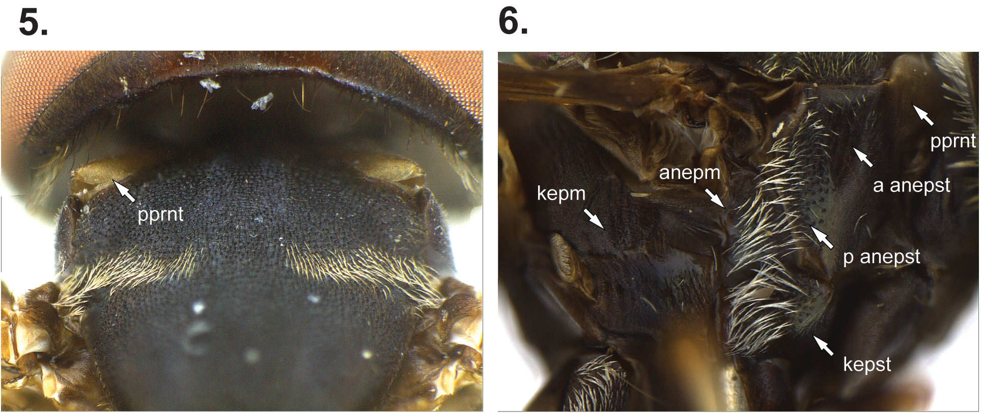
MALE. Head: Dark brown. Face convex and slightly expanded on the ventral 2/3, yellow with a dark median vitta on middle 1/3 that tapers toward anterior oral margin, face narrower than an eye, pile white. Oral margins slightly produced. Antennal fossa 2 times wider than high. Antenna longer than distance between antennal fossa and anterior oral margin; basoflagellomere oval and long, slightly expanded apically, 2.5 times longer than scape, bare. Dichoptic, eye margins slightly converging towards each other anterior to ocellar triangle ( Fig. 3 View FIGURES 1–4 ). Frons with white and appressed pile densely arranged on lateral margins and sparse medially, bare on lunule. Vertical triangle slightly produced; with sparse white pile. Eye bare, with linear depression, formed by enlarged ommatidia, at the level of the antennal insertion and extending 1/3 of the eye’s width from medial margin ( Fig. 3 View FIGURES 1–4 ). Occiput very narrow ventrally, slightly wider dorsally; with dark pile on dorsal 1/4, white elsewhere; pile distributed into 3–4 rows on ventral and dorsal 1/3, 2 rows on middle 1/3. Thorax: Dark brown. Postpronotum white and bare ( Fig. 5 View FIGURES 5–6 ). Scutum with very short dark pile, except slightly longer and white on suture ( Fig. 5 View FIGURES 5–6 ); transverse suture incomplete; notopleuron slightly expanded over the wing base as a thin triangular extension (the notal wing lamina). Scutellum semi-oval, small and without calcars; with very short dark and white pile. Anepisternum divided by shallow sulcus into anterior and posterior regions; anterior anepisternum with inconspicuous white pile antero-dorsally; posterior anepisternum with long white pile ( Fig. 6 View FIGURES 5–6 ). Katepisternum with long white pile postero-dorsally ( Fig. 6 View FIGURES 5–6 ). Anepimeron with long white pile anteriorly. Katepimeron flat ( Fig. 6 View FIGURES 5–6 ). Katatergum with bands of white microtrichia. Calypter white with dark margin; pile long on dorsal lobe and short on ventral lobe. Halter white. Metasternum developed, bare. Post-metacoxal bridge wide but narrowing towards middle. Legs: Anterior: Coxa, trochanter and basal 2/3 of femur brown, remaining of femur and basal 1/2 of tibia yellow, remaining of leg light orange; with short white pile, but inconspicuous on coxa. Medial: Coxa, trochanter and basal 2/3 of femur brown, remaining of femur and basal 1/2 of tibia yellow, remaining of leg light orange; with short white pile, inconspicuous on coxa, with some dark pile on femur. Posterior: Coxa, trochanter, most of the femur (except for the apex) and sub-apical ring on tibia brown, femur apex and basal ½ of tibia yellow, remaining of leg orange ( Fig. 1 View FIGURES 1–4 ); with short dark pile, except long and white on coxa, and more densely distributed on lateral ½ of coxa; femur with sub-basal ventral cicatrix; tibia with cicatrix on the sub-apical brown ring. Wing ( Fig. 2 View FIGURES 1–4 ): Hyaline, with slight darkening on apical 1/3 of cell c, around crossvein on cell sc, basal 1/3 of r 1, and antero-medially on r. With transversal vein between veins Sc and R 1, close to where vein Sc joins C. Vein r-m at basal 1/3 of cell dm. Vein R 4+5 with appendix into cell r 4+5. Vein M 1 perpendicular to vein R 4+5. Cell r 4+5 with round postero-apical corner. Wing bare on basal 2/3 of cell c, postero-basal margin of cell r 1, baso-anterior and baso-posterior margins of cell r 2+3, on cell r (except on spurious vein), basal 1/2 of cell r 4+5, cell bm, antero-basal and postero-basal margins of cell dm, cell cup (except the apex), antero-basal and postero-basal margins of cell cua 1 and antero-basal area on anal lobe; microtrichia sparse medial-basally on cell r 2+3. Alula convex, 2 times wider than cell c, with small bare region antero-medially. Abdomen ( Fig. 1 View FIGURES 1–4 ): Constricted and dark brown. Tergite 1 triangular; with white pile on basolateral ½, pile longer laterally. Tergite 2 very long and very narrow, 1.5 times longer than thorax, mostly pale except for apical ¼, apex yellow; bare on basal 1/3, with black pile elsewhere, except white on apex, sparse medially. Tergites 3 and 4 fused, but with sulcus visible between tergites; with black pile, except white on apical 1/4 of tergite 3, and baso-lateral corners and apical 1/6 of tergite 4. Sternite 1 bare. Genitalia ( Fig. 7 View FIGURES 7–10 ): Epandrium with ventrolateral ridge. Cercus pedunculate. Surstylus with wide base, tapering towards rounded apex, ventral margin convex medially, dorsal margin concave medially; with several spines on ventro-apical 2/3, with several pile on ventrobasal 2/3. Phallus furcate near to the apex, dorsal process long and whip-like, ventral process very short.
FEMALE ( Fig. 2 View FIGURES 1–4 ). Similar to male except: Eye margins do not converge towards each other ( Fig. 4 View FIGURES 1–4 ). Occipital dorsal pile mainly white, sometimes with some black pile intermixed. Scutum longer and white pile extends from one suture to the other, and white pile is also present on post-alar callus and anterior to scutellum. Calypter white to gray. Anterior and middle leg: Femur more extensively dark brown, usually just apex yellow. Posterior leg: Femur usually with basal 2/3 yellow, apex of tibia and tarsus dark orange; some specimens have the darker regions more distinct. Cell c sometimes bare only on basal 1/2 and cell r 2+3 bare on basal 1/3. Most of abdominal tergite 2 translucent brown; almost 2 times as long as thorax. Abdominal tergites 3–5 fused, but sulcus visible between tergites; white pile restricted to baso-lateral corners of tergites, white pile only on apico-lateral corners of tergites 4–5. Terminalia ( Figs 9–10 View FIGURES 7–10 ): Segments 7–8 elongated. Tergite 7 as a pair of apically tapering sclerotizations. Tergite 8 rectangular and long. Tergite 10 triangular and with pair of baso-lateral long apodemes (1/3 of the length of tergite 8, slightly longer than the length of tergite 10) ( Fig. 9 View FIGURES 7–10 ). Sternite 10 with pair of apico-lateral sclerotized stripes, each bearing a distinct long, apical, thin hair ( Fig. 10 View FIGURES 7–10 ). Cercus as large as tergite 10 and tapering basally.
Length: ♂ 9.0 mm, wing 5.9 mm; ♀ 10.7–12.6 mm, wing 7.4–9.0 mm.
Egg ( Figs 8 View FIGURES 7–10 , 11–16 View FIGURES 11–16 ): Length 0.43–0.46 mm (n = 3). Elongated-oval, rounded in one end, other end apparently truncate ( Fig. 8 View FIGURES 7–10 ). One of the ends of the egg ( Fig. 11 View FIGURES 11–16 ) has tuberculate structures that seem to bear orifices ( Fig. 12 View FIGURES 11–16 ) plus a pair of possible respiratory appendages ( Fig. 13 View FIGURES 11–16 ). Sculptures of the chorion form rectangular shapes, with paired papilla bearing possible aeropyles ( Fig. 14 View FIGURES 11–16 ). The putative micropyle apparatus is depicted in Fig. 15 View FIGURES 11–16 . The inner surface of the eggshell has a network of small tuberculate structures ( Fig. 16 View FIGURES 11–16 ).
Diagnosis: Ceriomicrodon may be confused with Mixogaster Macquart, 1842 and Rhopalosyrphus , but in Ceriomicrodon the postpronotum is bare ( Fig. 5 View FIGURES 5–6 ) and the second abdominal segment is very narrow and long (at least 1.5 times longer than thorax, Figs 1–2 View FIGURES 1–4 ).
Comments: Hull (1937) described the head as bluish black, eyes strongly converging at level of frons (also stated by Reemer & Stahls (2013a)), and second abdominal segment translucent yellow. Except for the second abdominal segment of the female, the other two characters from Hull’s description were not observed either in the studied specimens or in the pictures from the holotype (see redescription above) and are here considered a misinterpretation by Hull. The head shows some subtle bluish reflections when the specimens are held at certain angles, under fluorescent white light, but definitely not ‘bluish black’ as Hull stated. After comparing the male genitalia with the type (F.C. Thompson, personal communication), it was noticed that Reemer & Stahls (2013a) drawing of the male genitalia (Fig. 60, p. 158) underestimated the thickness of the spines on the surstylus ( Fig. 7 View FIGURES 7–10 ).
The specimen GFGM-MPEG 0009 is a variation, since it has the apical 1/2 of the middle tibia dark brown, a darker wing and is larger overall (body 12.6 mm, wing 9.0 mm).
The eggs depicted in Figs 8 View FIGURES 7–10 and 11–16 View FIGURES 11–16 were discovered accidentally, while preparing the female genitalia, in the abdomen of specimen INPA-DT0000003. All eggs went through the same maceration process, and the chorion was ruptured in the process of dissecting and preparing the eggs for MEV imaging. Although the description of the egg is still relevant to identify eggs of C. petiolatus , the description of the structures should be taken as preliminary until fresh material can be analyzed.
Material examined: Brazil, Amazonas, Parque Nacional do Jaú, Cachoeira, margem direita do rio Jaú , Igapó, Malaise trap, 22–29. VI.2003, D.M. Takiya (1 ♂, INPA-DT0000066 ); 25km NE de Manaus, Reserva Ducke, suspended trap at 20m, 01.xii.1988, J.A.Rafael (1 ♀, INPA-DT0000003 ); Maranhão, Bom Jardim, REBIO-Res. Biol. Gurupi , suspended trap, 17–27.I.2010, F. Limeira-de-Oliveira, J. T. Câmara & M.B.A. Neto (2 ♀, GFGMCZMA 0005 and 6); 02–11.IX.2010, F. Limeira-de-Oliveira, E.A.S. Barbosa & J.C. Silva (2 ♀, GFGMCZMA 0004 and 7); Rondônia, Ouro Preto do Oeste, suspended trap at 80m, 08–11.XI.1984 (1 ♀, GFGM-MPEG 0009 ); Roraima, Rio Uraricoera, Ilha de Maraca , Shannon trap, 02–13. V.1987, J.A. Rafael, J.E.B. Brasil & L.S. Aquino (1 ♀, INPA-DT0000741 ); suspended trap, 19–24.VII.1987, J.A. Rafael & L.S. Aquino (1 ♀, INPADT0000742 ) .


| VI |
Mykotektet, National Veterinary Institute |
| T |
Tavera, Department of Geology and Geophysics |
| V |
Royal British Columbia Museum - Herbarium |
No known copyright restrictions apply. See Agosti, D., Egloff, W., 2009. Taxonomic information exchange and copyright: the Plazi approach. BMC Research Notes 2009, 2:53 for further explanation.
|
Kingdom |
|
|
Phylum |
|
|
Class |
|
|
Order |
|
|
Family |
|
|
Genus |
Ceriomicrodon petiolatus Hull, 1937
| Miranda, Gil Felipe Gonçalves 2014 |
Ceriomicrodon petiolatus
| Thompson, F. C. & Vockeroth, J. R. & Sedman, Y. S. 1976: 60 |
| Hull, F. M. 1937: 25 |
| Hull, F. M. 1937: 25 |