
wasps
|
publication ID |
https://doi.org/ 10.11646/zootaxa.4885.4.5 |
|
publication LSID |
lsid:zoobank.org:pub:CBFB90F1-146A-4072-9685-7D7BA815FD0A |
|
DOI |
https://doi.org/10.5281/zenodo.4334016 |
|
persistent identifier |
https://treatment.plazi.org/id/312E8791-FF88-FFA0-FF20-FAB8279A7DFF |
|
treatment provided by |
Plazi |
|
scientific name |
wasps |
| status |
|
Key to species of the eusocial wasps View in CoL View at ENA occurring in Sulawesi Island
The present key is applicable to both females (“queens” and “workers”) and males unless the sexes are specified. Because of uncertainty of species identity and/or uncertainty of their establishment in Sulawesi Island as mentioned in “Taxonomic accounts and notes on distribution”, Ropalidia crassa van der Vecht, 1941 , R. jacobsoni (du Buysson, 1908) and R. variegata ( Smith, 1852) , are not included in this key. In addition to the figures given in this paper, the figures that can be referred to are available in van der Vecht (1941, 1962, and 1966), Archer (1989), Kojima (1999), Kojima et al. (2002) and Carpenter and Nguyen (2003).
1. Hind coxa with dorsal longitudinal carina on posterior face ( Fig. 13 View FIGURES 7–16 in Carpenter and Nguyen, 2003). Hind wing without jugal lobe. Metasoma sessile; T1 with anterior abrupt declivity ( Figs. 1, 3–4 View FIGURES 1–6 ; Figs. 14–15 View FIGURES 7–16 in Carpenter and Nguyen, 2003)....................................................................................... 2, Vespinae , genus Vespa View in CoL
- Hind coxa without carina (Figs. 38–39 in Carpenter and Nguyen, 2003). Hind wing with jugal lobe. Metasoma variable in shape, subsessile or petiolate ( Figs. 8, 11, 13 View FIGURES 7–16 , 17, 19–20, 22–25; Figs. 16–18 View FIGURES 7–16 View FIGURES 17–25 in Carpenter and Nguyen, 2003)...................................................................................................... 5, Polistinae
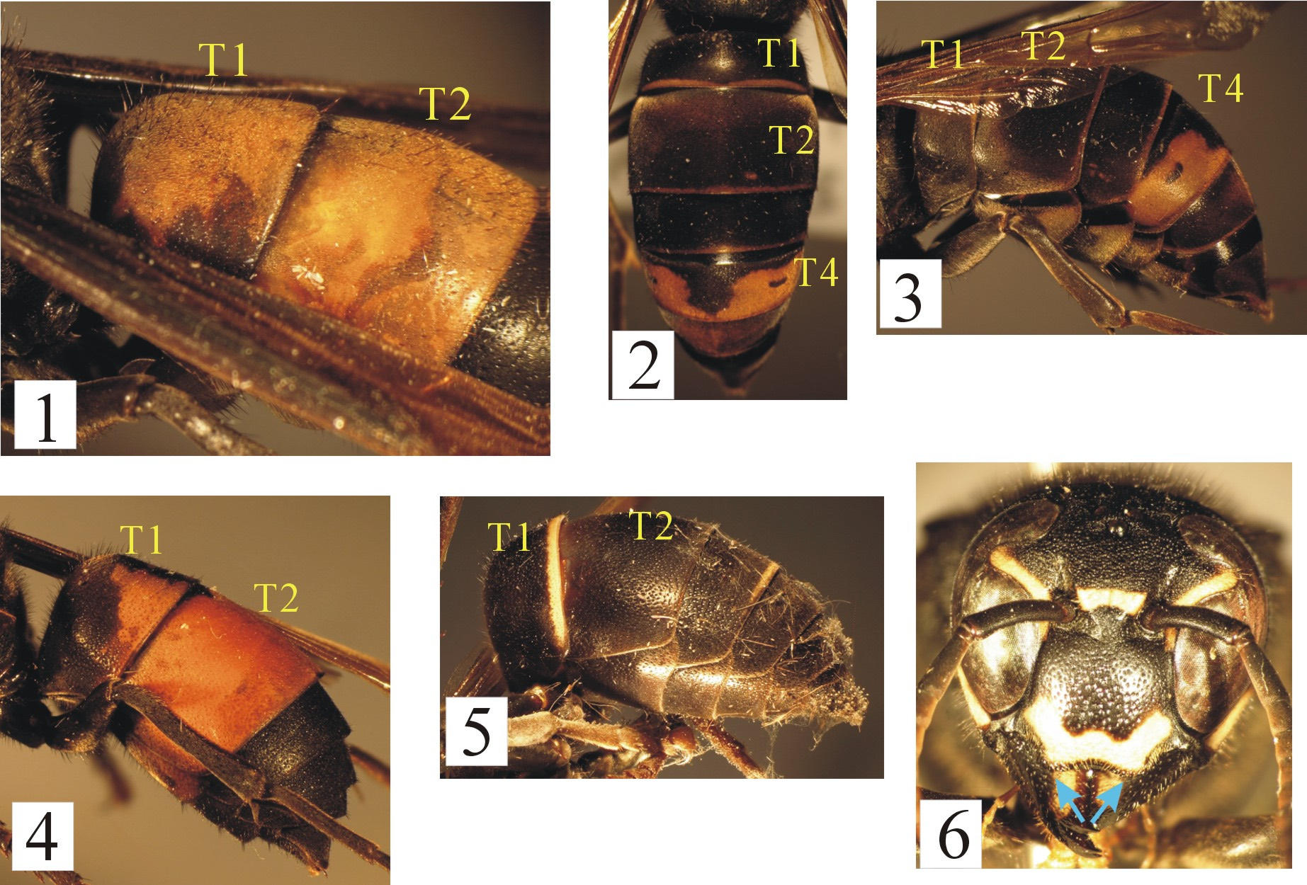
2. Female clypeus with bluntly triangular tooth on each side of ventromedian excision ( Fig. 27A View FIGURES 26–29 in Archer, 1989). Body covered with stiff hairs. T1 almost entirely colored orange (T1 and 2 each with apical yellowish orange band) ( Fig. 1 View FIGURES 1–6 )........................................................................................ Vespa tropica ( Linnaeus, 1758) View in CoL
- Female clypeus with short, broadly rounded lobe on each side of ventromedian excision ( Fig. 6 View FIGURES 1–6 ). T1 mostly black ( Figs. 2–3, 5 View FIGURES 1–6 ) or with dorsal face nearly entirely orange yellow ( Fig. 4 View FIGURES 1–6 )................................................... 3
3. Pronotal carina widely interrupted by pronotal fovea ( Fig. 29B View FIGURES 26–29 in Archer, 1989). Pretegular carina extending ventrally to the level of middle of pronotal tubercle ( Fig. 29B View FIGURES 26–29 in Archer, 1989). T4 with broad apical yellowish-brown band ( Figs. 2–3 View FIGURES 1–6 ).............................................................................. Vespa velutina Lepeletier, 1836 View in CoL
- Pronotal carina only narrowly interrupted by pronotal fovea ( Fig. 29A View FIGURES 26–29 in Archer, 1989). Pretegular carina extending ventrally beyond the level of ventral margin of pronotal tubercle ( Fig. 29A View FIGURES 26–29 in Archer, 1989)................................. 4
4. T1 relatively long, more than 2/3 as long as T2 length in dorsal view. Head and mesosoma black, sometimes with ferruginous or dull red markings, never with yellow markings. T1 with dorsal face largely orange yellow and T2 entirely orange yellow ( Fig. 4 View FIGURES 1–6 )......................................................................... Vespa affinis ( Linnaeus, 1764) View in CoL
- T1 short, transverse, about half as long as T2 length in dorsal view. Body black, with following parts cream-yellow: apical margin of clypeus, metanotum, apical band of T1 ( Figs. 5–6 View FIGURES 1–6 )............................. Vespa fervida Smith, 1859 View in CoL
5. First metasomal segment subsessile, not petiolate ( Figs. 8, 10–11, 13 View FIGURES 7–16 ); S1 bluntly angled basally ( Fig. 8 View FIGURES 7–16 ).............................................................................................. 6, Polistini , genus Polistes View in CoL
- First metasomal segment in most cases petiolate ( Figs. 17, 19–20, 22–23 View FIGURES 17–25 ); S1 flat throughout.............. 9, Ropalidiini
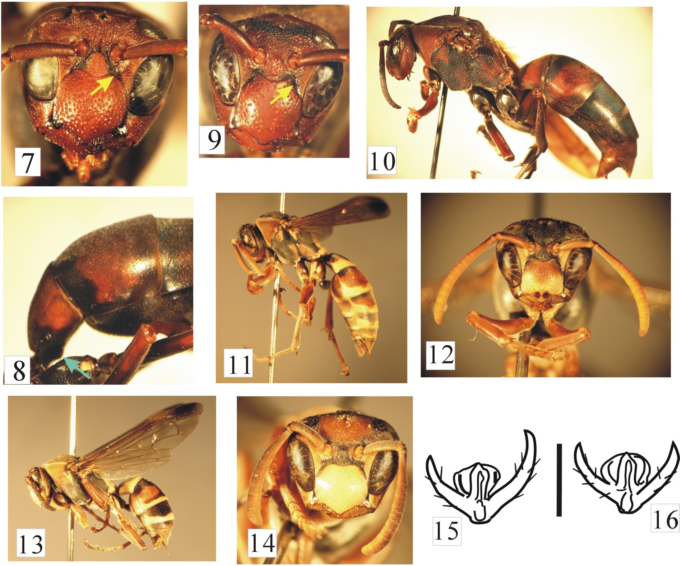
6. Clypeus not produced dorsally beyond the level of anterior tentorial pits ( Fig. 7 View FIGURES 7–16 ). Pronotal fovea present. Mesepisternum with dorsal groove, without epicnemial groove. Body brown to dark brown; T1 and S1 brown or blackish brown ( Figs. 7–8 View FIGURES 7–16 )............................................................... Polistes (Gyrostoma) tenebricosus Lepeletier, 1836 View in CoL
- Clypeus produced dorsally beyond the level of anterior tentorial pits ( Figs. 9, 12, 14 View FIGURES 7–16 ). Pronotal fovea, epicnemial carina and dorsal groove, all absent. Body color and markings similar to P. tenebricosus View in CoL ( Figs. 9–10 View FIGURES 7–16 ; P. sagittarius View in CoL ) or much brighter ( Figs. 11–14 View FIGURES 7–16 )............................................................................ 7, subgenus Polistella
7. Larger species; forewing length 15-20 mm. Gena wide, in profile about 0.7-0.9 times as wide as eye in female ( Fig. 10 View FIGURES 7–16 ). Fore wing without a dark spot. Propodeum with strong transverse striae. Body ferruginous to dark brown, sometimes T1 and/or T2 with orange-yellow or dark brownish bands ( Fig. 8 View FIGURES 7–16 ).................. Polistes (Polistella) sagittarius de Saussure, 1853 View in CoL
- Smaller species, forewing length 11-14 mm. Gena about 0.6-0.7 times as wide as eye in female ( Figs. 11, 13 View FIGURES 7–16 ). Fore wing with apical dark spot ( Figs. 11, 13 View FIGURES 7–16 ). Propodeum shallowly excavated medially and with weak transverse striae. Body with extensive yellow and reddish-brown markings ( Figs. 11–14 View FIGURES 7–16 )............................... 8, Polistes (Polistella) stigma View in CoL group
8. Hind tarsal claw asymmetrical ( Fig. 15 View FIGURES 7–16 ). Clypeus reddish-brown in basal half and yellow in apical half, with a small pair reddish-brown spots on the apex, scattered punctures, rather deep punctures ( Fig. 12 View FIGURES 7–16 ). T1-4 with apical yellow bands; S2-4 with broad apical yellow bands ( Fig. 11 View FIGURES 7–16 )..................................... Polistes (Polistella) celebensis Selis, 2018
- Hind tarsal claws more or less symmetrical ( Fig. 16 View FIGURES 7–16 ). Clypeus entirely yellow, with shallow punctures ( Fig. 14 View FIGURES 7–16 ). T4 without apical yellow band, S2 with posterolateral spots and S3 with narrow apical yellow band ( Fig. 13 View FIGURES 7–16 )................................................................................ Polistes (Polistella) stigma View in CoL stigma ( Fabricius, 1793) View in CoL
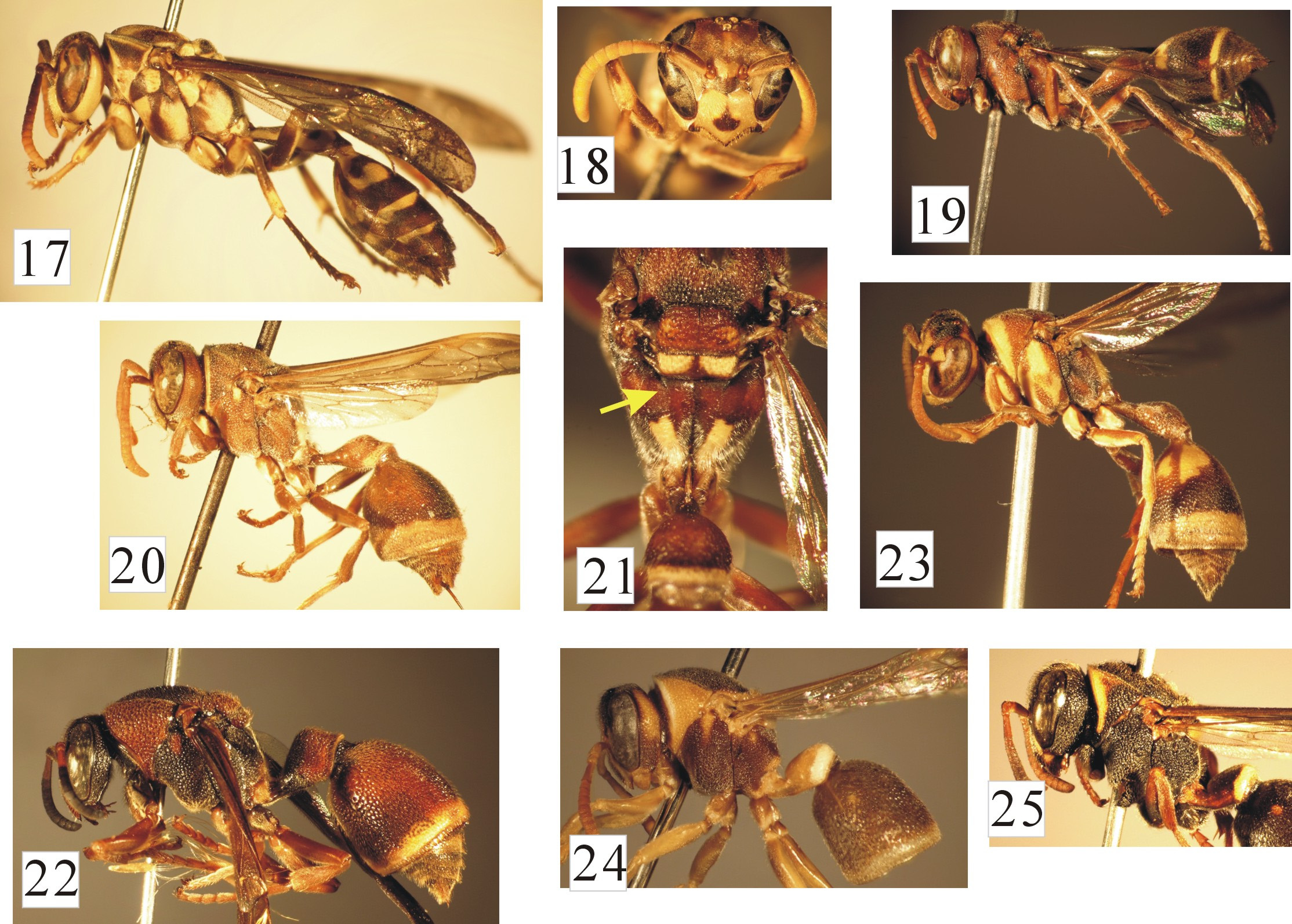
9. Pronotal fovea, dorsal groove of mesepisternum, pretegular carina, all present ( Fig. 17 View FIGURES 17–25 ; Fig. 39 in Carpenter and Nguyen, 2003). T2 and S2 not fused, overlapping ( Fig. 17 View FIGURES 17–25 ). Body mostly brown with yellow markings ( Figs. 17–18 View FIGURES 17–25 ; Fig. 12 View FIGURES 7–16 in van der Vecht, 1966).................................................................. Parapolybia varia ( Fabricius, 1787) View in CoL
- Pronotal fovea, dorsal groove of mesepisternum, pretegular carina, all absent ( Figs. 19–20, 22–25 View FIGURES 17–25 ; Fig. 38 in Carpenter and Nguyen, 2003). T2 and S2 fused, leaving at most suture ( Figs. 19–20, 22–24 View FIGURES 17–25 )...................... 10, genus Ropalidia View in CoL
10. Suture between T2 and S2 more or less distinct, visible from base to apex of the segment ( Fig. 19 View FIGURES 17–25 ; Fig. 19 View FIGURES 17–25 in van der Vecht, 1941). First metasomal segment elongate, flask-shaped, more than twice as long as wide in dorsal view. T1 laterally rufous, rarely with yellow spots at base; S2 rarely with yellow spots ( Fig. 19 View FIGURES 17–25 )............. Ropalidia mathematica (Smith, 1860) View in CoL
- Suture between T2 and S2 indistinct, hardly visible in posterior part ( Figs. 20, 22–24 View FIGURES 17–25 ). First metasomal segment shorter, less than twice as long as wide in dorsal view ( Fig. 21 View FIGURES 17–25 )......................................................... 11
11. Propodeum with paired basal longitudinal carinae, running from posterolateral angles of metanotum ( Fig. 21 View FIGURES 17–25 ).......... 12
- Propodeum without carinae............................................................................ 13
12. In dorsal view head narrower than mesosoma excluding tegulae; female gena narrow, less than half as wide as eye in lateral view ( Fig. 22 View FIGURES 17–25 ). Apical margin of T2 and S2 armed with short, blunt teeth ( Fig. 22 View FIGURES 17–25 ; Fig. 9 View FIGURES 7–16 in van der Vecht, 1941). Body coarsely punctate, clypeus with scattered punctures......................................... Ropalidia pilosa (Smith, 1858) View in CoL
- In dorsal view head wider than mesosoma excluding tegulae; female gena as wide as eye in lateral view ( Fig. 20 View FIGURES 17–25 ). Apical margin of T2 and S2 without teeth ( Fig. 20 View FIGURES 17–25 ). Body not strongly punctate, but clypeus densely punctate............................................................................................ Ropalidia marginata ( Lepeletier, 1836) View in CoL
13. T1 elongate ( Fig. 23 View FIGURES 17–25 ), in dorsal view gradually swollen posteriorly and narrowed near apical margin. Second metasomal seg-ment in lateral view obliquely cut off apically, T2 longer than S2 ( Fig. 23 View FIGURES 17–25 ). Male terminal antennal flagellomere bullet-shaped ( Fig. 3 View FIGURES 1–6 in Kojima, 1999)................................................... Ropalidia impetuosa (Smith, 1860) View in CoL
- T1 short ( Figs. 24–25 View FIGURES 17–25 ), in dorsal view gradually swollen posteriorly toward apical margin. Male terminal antennal flagellomere slightly curved...................................................................................... 14
14. T1 gradually swollen dorsally from the posterior end of basal slit for the ligament connecting propodeum and T1, then strongly widened laterally as long as less than 1.5 times high (Fig. 43 in van der Vecht, 1941). Median excavation of propodeum rather shallow............................................................. Ropalidia cyathiformis ( Fabricius, 1804) View in CoL
- T1 barely petiolate basally, in lateral view abruptly swollen dorsally at the posterior end of basal slit to receive propodeal liga-ment ( Figs. 24–25 View FIGURES 17–25 ). Median excavation of propodeum rather wide and deep................ 15, Ropalidia plebeja View in CoL group
15. T 1 in lateral view strongly swollen dorsally just behind basal slit to receive propodeal ligament, with dorsal surface relatively broadly curved down toward posterior margin from its highest point, in dorsal view distinctly narrowing posteriorly near pos-terior margin ( Fig. 24 View FIGURES 17–25 ; Figs. 8–9 View FIGURES 7–16 in Kojima et al., 2002). In female, POD less than 3 times Od ( Fig. 13 View FIGURES 7–16 in Kojima et al., 2002); gena in lateral view less than 0.9 times as wide as eye ( Fig. 24 View FIGURES 17–25 ; Fig. 2 View FIGURES 1–6 in Kojima et al., 2002)............................................................................................... Ropalidia plebeja (de Saussure, 1862) View in CoL
- T 1 in lateral view less strongly swollen dorsally at posterior margin, with dorsal face broadly and weakly curving down toward posterior margin from its highest point, in dorsal view barely narrowing posteriorly ( Fig. 25 View FIGURES 17–25 ; Figs. 55–56 in Kojima et al., 2002). In female, POD more than 3 times Od (Fig. 50 in Kojima et al., 2002); gena in lateral view nearly as wide as eye ( Fig. View FIGURES 17–25 25; Fig. 49 in Kojima et al., 2002)..................................... Ropalidia celebensis (van der Vecht, 1941) View in CoL

No known copyright restrictions apply. See Agosti, D., Egloff, W., 2009. Taxonomic information exchange and copyright: the Plazi approach. BMC Research Notes 2009, 2:53 for further explanation.