
Arjuna, MUIR, 1934
|
publication ID |
https://doi.org/ 10.1111/zoj.12319 |
|
DOI |
https://doi.org/10.5281/zenodo.10543617 |
|
persistent identifier |
https://treatment.plazi.org/id/316487FC-FFFA-FFD8-FEC7-4C77FA449D0D |
|
treatment provided by |
Marcus |
|
scientific name |
Arjuna |
| status |
|
Arjuna Muir, 1934: 583 View in CoL ; Metcalf 1946: 90; Emeljanov 2008: 311; Emeljanov 2011: 1124.
Type species
Arjuna dohertyi Muir, 1934 View in CoL ; by original designation.
Diagnosis
The genus can be distinguished by the following combination of characters: cephalic process curved downwards before eyes; vertex with a secondary longitudinal carina bifurcating from subapex of lateral carinae; frons with lateral carinae well developed from clypeus, but obscure and gradually disappearing anterad to eyes, only a pale trace extending forwards; intermediate carinae extending before eyes; median carina complete, robust, and strongly convex; and aedeagus with a pair of ventrolateral lobes twining around endosomal processes.
Redescription
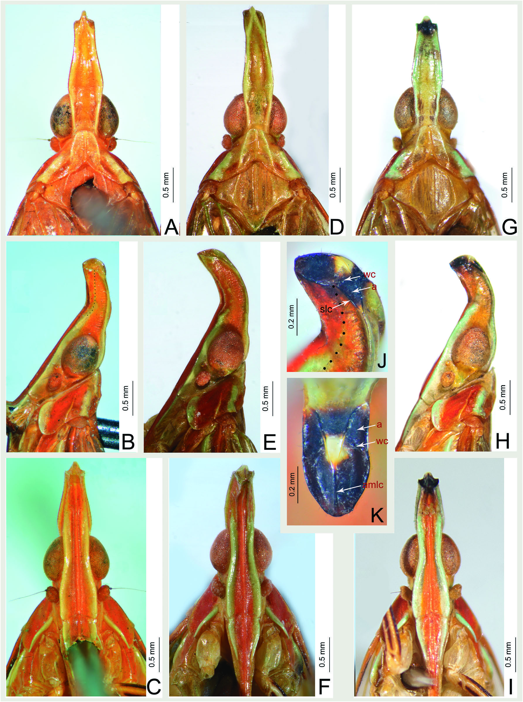
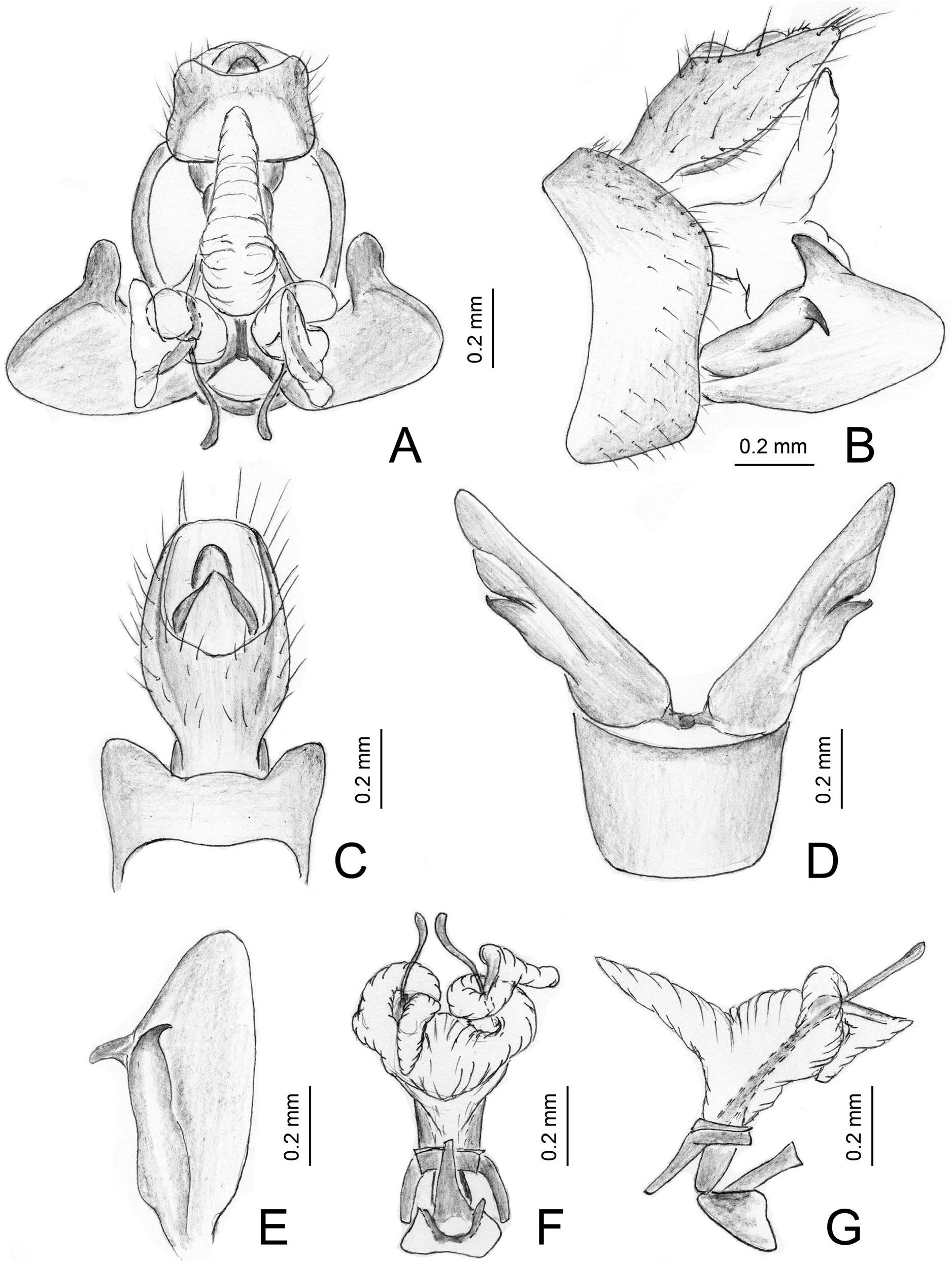
Head ( Fig. 14A–I View Figure 14 ) moderately elongate, as long as or slightly longer than pronotum and mesonotum combined, and curved downwards before eyes. Vertex ( Fig. 14A, D, G View Figure 14 ) with lateral carinae distinctly thickened, gradually widened from base, widest before eyes, gradually narrowed towards subapex, then abruptly converging to apex; surface between lateral carinae more or less convex, median carina absent, a transverse depression present at widest area; apex acute; posteri- or margin weakly ridged, arcuately concave. Apex of cephalic process with sharp secondary longitudinal carina bifurcating from subapex of lateral carinae of vertex, extending to the dorsal branch of lateral carinae of frons forming areolets (lanceolate cells in Emeljanov, 2008: 311), then joining to apex of intermediate carinae of frons ( Fig. 14J, K View Figure 14 ). Frons ( Fig. 14C, F, I View Figure 14 ) widest below antennae, slightly constricted between eyes and narrowed forwards; lateral carinae well developed from clypeus, but obscure and gradually disappearing anterad to eyes, only a pale trace extending forwards ( Fig. 14B, J View Figure 14 ); a weak carina crossed areolets, sometimes too weak to be visible ( Emeljanov, 2008); intermediate carinae converging posteriorly and extending before eyes; median carina complete, robust, and strongly convex. An apical median longitudinal carina between anterior margins of frons and vertex very sharp and convex ( Fig. 14K View Figure 14 ). Eyes nearly rounded, callus postocularis reduced, forming a triangular process projecting posteriorly. Antennae shifted backwards, pedicel globose, with ∼40 distinct sensory plaque organs distributed over the entire surface.
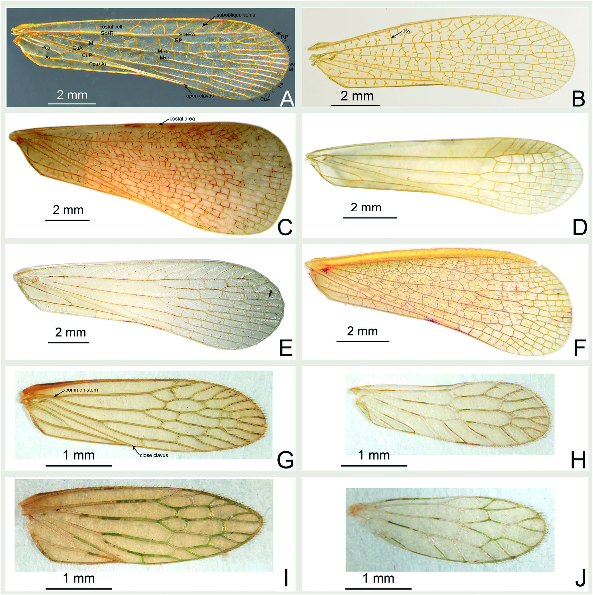
Pronotum ( Fig. 14A, D, G View Figure 14 ) relatively large, longer than half of mesonotum; anterior margin centrally arcuately convex, lateral marginal areas straight and sloping, with two lateral carinae on each side; upper carinae narrower than the lower carinae, so in dorsal view the lower carinae are distinctly visible ( Fig. 14A, D, G View Figure 14 ); posterior margin broadly angularly concave; disc flat and tricarinate, median and intermediate carinae sharp and high, with a lateral pit on each side. Mesonotum ( Fig. 14A, D, G View Figure 14 ) transversely broad, short, flat, and tricarinate; lateral carinae weakly tortuous and abruptly incurved apically, slightly converging forwards. Tegmina ( Fig. 2G View Figure 2 ) nearly three times as long as broad; apical area with transverse veins sparse. Legs elongate and slender, fore and middle femora and tibiae distinctly elongate, hind tibiae more than twice as long as hind femora; fore and middle femora slightly thickened at apex, without spine; hind tibiae with four lateral spines and seven apical teeth; hind tarsomeres I and II with between eight and 13 apical teeth, respectively.
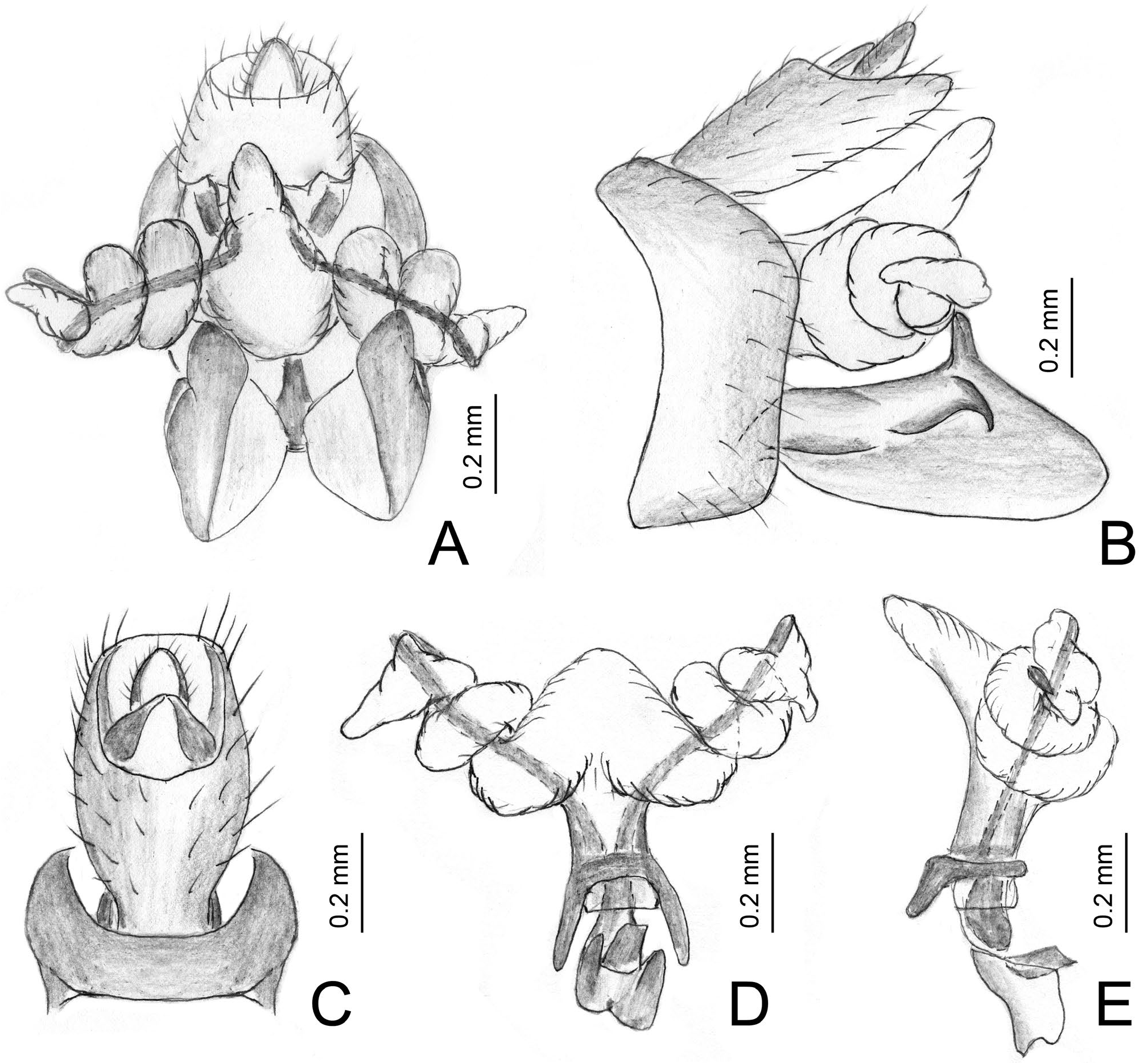
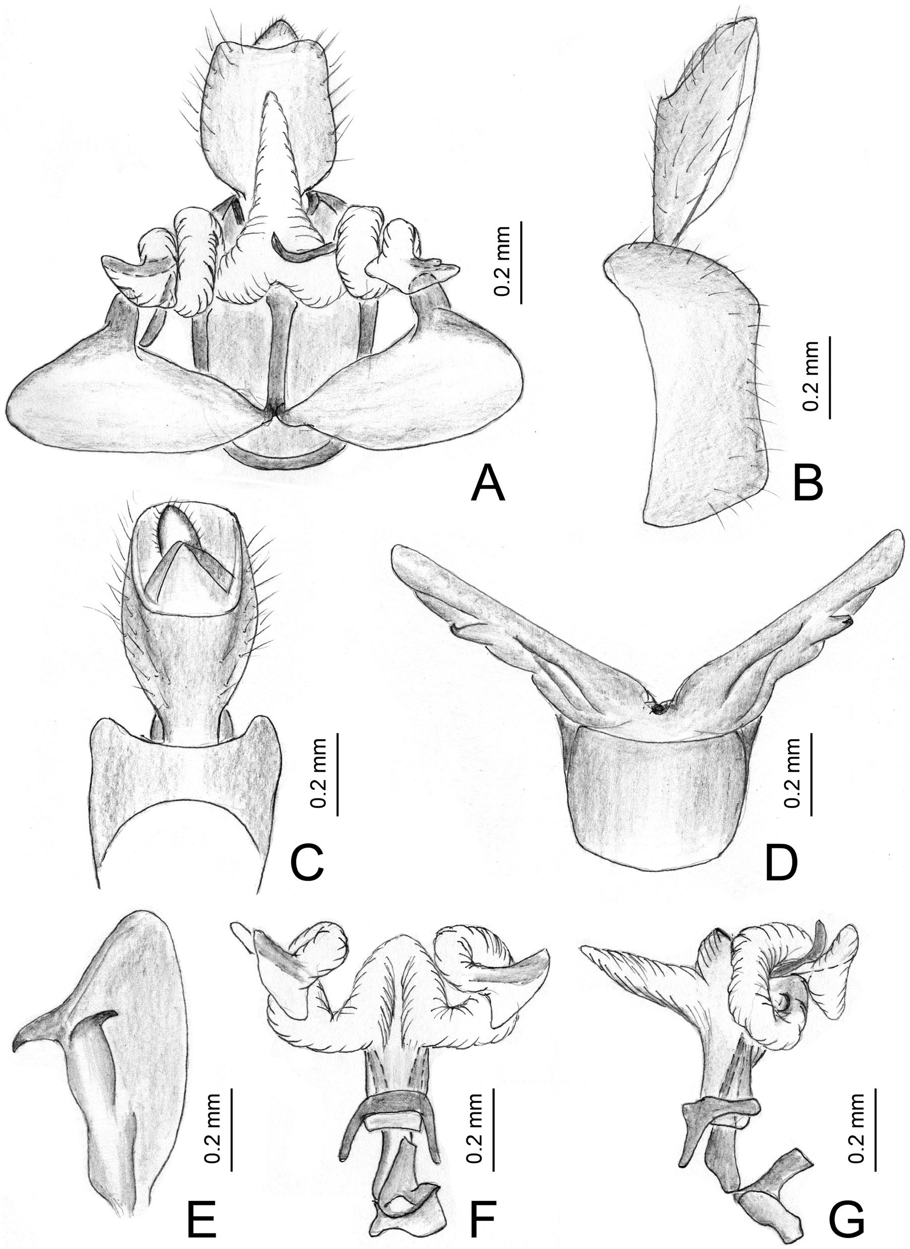
Male genitalia with pygofer distinctly narrow and elongate, more than twice ventral width, distinctly wider ventrally than dorsally, in lateral view posterior margin more or less projecting near upper middle ( Figs 15B View Figure 15 , 16B View Figure 16 , 17B View Figure 17 ). Gonostyles symmetrical; base narrow, expanded towards middle, then gradually narrowed towards apex; apex bluntly rounded and projecting backwards; upper process elongate, obtuse, and compressed dorsoventrally ( Figs 16E View Figure 16 , 17E View Figure 17 ). Aedeagus ( Figs 15A, D, E View Figure 15 , 16A, F, G View Figure 16 , 17A, F, G View Figure 17 ) with a pair of slender and moderately long endosomal processes extended from phallotheca: pigmented, sclerotized, nearly straight, and obtuse apically; phallobase sclerotized and pigmented at base, with inflated membranous apical lobes, without spines; dorsal lobe directed posteriorly and two ventrolateral lobes twining around endosomal processes, directed laterally. Segment X in dorsal view long, oval, dorsal margin deeply excavated to accommodate anal style, with ratio of length to width at base
No known copyright restrictions apply. See Agosti, D., Egloff, W., 2009. Taxonomic information exchange and copyright: the Plazi approach. BMC Research Notes 2009, 2:53 for further explanation.
|
Kingdom |
|
|
Phylum |
|
|
Class |
|
|
Order |
|
|
Family |
Arjuna
| Song, Zhi-Shun, Szwedo, Jacek, Wang, Rong-Rong & Liang, Ai-Ping 2016 |
Arjuna
| Emeljanov AF 2008: 311 |
| Metcalf ZP 1946: 90 |
| Muir F 1934: 583 |