
Domorganus gigas, Gagarin, Vladimir G. & Naumova, Tatyana V., 2015
|
publication ID |
https://doi.org/10.11646/zootaxa.4052.4.9 |
|
publication LSID |
lsid:zoobank.org:pub:B74818AF-5673-47E8-B648-85CC19131085 |
|
DOI |
https://doi.org/10.5281/zenodo.6106885 |
|
persistent identifier |
https://treatment.plazi.org/id/3171FB30-2D69-5855-FF22-FB682936F8FF |
|
treatment provided by |
Plazi |
|
scientific name |
Domorganus gigas |
| status |
|
Genus Domorganus Goodey, 1947
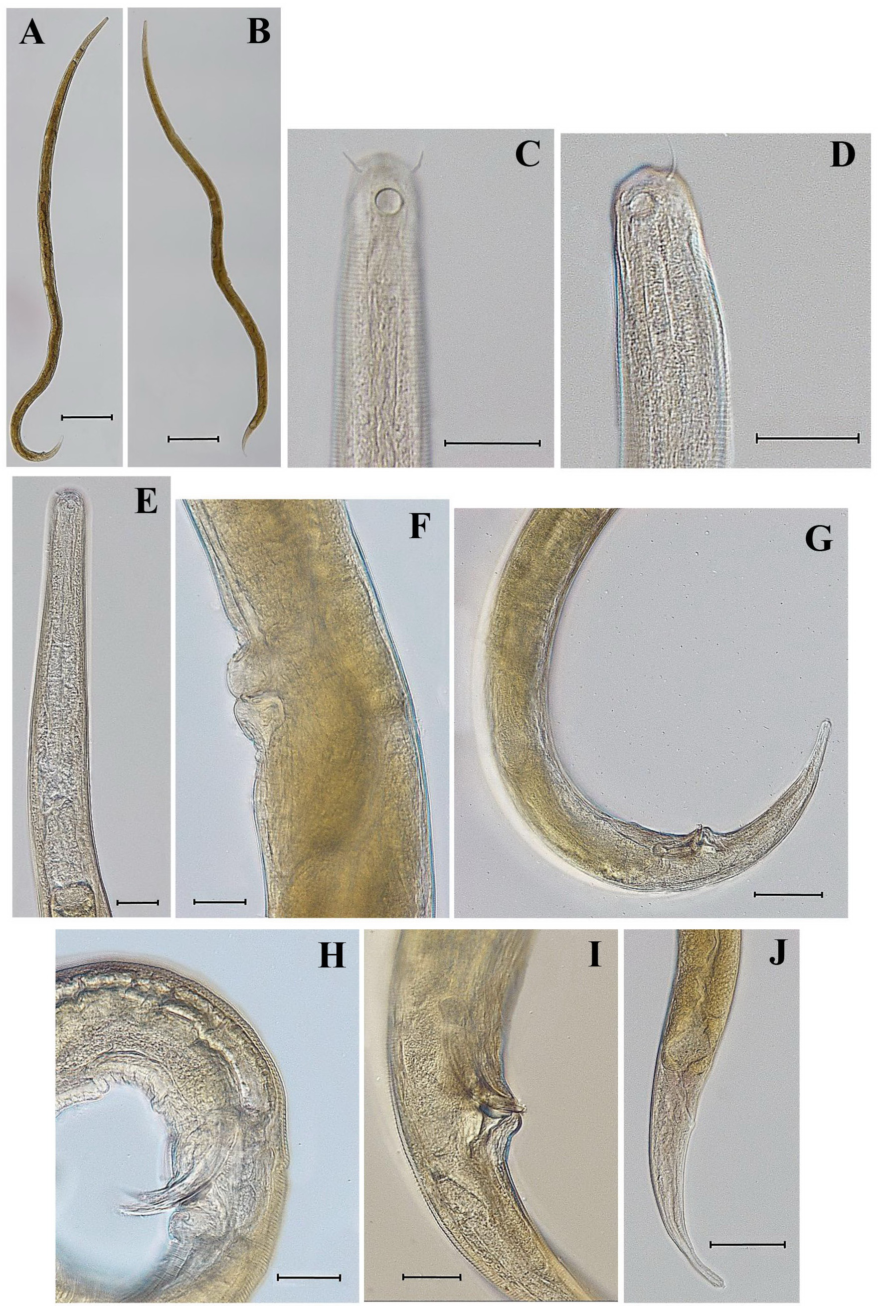
Domorganus gigas sp. n. ( Figs 1 View FIGURE 1 , 2 View FIGURE 2 ; Table)
Type material. Holotype male, slide reference number 102/49, deposited in the Helminthological Museum RAS, Institute of Ecology and Evolution, Center of Parasitology, Russian Academy of Sciences, Moscow, Russia. Paratypes: 10 ♂♂, 10 ♀♀ deposited in the collection of the Limnological Institute, Siberian Branch of Russian Academy of Sciences, Irkutsk, Russia.
Measurements. Table 2.
Type habitat and locality. Bolshie Koty Bay (near Dva Brata rock), Lake Baikal, Siberia, Russia, depth 3 m, sand.
Etymology. The specific epithet “ gigas ” means “very big”, “gigantic”.
Description. Male. Body comparatively long and thin. Anterior and posterior ends of body narrowed. Cuticle finely annulated, 2–3 µm thick. Lateral alae in the shape of narrow, smooth bands demarcated by two straight lines beginning at the level of middle of the pharynx and terminating at middle of the tail. Lateral alae about 0.3 times as wide the corresponding diameter at mid-body, narrower at the level of pharynx and on tail. Hypodermal somatic setae, glands and body pores absent. Anterior edge of head truncated. Labial sensillae very small, not seen under light microscope. Cephalic sensillae in the shape of thin setae 8–11 µm long (51–55% of labial region diameter). Stoma very small, its walls not cuticularized. Amphidial fovea circular, open in its posterior part, occupying 30–35% of the corresponding body diameter, situated about 0.4 labial region diameter from the anterior body end. Pharynx muscular with metacorpal and basal bulbs. Valvular apparatus in basal bulb absent. Cardia small, oval in shape, muscular, surrounded by intestinal tissue. Ventral gland not visible. Excretory pore situated 76–98 µm behind cardia. Excretory canal is not cuticularized.
Testes paired, outstretched, opposed. Anterior testis situated on the left side and posterior testis on the right side of the intestine. Spicules comparatively short, curved, cephalated, 1.3–1.5 times as long as cloacal body diameter. Gubernaculum short, in the shape of a curved plate with long caudal apophysis. One precloacal supplement appearing as a large papilla and located 43–55 µm from cloaca (at the level of proximal end of spicules). Tail slender, comparatively long, gradually narrowing to the terminus. Caudal setae absent. Three caudal glands open through a terminal spinneret appearing like a short tube.
TABLE. Morphometrics of Domorganus gigas sp. n.
Female. General morphology, structure of the cuticle and anterior portion of the body similar to that of male. Reproductive system didelphic, amphidelphic. Anterior ovary situated on the left side of intestine, posterior ovary on right side of intestine. Ovaries comparatively long, often with two flexures and reaching almost to vulva. Vulva a transverse slit, situated about mid-body. Vulva lips sclerotized, protruding beyond body contour. Vagina short, straight, with muscular walls. Numerous oogonia present in blind end of ovaries, followed by an oocyte zone, from which extends the thin oviduct made up of one row of small cells. Uterus spacious with one-two mature eggs measuring 93–122 x 37 – 48 µm. Tail slender, comparatively long, gradually narrowing to terminus. Three caudal glands open through the short, tube-shaped spinneret.
Diagnosis. Body comparatively long and thin (males: L = 2258–3084 µm, a = 39–56; females L = 2648–3282 µm, a = 34–44). Cuticle finely annulated, with lateral alae in the shape of narrow, smooth bands. Somatic setae, hypodermal glands and body pores absent. Labial sensillae very small, not seen under light microscope. Cephalic sensillae in the shape of thin setae, 9–11 µm or 51–55% of labial region diameter. Stoma very small. Amphidial fovea circular, occupying 30–35% of the corresponding body diameter, situated about 0.4 labial region diameter from the anterior body end. Pharynx muscular with metacorpal and cardial bulbs. Excretory pore situated 76–98 µm behind cardia. Female reproductive system didelphic, amphidelphic. Vulva equatorial. Ovaries comparatively long, often with two bends. One or two mature eggs measuring 93–122 x 37 –48 µm. Testes paired, outstretched, opposed. Spicules curved, cephalated, 50–54 µm long. Gubernaculum short, plate-shaped with long caudal apophysis. One precloacal papilliform supplement. Tail slender, comparatively long, gradually narrowing to the terminus. Three caudal glands open through short, tubeshaped spinneret.
Differential diagnosis. At present, the genus Domorganus Goodey, 1947 contains nine valid species: D. macronephriticus Goodey, 1947 ( type species); D. delgadoi Hernandez & Jordana, 1990 ; D. navarrensis Hernandez & Jordana, 1990 ; D. oligochaetophilus Thun, 1967 ; D. beklemishevi Valovaya, 1989 ; D. bathybius ( Schneider, 1943) Lorenzen, 1981 ; D. acutus ( Tsalolikhin, 1977) Lorenzen, 1981 ; D. subtilis ( Tchesunov, 1978) Lorenzen, 1981 ; and D. suecicus Holovachov, 2012 ( Holovachov 2012) . D. gigas sp. n. is morphologically close to D. acutus , also found in Lake Baikal ( Tsalolikhin 1977, Gagarin & Naumova 2011), but differs in the longer body (L = 2258–3282 µm versus L = 1439–2200 µm in D. acutus ), position of vulva (V = 47.9–52.0% versus V = 54–60% in D. acutus ), longer cephalic setae (9–11 µm long, 51–55% of labial region diameter versus 1.0–1.5 µm long, 10–15% of labial region diameter, position of amphidial fovea (0.4 labial region diameter from anterior body end versus 0.7 labial region diameter in D. acutus ), longer spicules (50–54 µm long versus 36–41 µm long in D. acutus ) and location of excretory pore (at distance 76–98 µm behind cardia versus 12–14 µm behind cardia in D. acutus ( Tsalolikhin 1977, Gagarin & Naumova 2011).

No known copyright restrictions apply. See Agosti, D., Egloff, W., 2009. Taxonomic information exchange and copyright: the Plazi approach. BMC Research Notes 2009, 2:53 for further explanation.
|
Kingdom |
|
|
Phylum |
|
|
Class |
|
|
Order |
|
|
Family |
|
|
Genus |