
Simulium lobatoi Luna Dias, Hernández, Maia-Herzog & Shelley
|
publication ID |
https://doi.org/ 10.5281/zenodo.274211 |
|
DOI |
https://doi.org/10.5281/zenodo.6229785 |
|
persistent identifier |
https://treatment.plazi.org/id/320387A6-FF95-FFF3-83DD-652845986794 |
|
treatment provided by |
Plazi |
|
scientific name |
Simulium lobatoi Luna Dias, Hernández, Maia-Herzog & Shelley |
| status |
|
Simulium lobatoi Luna Dias, Hernández, Maia-Herzog & Shelley View in CoL
( Figs. 1–10 View FIGURES 1 – 5 View FIGURES 6 – 10 )
Simulium lobatoi Luna Dias, Hernández, Maia Herzog & Shelley, 2004: 37 View in CoL . HOLOTYPE Ψ (reared), BRAZIL: Mato Grosso State, Tangará da Serra, Estância Primavera, Cachoeira I, (site 1053); 26.v.1995, (A.P.A. Luna Dias, P.R. Garritano, M.M. Elázaro & M. Leila) (IOC) [Examined.]
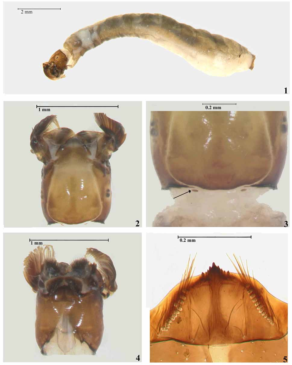
Mature larva. Body length 8.7–10.6 mm (n=4); length of head capsule 0.9–1.0 mm (n=4); width of head capsule 0.7– 0.8 (n=4). Body colour dark grey dorso-laterally, whitish ventrally (specimens fixed in Carnoy’s solution and/or alcohol). General body form as in Fig. 1 View FIGURES 1 – 5 .
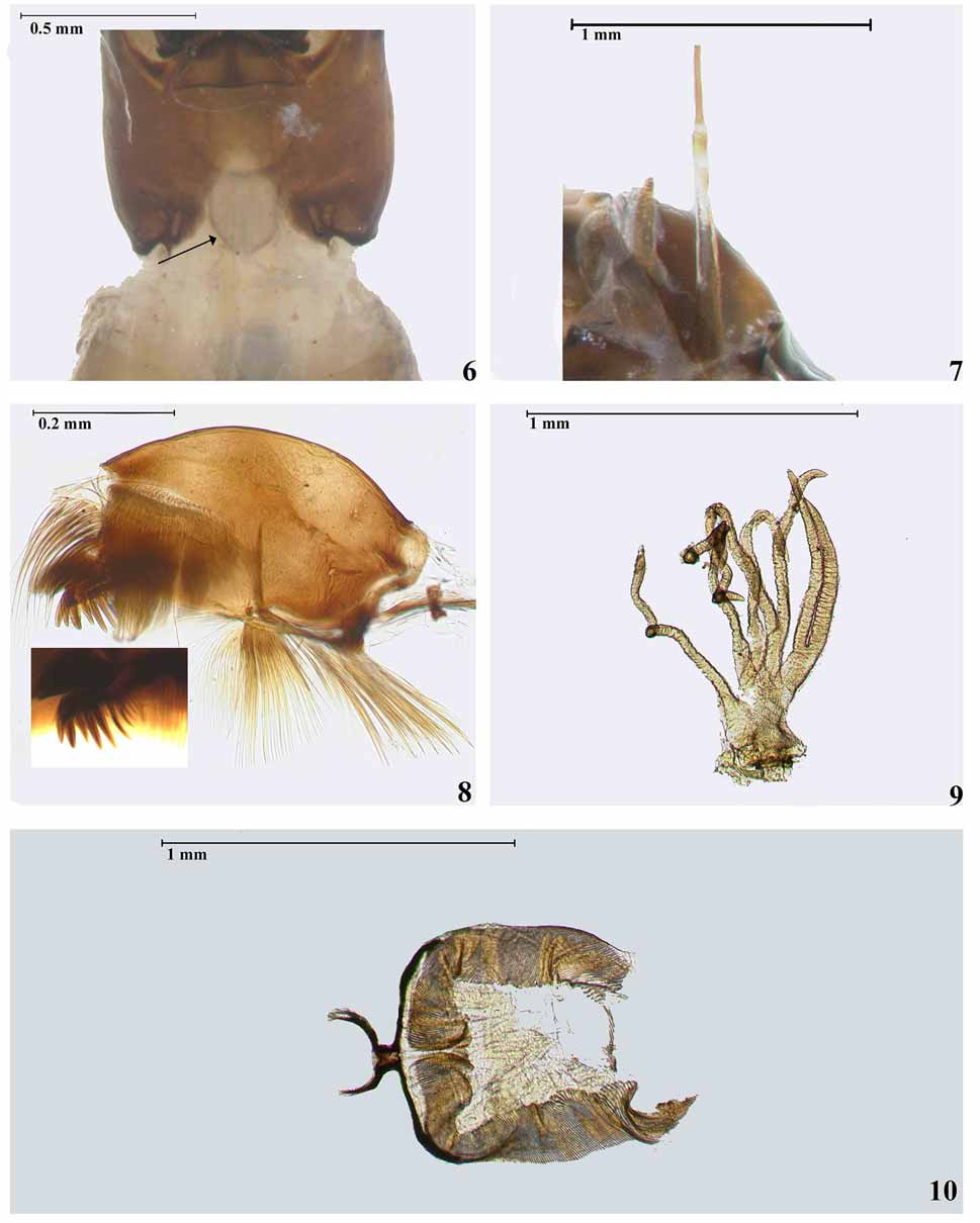
Head— mainly dark brown, anterior region of cephalic apotome yellowish. Numerous small setae present on all surfaces and head capsule slightly wrinkled. Head pattern positive ( Fig. 2 View FIGURES 1 – 5 ). Cervical sclerites small, elliptical, free in membrane ( Fig. 3 View FIGURES 1 – 5 ). Postgenal cleft deep, bell-shaped, subtriangular apically; postgenal bridge nearly as long as hypostoma ( Fig. 4 View FIGURES 1 – 5 ). Hypostoma strongly pigmented on anterior margin, with approximately nine apical teeth distinctly protruding in central region; median tooth sharp, well-developed and most prominent; 3+3 sublateral teeth, with the pair adjacent to base of median tooth longer than remainder; 1+1 lateral tooth, longer than basal sublateral tooth; 2+2 small, lateral serrations; 1+1 lines of approximately fourteen hypostomial setae parallel to lateral margin; 1+1 long, simple setae in posterior half of hypostoma ( Figs. 5 View FIGURES 1 – 5 ). Subesophageal ganglion lightly pigmented ( Fig. 6 View FIGURES 6 – 10 ). Antenna longer than labral fan stalk, segment I, apex of segment II and segment III dark brown, two thirds of segment II pale whitish ( Fig. 7 View FIGURES 6 – 10 ), length of antennal segments I–III excluding the sensillum 0.1:0.1–0.2:0.1 (n=6). Mandible with three apical teeth, first one longer than second and third apical teeth; mandibular comb with approximately eleven teeth, first fourteen more prominent than remainder, third, fourth and fifth comb teeth longer and more prominent than first, and sixth to eleventh mandibular comb teeth; two mandibular serrations, anterior more prominent and longer than posterior ( Fig. 8 View FIGURES 6 – 10 ). Lateral mandibular process absent. Maxillary palps heavily pigmented; one and a half times as long as wide at base. Labral fan with 49 primary rays (n=4) with fine, single line of microtrichia in a row.
Thorax — grey dorsally and whitish ventrally. Cuticle without setae. Proleg with plate heavily sclerotised with band of approximately 41–50 processes (n=4). Pupal respiratory gill histoblast dark brown; dissected gill histoblast with 8 filaments, all branching from a common trunk and pointed apically ( Fig. 9 View FIGURES 6 – 10 ).
Abdomen — usually grey dorsally, progressively paler ventrally, especially towards posterior where last segments are white, gradually expanded posteriorly. Ventral nerve cord greyish. Ventral tubercles absent. Cuticle lacking setae except area around anal sclerite and anal gills. Anal sclerite well sclerotised with anterior arms extending one third of diameter of posterior circlet anteriorly; no sclerotised areas between arms ( Fig. 10 View FIGURES 6 – 10 ). Posterior circlet with 251–290 rows of 45–46 simple hooks (n= 3). Rectal papillae with two lobes of approximately 24 small, finger-like lobes (n=1).
Taxonomic Discussion. Luna Dias et al. (2004) did not assign S. lobatoi to subgenus because of their morphological resemblance to adults and pupal exuviae of species assigned to the subgenera Hemicnetha and Trichodagmia . Therefore, we here compare the larva of S. lobatoi with those currently included in the latter two subgenera.
The larva of S. lobatoi can be distinguished readily from those of Brazilian species of Trichodagmia and Hemicnetha by the gill histoblast possessing eight filaments, all branching basally ( Fig. 9 View FIGURES 6 – 10 ). In this respect, S. lobatoi is similar to S. rubrithorax Lutz , but in the latter species the gill histoblast filaments are rounded apically ( Simuliidae Digital Image Archives , BMNH; Shelley et al., 1997), while in S. lobatoi they are pointed ( Fig. 9 View FIGURES 6 – 10 ). The best character for distinguishing the larva of S. lobatoi from that of S. rubrithorax is the structure of the hypostomal teeth. In S. lobatoi the teeth of the hypostoma are produced anteromedially, with the median tooth longer than the remaining teeth, only three sublateral teeth and 1+1 lateral teeth that are slightly longer than the posterior sublateral tooth; the hypostoma has approximately 14 hypostomal setae per side ( Fig. 5 View FIGURES 1 – 5 ). The hypostoma of S. rubrithorax has all teeth at the same level, except that the median tooth is longer, and there are only 2+2 sublateral teeth and 1+1 lateral teeth are all projected to nearly the same level anteromedially; the hypostoma has approximately nine hypostomal setae ( Shelley et al., 1997).
The general morphology of the postgenal cleft and mandibular teeth of S. lobatoi largely agree with the variation found in species of Hemicnetha . Therefore, we assign S. lobatoi to the subgenus Hemicnetha . Because of its overall similarity with S. rubrithorax we place this species in the paynei— species group of Crosskey & Howard (2004).
Bionomics. Larvae and pupae of S. lobatoi were collected in the municipality of Formosa, Goiás state, below the waterfall named Salto do Itiquira. The stream was approximately 10m wide with a bed of boulders and sand. The water had a temperature of 17o C, a pH of 7.9 and an electrical conductivity below 30 µS/cm. All larvae were collected in large numbers, mainly on stones, where the water current was very fast.
Acknowledgments. The first author would like to thank The Natural History Museum (BMNH) for providing funds for collecting and curating Neotropical Simuliidae and A.J.Shelley for critically reviewing the manuscript. The second author would like to thank the Instituto Nacional de Pesquisas da Amazônia (INPA) and Conselho Nacional de Desenvolvimento Científico e Tecnológico (CNPq) for providing funds to carry out this research. We thank Peter H. Adler and an anonymous reviewer for reading and commenting on the manuscript.
No known copyright restrictions apply. See Agosti, D., Egloff, W., 2009. Taxonomic information exchange and copyright: the Plazi approach. BMC Research Notes 2009, 2:53 for further explanation.
|
Kingdom |
|
|
Phylum |
|
|
Class |
|
|
Order |
|
|
Family |
|
|
Genus |
Simulium lobatoi Luna Dias, Hernández, Maia-Herzog & Shelley
| Hernández, Luis Miguel, Hamada, Neusa, Pepinelli, Mateus & Kikuchi, Regina Mayumi 2008 |
Simulium lobatoi Luna Dias, Hernández, Maia Herzog & Shelley, 2004 : 37
| Shelley 2004: 37 |