
Rhodnius montenegrensis, Rosa, João Aristeu Da, Rocha, Cláudia Solano, Gardim, Sueli, Mendonça, Mara Cristina Pinto Vagner José, Filho, Júlio César Rente Ferreira, Carvalho, Elaine Oliveira Costa De, Camargo, Luís Marcelo Aranha, Oliveira, Jader De & Damieli, Juliana, 2012
|
publication ID |
https://doi.org/ 10.5281/zenodo.212770 |
|
DOI |
https://doi.org/10.5281/zenodo.5672520 |
|
persistent identifier |
https://treatment.plazi.org/id/33070760-FFE9-FF8B-8AAA-92BDFA5FF840 |
|
treatment provided by |
Plazi |
|
scientific name |
Rhodnius montenegrensis |
| status |
sp. nov. |
Rhodnius montenegrensis View in CoL n. sp.
Holotype. Ƥ, BRAZIL: Triatominae Insectarium of the Faculty of Pharmaceutical Sciences, Unesp, Araraquara, SP, Rodovia Araraquara-Jaú km 1. 2012.
Paratypes. BRAZIL: 8Ƥ, 83, Oswaldo Cruz Institute, Rio de Janeiro, RJ, Avenida Brasil, 4365. 2012.
Etymology. The name Rhodnius montenegrensis was chosen because this species was found in the municipality of Monte Negro, state of Rondônia, Brazil.
Description. Length of male 18.49 ± 0.82 mm, of female 21.54 ± 0.75 mm, width of pronotum of male 3.99 ± 0.32 mm, of female 4.54 ± 0.26 mm; width of abdomen of male 6.16 ± 0.39 mm, of female 6.95 ± 0.39 mm ( Table 1 View TABLE 1 ).
Female Male
HL, head length; IE, inner distance between eyes; AO, anteocular distance; PO, postocular distance (excluding neck); DE, diameter of the eye; R1, R2, and R3, lengths of first, second, and third rostral segments, respectively; TL, Total length of the Triatominae ; MWT, maximum width of the thorax; MWA, maximum width of the abdomen; A1, A2, A3 and A4, 1st, 2nd, 3rd, and 4th antennal segments, respectively. The values in bold were significant at α = 0.05, using unpaired t-test.
General color yellow with longitudinal dorsal black stripes on pronotum, wings, and connexivum ( Fig. 2 View FIGURE 2. R A).
Head of male and female with no spots, from clypeus to neck, with a central yellow stripe located between two (1+1) continuous brown stripes. Neck with a brown mark and a yellow central circular mark ( Fig. 2 View FIGURE 2. R A). Head length, inner distance between eyes, anteocular distance, postocular distance (excluding neck), diameter of the eye, lengths of 1st, 2nd, and 3rd -rostral segments, respectively, and 1st, 2nd, 3rd, and 4th -antennal segments, respectively, are shown in Table 1 View TABLE 1 .
Mesothoracic wings with well-demarcated veins, notably subcostal (Sc) vein with its yellow color ( Fig. 4 View FIGURE 4 ). Legs: yellow, except for a black stripe posteriorly to tibia, near tarsus ( Fig. 2 View FIGURE 2. R ). Mesosternum with lateral stripes and one central stripe that delimits two dark areas. Metasternum with two yellow stripes between coxa that delimit a dark central area ( Fig. 2 View FIGURE 2. R ). First urotergite: brown and with distinct transverse groove ( Fig. 5 View FIGURE 5 A). Scutellum with a long glabrous space between pronotum and beginning of a semi-circular base that supports central depression with sensillae. Lateral edges of scutellum slightly curved and with fine apex ( Fig. 6 View FIGURE 6 A).
Ventral abdomen with yellow and dark areas anteriorly to posteriorly. Dorsal connexivum with a dark spot occupying about 1/3 of each segment, wider on anterior portion, and narrowing towards posterior portion. Internal and external borders of connexivum with a noncontinuous yellow stripe. Externally yellow stripe not continuous. First abdominal segment resembling metasternum, but smaller ( Fig. 2 View FIGURE 2. R ).
Median length of the basal plate (EPlb) does not involve endosomes. Basal plate (Plb) has a regular arc shape. Process of phallosoma (PrPh) is the most intensely chitinized structure of phallus, contains two cylindrical pieces with well-defined contours ( Fig. 7 View FIGURE 7 A, B, C) Median process of the pygophore inserted in a wide triangular base surrounded by a groove-shaped cuticle, anterior 2/3 wider than the posterior 1/3, which narrows to a gross point ( Fig. 8 View FIGURE 8 A).
Ventral view: female external genitalia with two small lateral saliencies on 9th sternite and a circular line ( Fig. 9 View FIGURE 9 A) on 7th sternite, which borders gonocoxites. Dorsal view: 7th tergite with a trapezoidal shape ending on two tips on posterior portion ( Fig. 10 View FIGURE 10 A). Posterior view: female external genitalia with small 9th tergite; large opening limiting X segment with gonapophyses VIII and gonocoxites VIII ( Fig. 11 View FIGURE 11 A).
Eggshell length 1.58 ± 0.080 mm, width 0.99 ± 0.035 mm, area 1.26 ± 0.061 mm. Egg featuring a collar, an exochorion with a grainy surface, tapered holes regularly distributed and no cell demarcation ( Figs. 12 View FIGURE 12 A, 13A).
Classic and geometric morphometrics. Statistical analyses based on unpaired t-test show significant values (α=0.05) for some parameters between females (HL, IE, AO, PO, DE, R1, R2, TL, MWT, MWA, A2 and A3) and between males (HL, AO, PO, DE, R2, R3, MWT, A1, A2 and A3) ( Table 1 View TABLE 1 ).
Eggshell measurements and the statistical analyses based on unpaired t-test show significant values (α=0.0001) for eggshell area and length, whereas the others parameters were not significant.
For the geometric morphometrics the factorial map built with 18 specimens of R. robustus and 23 specimens of R. montenegrensis n. sp. distinguished both species in well-defined groups. The Mahalanobis distance between the two species was 3.85. Considering the shape variation components, the contribution of the first principal (PC1) component accounted for 39% of the total variation, whereas the second principal component (PC2) accounted for 35% ( Fig. 14 View FIGURE 14 ).
Molecular analysis. For the Bayesian analysis, the alignment of a 369pb of Cyt b gene was obtained, and the phylogenetic tree showed that the R. montenegrensis n. sp. is a sister to R. robustus . This tree also included other six Rhodnius species ( R. prolixus , R. neglectus , R. pallescens , R. ecuadoriensis , R. pictipes , R. brethesi ). Phylogenetic reconstruction also certified the taxonomic status of insects belonging to CTA 88 ( Fig. 15 View FIGURE 15 ).
Based on this, an additional molecular methodology was applied to differentiate both species. Therefore, 5.8S / ITS-2 genes from nuclear DNA were amplified by PCR and generated a fragment of 1,200 base pairs ( Marcilla et al. 2001). This gene were treated by restriction enzyme BstUI by PCR-RFLP ( Naegele et al. 2006) and generated two fragments of 1,000 and 150 pair of bases for R. robustus and there was no digestion for R. montenegrensis ( Fig. 16 View FIGURE 16 ).

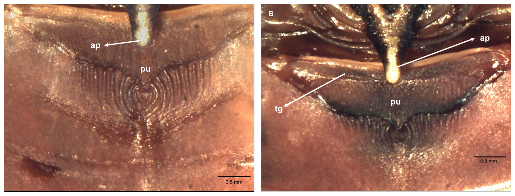
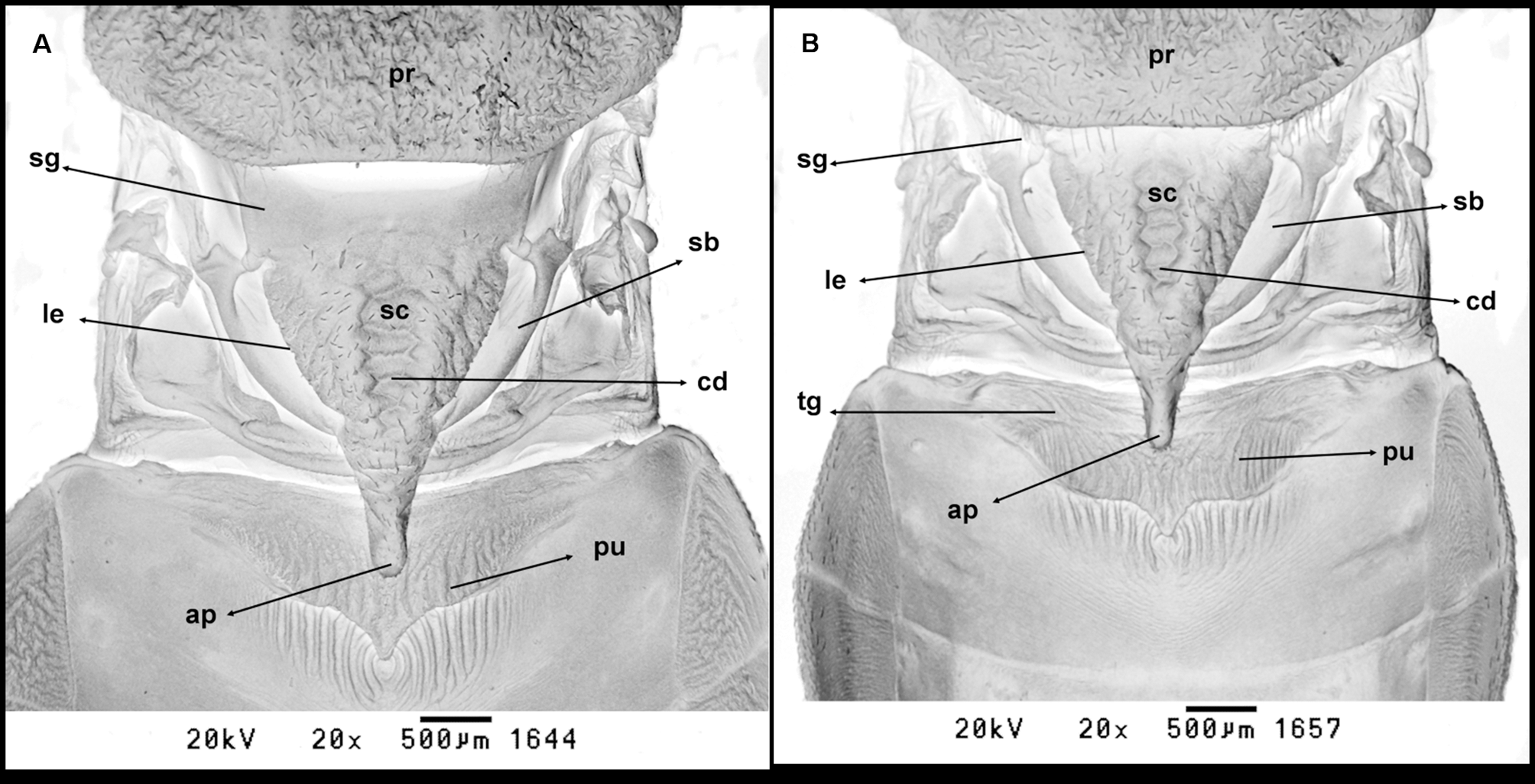
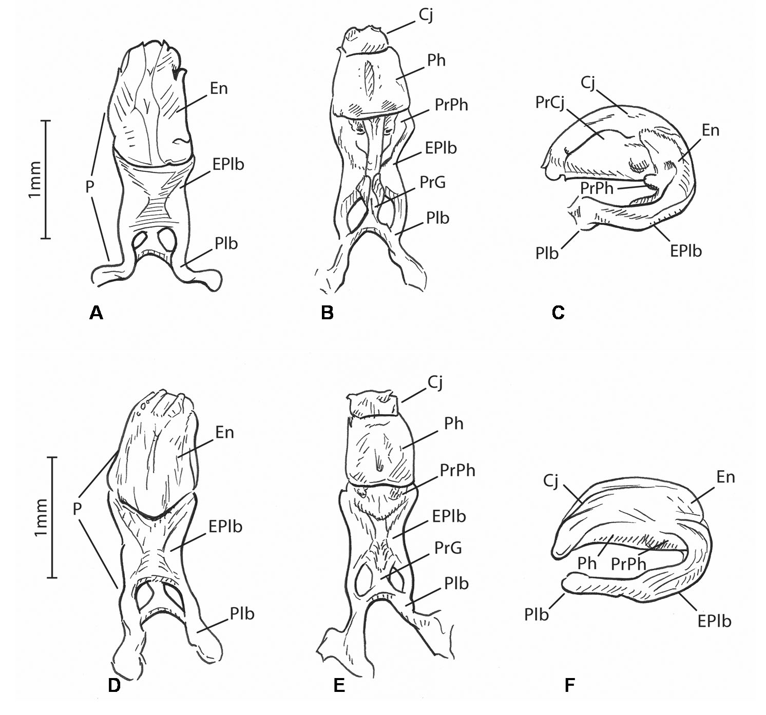
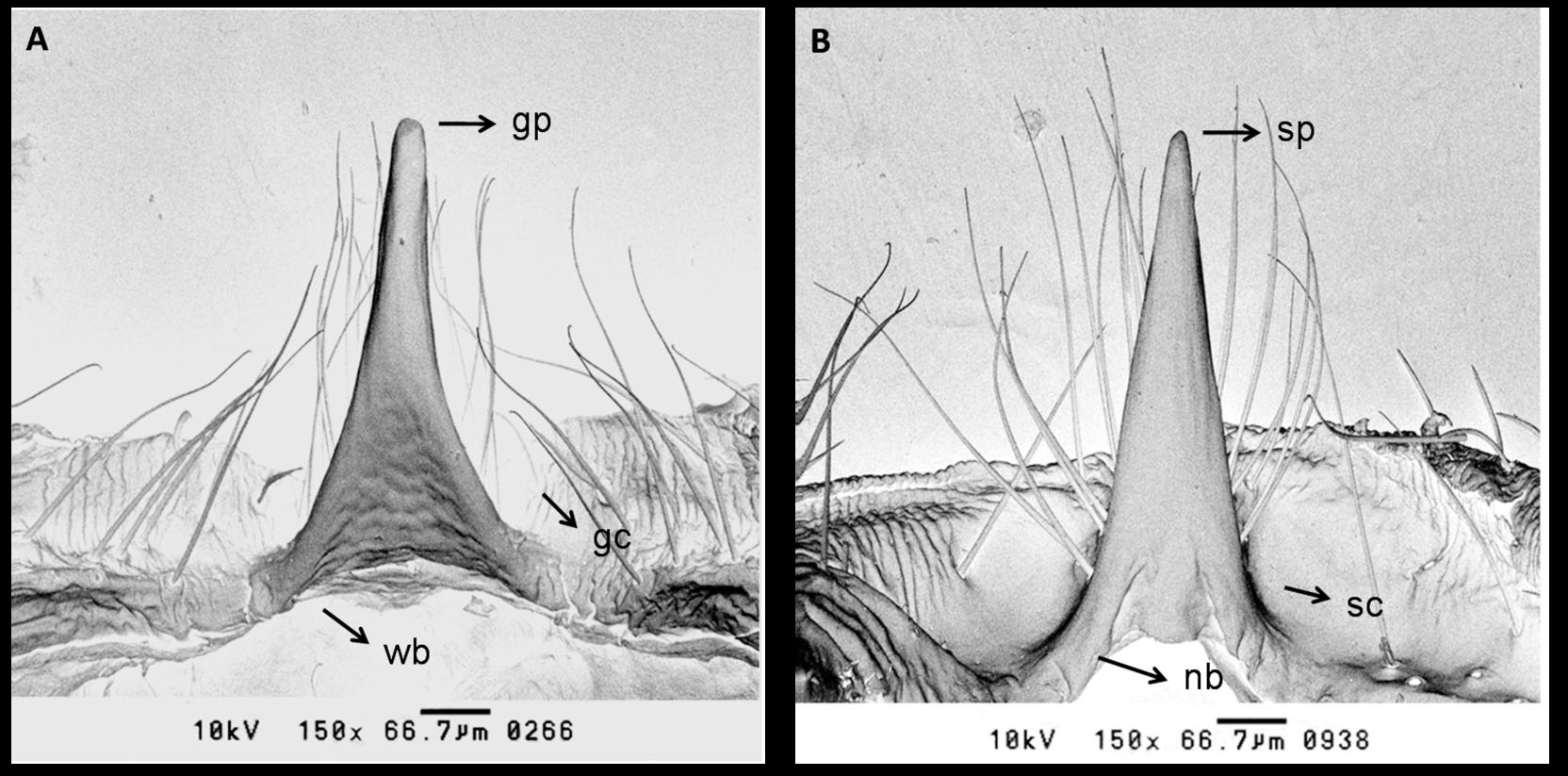

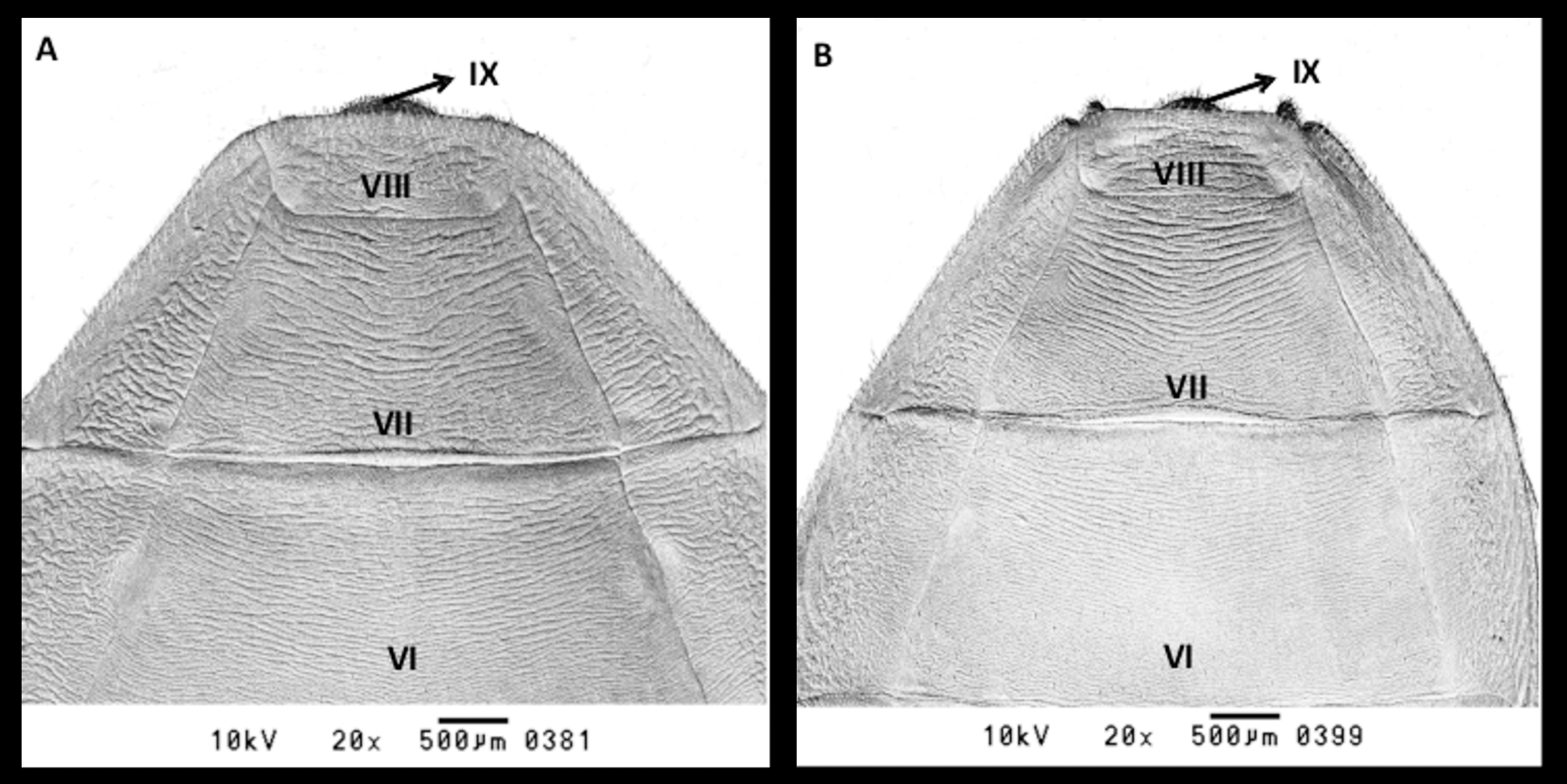
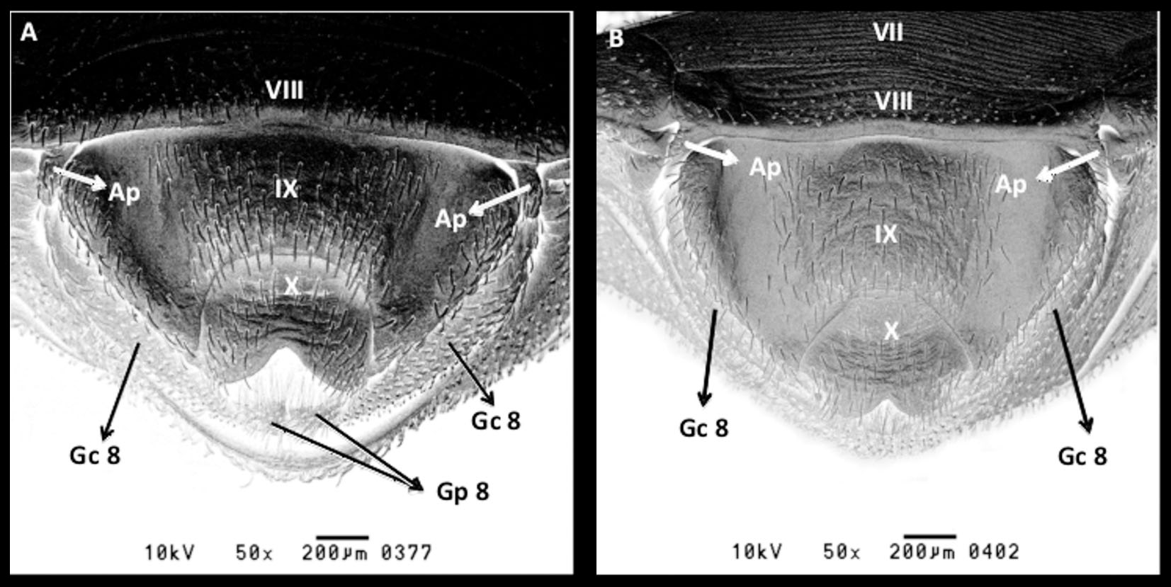
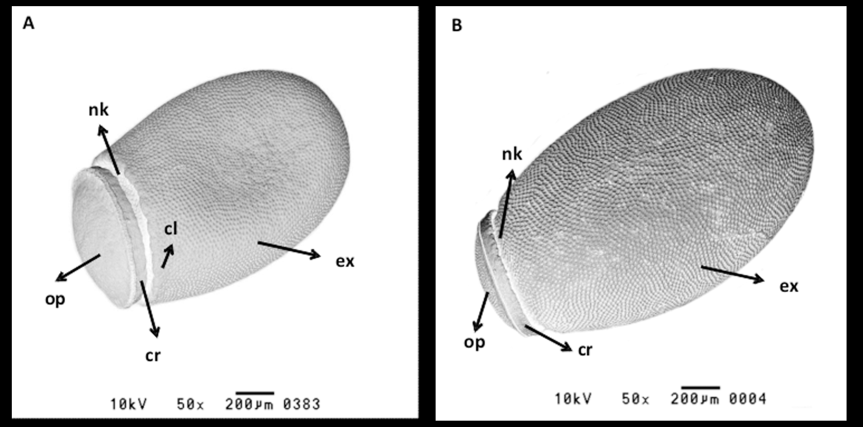
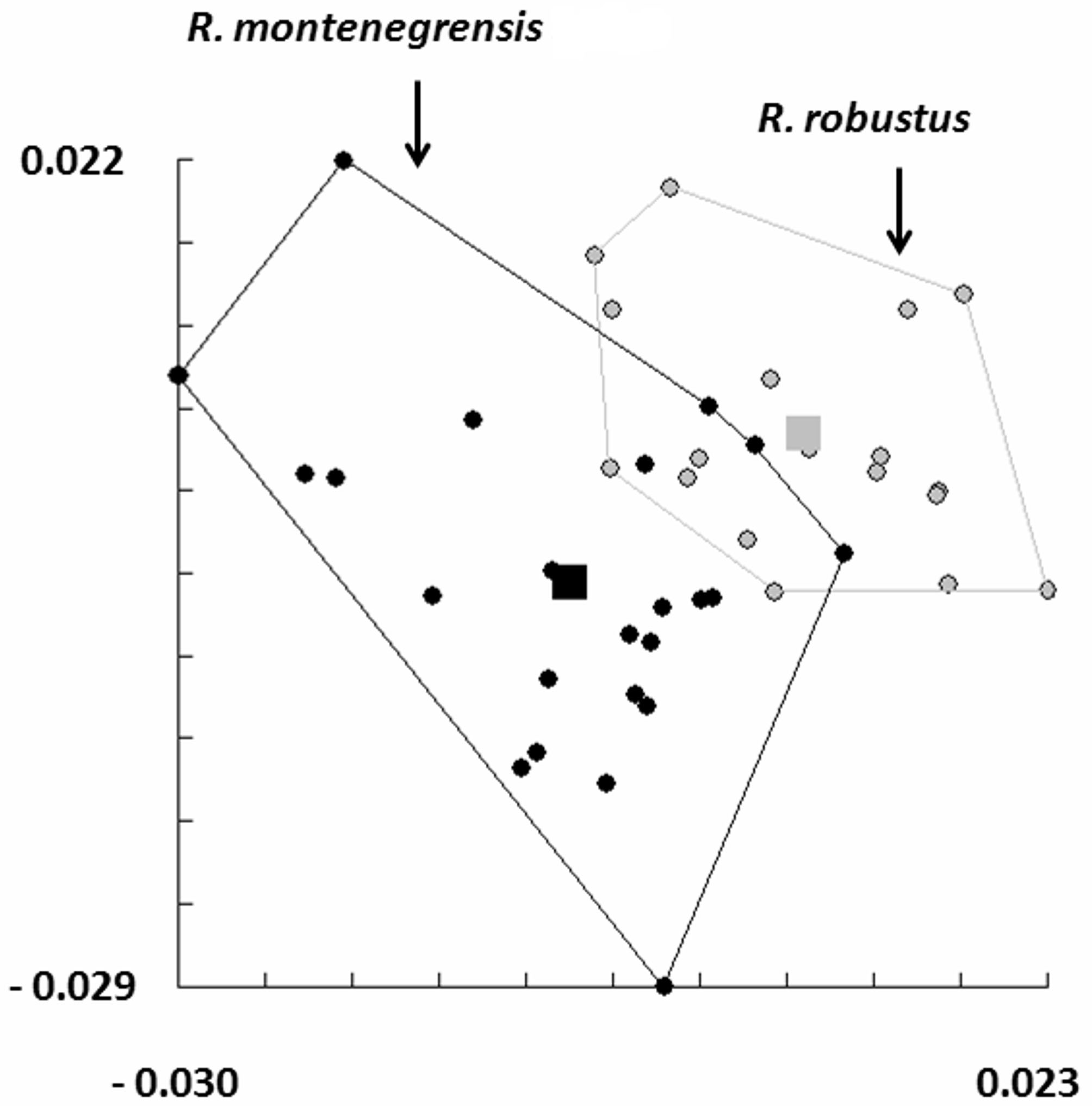
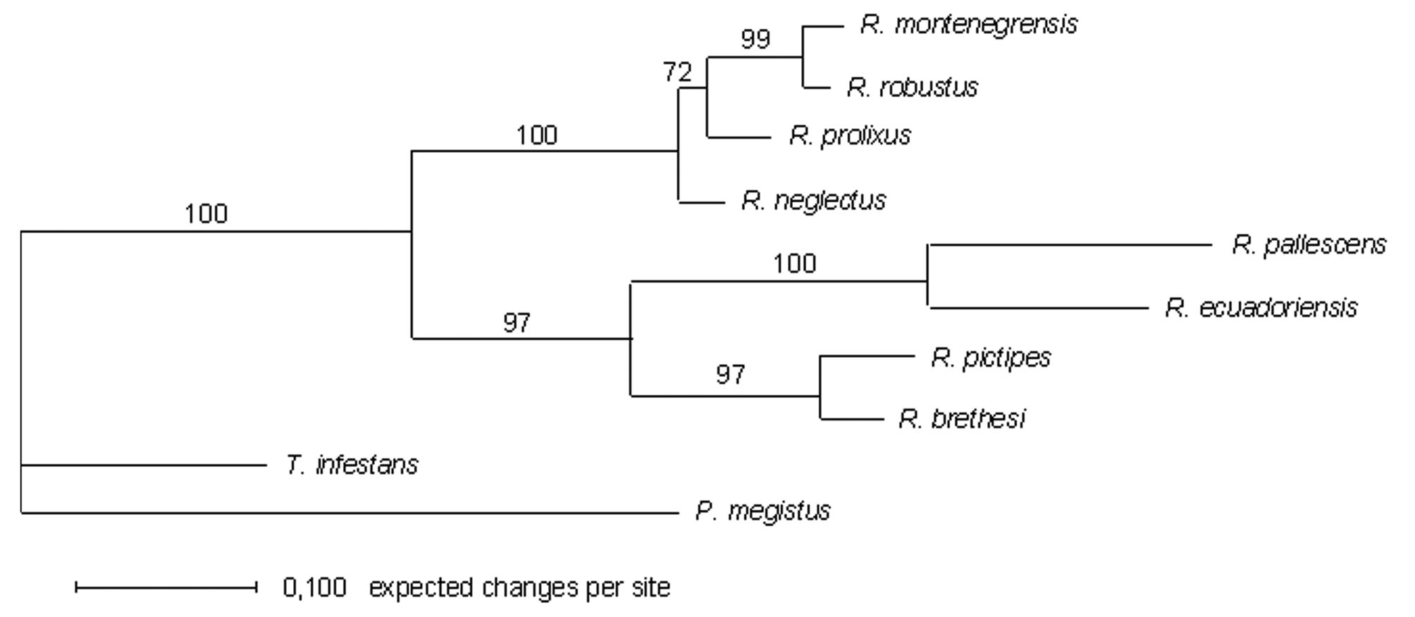
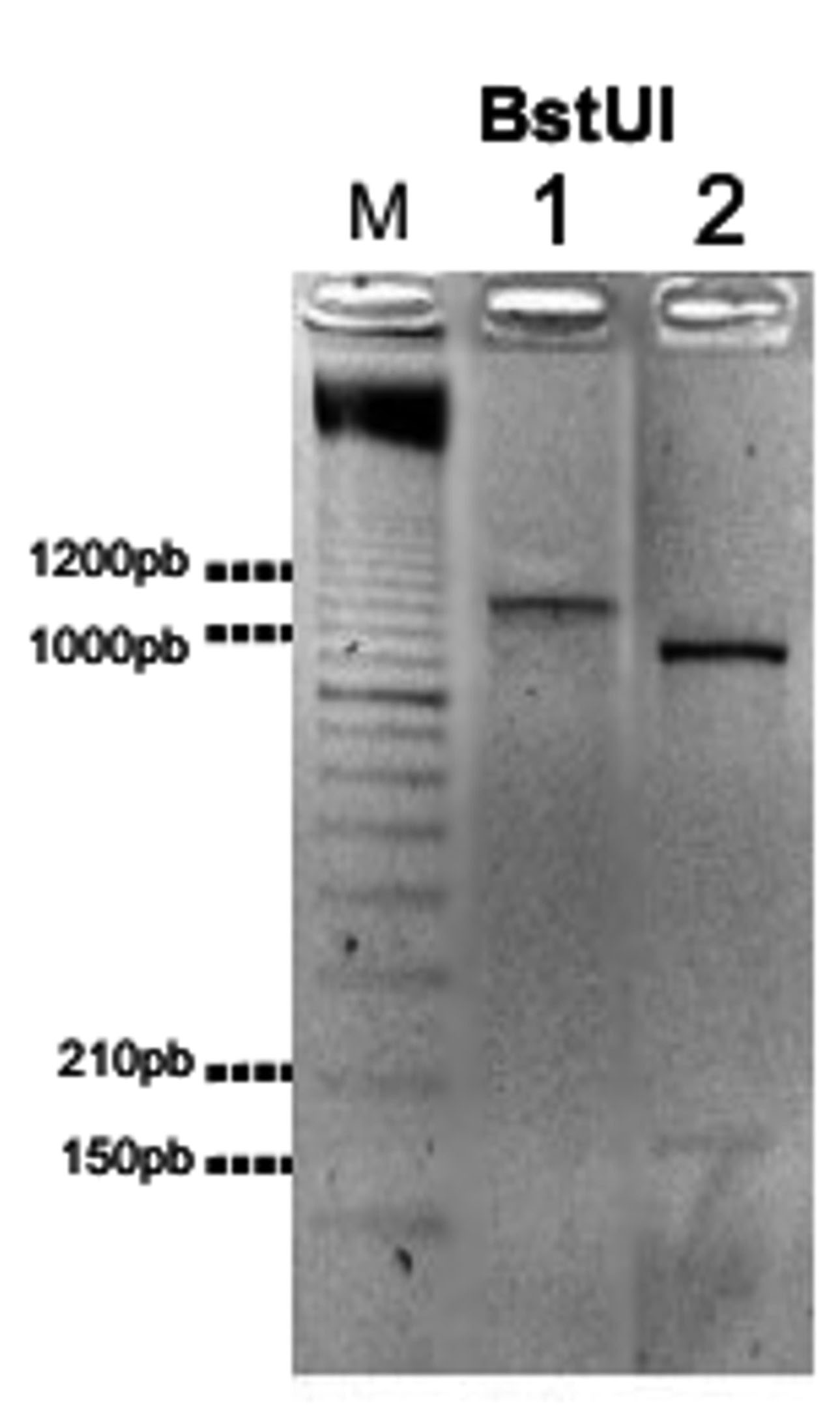
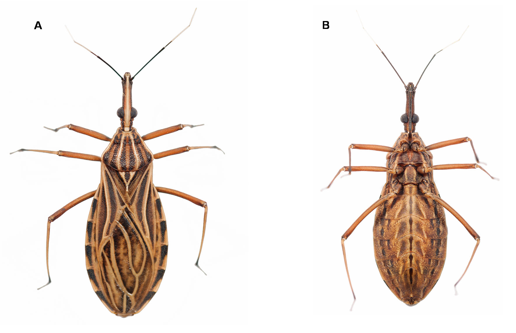
TABLE 1. Mean of measurement (mm) of 15 specimens of Rhodnius montenegrensis and Rhodnius robustus, females and males.
| R. montenegrensis | R. robustus | R. montenegrensis | R. robustus | |
|---|---|---|---|---|
| HL | 4.81 | 4.06 | 4.45 | 3.80 |
| IE | 0.63 | 0.68 | 0.59 | 0.61 |
| AO | 2.97 | 2.39 | 2.73 | 2.23 |
| PO | 0.83 | 0.79 | 0.78 | 0.73 |
| DE | 1.22 | 1.08 | 1.16 | 0.99 |
| R1 | 0.82 | 0.97 | 0.85 | 0.90 |
| R2 | 3.81 | 3.29 | 3.56 | 3.03 |
| R3 | 0.93 | 0.96 | 0.85 | 0.92 |
| TL | 21.54 | 18.96 | 19.29 | 18.49 |
| MWT | 4.54 | 4.14 | 3.99 | 3.99 |
| MWA | 6.95 | 6.56 | 6.16 | 6.09 |
| A1 | 0.44 | 0.38 | 0.49 | 0.37 |
| A2 | 3.94 | 3.19 | 4.00 | 3.28 |
| A3 | 2.63 | 2.39 | 2.54 | 2.30 |
| A4 | 1,53 | 1.61 | 1.53 | 1.54 |
No known copyright restrictions apply. See Agosti, D., Egloff, W., 2009. Taxonomic information exchange and copyright: the Plazi approach. BMC Research Notes 2009, 2:53 for further explanation.
|
Kingdom |
|
|
Phylum |
|
|
Class |
|
|
Order |
|
|
Family |
|
|
Genus |