
Nipponodipogon sudai Shimizu
|
publication ID |
https://doi.org/ 10.11646/zootaxa.3948.3.6 |
|
publication LSID |
lsid:zoobank.org:pub:876F1196-2BA6-466E-9BD6-14374AC070FE |
|
DOI |
https://doi.org/10.5281/zenodo.6100506 |
|
persistent identifier |
https://treatment.plazi.org/id/33328787-FF98-FB56-FF70-1691FE67F8F8 |
|
treatment provided by |
Plazi |
|
scientific name |
Nipponodipogon sudai Shimizu |
| status |
sp. nov. |
7. Nipponodipogon sudai Shimizu View in CoL , sp. nov.
( Figs 7 View FIGURE 7 , 8 View FIGURE 8 E, I, 9J, K)
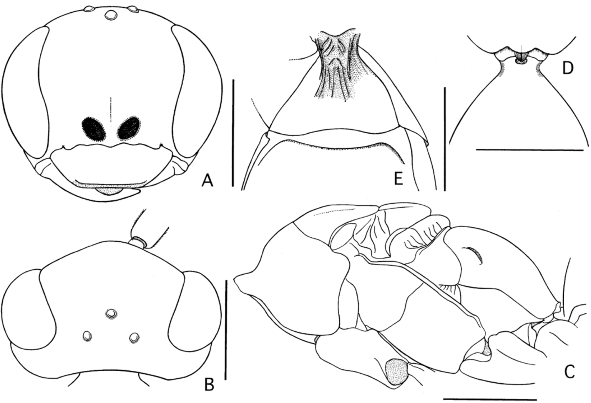
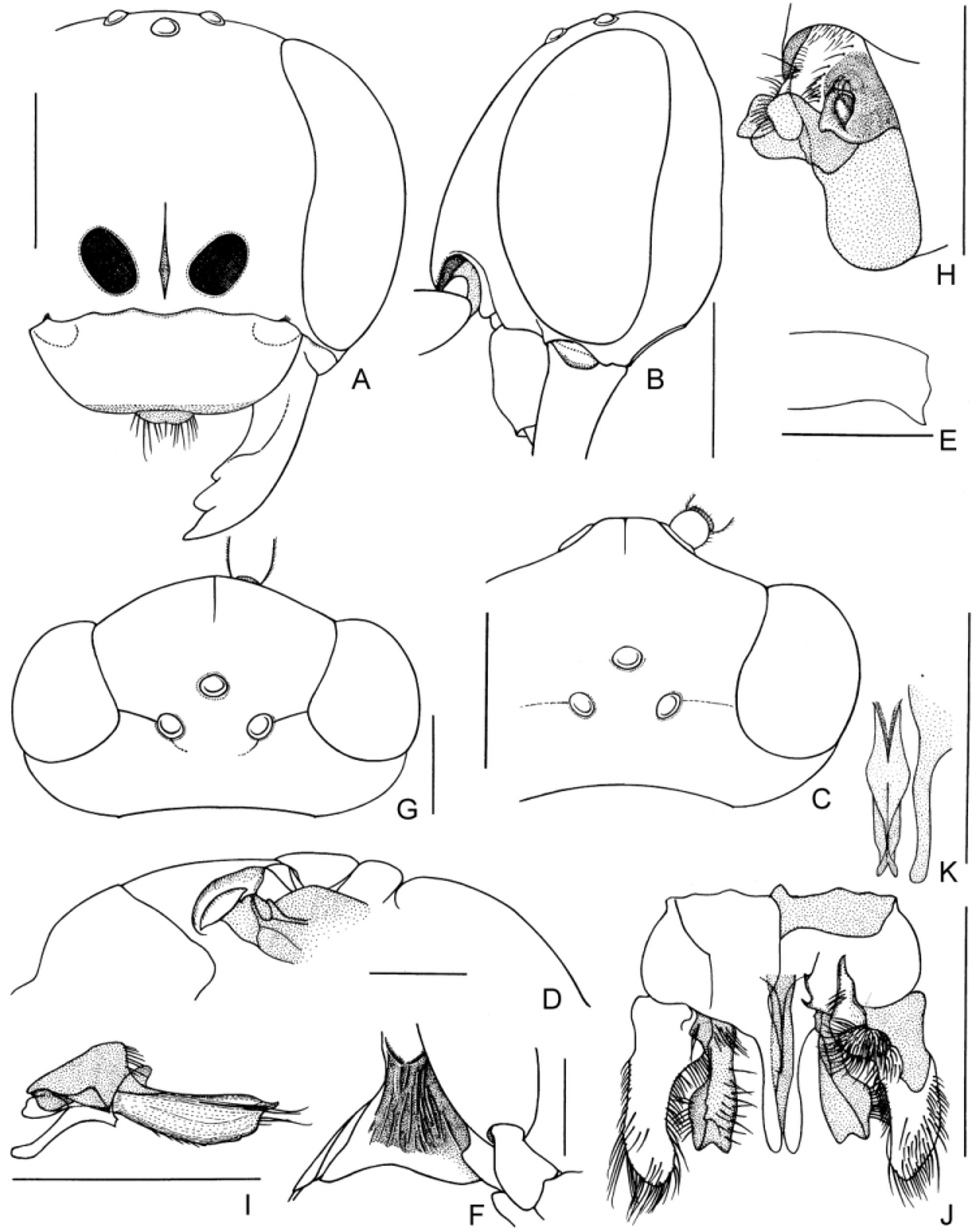
Diagnosis. FEMALE. The female is similar to that of N. kurilensis by having the outer apicoventral corner of the hind femur produced triangularly ( Fig. 7 View FIGURE 7 E) and T1 petiolate basally ( Fig. 8 View FIGURE 8 E), but differs from the latter by having the following characteristics: T1 with a long petiole ( Fig. 8 View FIGURE 8 E); the vertex between the eye tops not strongly convex ( Fig. 7 View FIGURE 7 A vs. Fig. 3 View FIGURE 3 A); and the mesoscutum not raised along the midline. MALE. The male differs from those of the other species by having the petiolate T1 and by characteristic configurations of the lateral hooks on S6 ( Fig. 7 View FIGURE 7 H), subgenital plate ( Fig. 7 View FIGURE 7 I) and genitalia ( Fig. 7 View FIGURE 7 J).
Description. FEMALE (measurements of the holotype are given in parentheses.) Length: body 4.2−7.0 mm; fore wing 3.8–5.9 mm. Head and mesosoma matte, metasoma subpolished. Body black; following brown to ferruginous: labrum, apical half of mandible, palpi, antenna beneath, lateral and posterior margins of pronotum, posterolateral margin of metapostnotum, tegula, posterior and lateral margins of metasomal terga and sterna and legs (coxae distally, trochanters and fore tibia sometimes, tibial spurs always yellowish brown). Fore wing with distinct two fuscous fasciae; inner fascia broad ( Fig. 9 View FIGURE 9 J).
Frons, vertex and mesosoma except propodeum finely and densely punctate (mesoscutum sometimes impunctate along midline). Mesepisternum and lower metapleuron scattered with large punctures. Upper metapleuron (occasionally lower metapleuron also) finely and densely striate, interspace of striae punctate, without posterior impunctate rim. Lateral side of metanotum with several regular oblique striae. Propodeum except anterolateral and median portions strongly and densely punctate, transversely rugulose posteriorly.
Body with pubescence mostly short but longer on lower frons, clypeus, propleuron, mesepisternum, mesosternum, propodeum posterolaterally and coxae.
Head in frontal view 1.2 × as wide as long. Vertex moderately convex between eye tops ( Fig. 7 View FIGURE 7 A). Upper frons gently convex ( Fig. 7 View FIGURE 7 B); frontal line finely impressed on its lower third. Inner orbits weakly convergent above and subparallel below. Ratio of UID:MID:LID = 7.3–8.4:10:9.1–9.4. Half of MID 1.6–1.9 × eye width. Ocelli large; ocellar triangle acute- or right-angled, slightly raised. Ratio of POL:OOL = 1:0.90–1.2. Posterior margin of vertex weakly concave. Clypeus 2.5–2.9 × as wide as long, convex medially with distinct elliptical depression basilaterally; anterolateral corner broadly rounded; apical rim scarcely depressed and alutaceous; apical margin nearly truncate but weakly emarginate medially ( Fig. 7 View FIGURE 7 A). Apical margin of labrum almost straight or barely emarginate. Mandible with basal tooth small, sometimes indistinct. Malar space short. Gena in dorsal view strongly narrowing posteriorly ( Fig. 7 View FIGURE 7 C), in profile 0.4–0.6 × eye width, broadest below middle. Ratio of F1 and F2 = 1:1.0– 1.2; F1 2.3–2.7 × as long as thick, 0.45–0.58 × as long as UID.
Pronotum with anterior declivity flattened or slightly concave, not distinctly differentiated from dorsum; dorsum in dorsal view slightly narrowing anteriorly; shoulder gently rounded; juncture between dorsal and lateral faces narrowly and roundly raised; lateral face finely striate medially; posterior margin arcuately emarginate. Mesoscutum slightly sloped anteriorly; posterolateral margin not reflexed; parapsidal sulcus finely impressed. Discs of scutellum and metanotum barely raised above level of mesoscutum and propodeum ( Fig. 7 View FIGURE 7 D). Metanotum with about 10 regular strong striae laterally. Propodeum evenly convex with posterior declivity not well differentiated from dorsum, scarcely flattened unlike in N. iwatai ; median groove obscure, if any.
Fore wing ( Fig. 9 View FIGURE 9 J) with SMC2 receiving crossvein 1m-cu at basal 0.41–0.63. SMC3 0.97–1.4 × as long as SMC2 on vein M, 0.73–1.3 × as long as SMC2 on vein Rs, narrowed on vein Rs by 0.43–0.53 × its own length on vein M, receiving crossvein 2m-cu at basal 0.37–0.55. Crossvein 2rs-m barely curved. Crossvein 3rs-m gently or moderately curved. Crossvein cu-a originating at or a little posteriorly to fork of vein M+CuA.
T1 distinctly petiolate, petiole comparatively long ( Fig. 8 View FIGURE 8 E). S1 with several longitudinal rugae medially ( Fig. View FIGURE 7
7F), their interspace punctate and subpolished. Transverse groove on S2 gently arcuate. S6 with longitudinal median carina posteriorly. Outer apicoventral corner of hind femur produced triangularly ( Fig. 7 View FIGURE 7 E).
MALE. Length: body 3.5–5.7 mm; fore wing 3.3–5.0 mm. Similar to female in coloration; apical portions of fore coxa, fore trochanter and fore femur, fore tibia, fore tarsomeres 1–4, tibial spurs and sometimes mid and hind trochanters yellowish brown. Clypeus sometimes light brown apically. Fore wing fasciae almost obsolete.
Punctures on body stronger and coarser than in female. Pronotum distinctly punctate; striae on lateral side more distinct than in female.
Head in frontal view 1.2–1.3 × as wide as long. Ratio of UID:MID:LID = 8.3–9.0:10:8.3–9.1. Half of MID 1.6–1.9 × eye width. Ocellar triangle right- to obtuse-angled. Ratio of POL:OOL = 1:0.71–0.93. Oblique furrow running from lateral ocellus to eye sharp. Clypeus 2.3–2.8 × as wide as long; apical rim often slightly depressed but not distinctly set off from main portion. Gena in dorsal view more roundly narrowing posteriorly than in female ( Fig. 7 View FIGURE 7 G vs. Fig. 7 View FIGURE 7 C), in profile 0.4–0.6 × eye width. Ratio of F1 and F2 = 1:1.0–1.4; F1 1.5–1.9 × as long as thick and 0.27–0.37 × as long as UID.
Dorsum of pronotum longer and more strongly narrowing anteriorly than in female; anterior declivity nearly vertical and flattened; juncture between dorsal and lateral faces carinate. Dorsum of mesoscutum more or less raised anteromedially, slightly reflexed posterolaterally; parapsidal sulcus sharply impressed. Discs of scutellum and metanotum much more raised above level of mesoscutum and propodeum than in female.
Fore wing SMC2 receiving crossvein 2m-cu at basal 0.35–0.64. Fore wing SMC3 0.89–1.6 × as long as SMC2 on vein M, 0.55–1.2 × as long as SMC2 on vein Rs, narrowed on vein Rs by 0.38–0.59 × its length on vein M, receiving crossvein 2m-cu at basal 0.45–0.67.
T1 barely petiolate, its petiole short. Transverse groove on S2 sometimes weak and indistinct. Lateral hook on S6 ( Fig. 7 View FIGURE 7 H) large and characteristically shaped, i.e., broad basally, triangular apically, thin; posterior margin of S6 between hooks weakly emarginate. Exposed portion of subgenital plate arcuately convex ventrally and flattened dorsally, compressed laterally with median sharp carina ventrally and two pairs of strong curved setae distally ( Fig. 7 View FIGURE 7 I); subbasal portion triangularly extended laterally, without lateral process directed posterolaterad ( Fig. 8 View FIGURE 8 I).
Genitalia ( Figs 7 View FIGURE 7 J–K): paramere broad basally, wedge-shaped apically, extending far beyond apex of digitus volsellaris, with numerous setae on outer face; digitus volsellaris broadened and spoon-shaped apically, apical margin notched; parapenial lobe slender, slightly curved downward apically; aedeagus not extending beyond apex of digitus volsellaris, broadest at middle, tapering toward apex, with small fan-shaped apex.
Material examined. Holotype, ♀ (TMUZ-TYPE-2014-001), JAPAN, Honshu, “Satsura Izumi, Fukui 17.ix.1983 Y. HANEDA” “ HOLOTYPE Nipponodipogon sudai Shimizu ♀”. Paratypes. JAPAN. Hokkaido: 1 ♀, Hyakumatsu-zawa, Sapporo-shi, 12–28.vii.2001 (T. Yoshida) (M.T.). Honshu: 4 ♂, Kawaratai, Nishimeya-mura, Aomori Pref., 3–16.viii.2010 (T. Nakamura). 1 ♀ 5 ♂, Mt. Hakase, beech forest, 1000 m, Showa-mura, Fukushima Pref., 29.vi–26.vii.1998 (T. Muroi) (M.T.). 1 ♀ 2 ♂, same locality and collector, 27.vii–23.viii.1998. 1 ♂, same locality and collector, 24.viii–19.ix.1998. 1 ♂, Mt. Komagatake, Hakone-machi, Kanagawa Pref., 18.vii.2001 (A. Shimizu). 2 ♀, same locality and collector, 1.viii.2001. 1 ♂, Heikedaira, Ôno-shi, Fukui Pref., 4.vii.1998 (S. Inoue). 1 ♂, same locality and collector, 1.ix.1998. 1 ♀, Nakahora, Ôno-shi, Fukui Pref., 30.viii.1988 ( Y. Haneda). 1 ♀, same locality and collector, 2.x.1983. 1 ♀, Suwara, Ôno-shi, Fukui Pref., 5.ix.1986 ( Y. Haneda). 1 ♀, same locality and collector, 26.vii.1999. 1 ♀, same locality and collector, 6.ix.1999. 1 ♀, Nigure, Izumi-mura, Fukui Pref., 26.viii.1982 (C. Nozaka). 1 ♀, Mt. Heko-san, Ikeda-chô, Fukui Pref., 20.vii.1998 (S. Inoue). 1 ♀, Mt. Ochisan, Fukui-shi, Fukui Pref., 11.ix.2002 (C. Nozaka). 1 ♀, Shirabidaira, Miyada-mura, Nagano Pref., 5.viii.1980 ( Y. T. & H. Suda). 6 ♀, Suhara, Decid. forest, Ôkuwa-mura, Nagano Pref., 1–7.ix.1996 ( Y. Jishage) (M.T.). 1 ♀, same locality and collector, Japanese cedars, 15–21.ix.1996 ( Y.P.T.). 1 ♂, Jukkoku-tôge, Kannami-chô, Shizuoka Pref., 1.viii.2001 (A. Shimizu). 1 ♀, Uradani, Beech forest, 900 m, Shitara-chô, Aichi Pref., 12–18.ix.1994 (K. Yamagishi).
Etymology. Named after H. Suda, the provider of a paratype specimen.
Distribution. Japan (Hokkaido and Honshu).
Biology. Unknown.


No known copyright restrictions apply. See Agosti, D., Egloff, W., 2009. Taxonomic information exchange and copyright: the Plazi approach. BMC Research Notes 2009, 2:53 for further explanation.