
Polyarthra platensis, José, Susana B., Paggi, De & Paggi, Juan C., 2011
|
publication ID |
https://doi.org/ 10.5281/zenodo.203769 |
|
DOI |
https://doi.org/10.5281/zenodo.6192014 |
|
persistent identifier |
https://treatment.plazi.org/id/33420333-FFB4-FFD7-1CB1-B7E4FC71FE51 |
|
treatment provided by |
Plazi |
|
scientific name |
Polyarthra platensis |
| status |
sp. nov. |
Polyarthra platensis sp. nov.
Type locality. Shallow lake in Reserva Ecológica U.N.L., Santa Fe (floodplain of Paraná River), Santa Fe Province, Argentina (31° 38’ 16” S 60° 40’ 21” W; temperature 27 ºC, conductivity 1065 µS.cm -1, pH 8, dissolved oxygen 9 mg.l -1). February 11th, 2009.
Type material. Holotype: 1 parthenogenetic Ψ, complete, mounted on a slide, Accession Number MACN-In 38202. Paratypes: two parthenogenetic Ψ, dissected and mounted on two slides, from type locality, Accession numbers MACN-In 38203–38204; two parthenogenetic Ψ, complete, mounted on two slides, all from type locality and on the same date. Accession number MFA/Z 8 – 9.
Additional material. Numerous specimens in liquid samples, preserved in formalin deposited in INALI. Shallow lake in Reserva Ecológica U.N.L., Santa Fe (type locality), February 24th, 2009. Laguna Gonzalez, near Santa Fe (floodplain of Paraná River), Santa Fe Province, Argentina (31° 40’ 28” S 60° 34’ 11” W), November 5th, 2009. Pond situated one Km south of Cuay Grande River, Corrientes Province, Argentina (28° 41’ 41” S 56° 14’ 30” W), November 16th, 2004. Swamp at Garrucho, Corrientes Province, Argentina (28° 7’ 50” S 55° 42’ 30” W), November 16th, 2004. Pond in Saenz Peña, Formosa Province (26º 54’ 26” S 60º 27’ 37” W), December 1, 2007. The environmental conditions at all five listed sites combined were: temperature 25–27.5 ºC, conductivity 292–1065 µS.cm -1, pH 7.5–9, dissolved oxygen 4.26–6.90 mg.l -1.
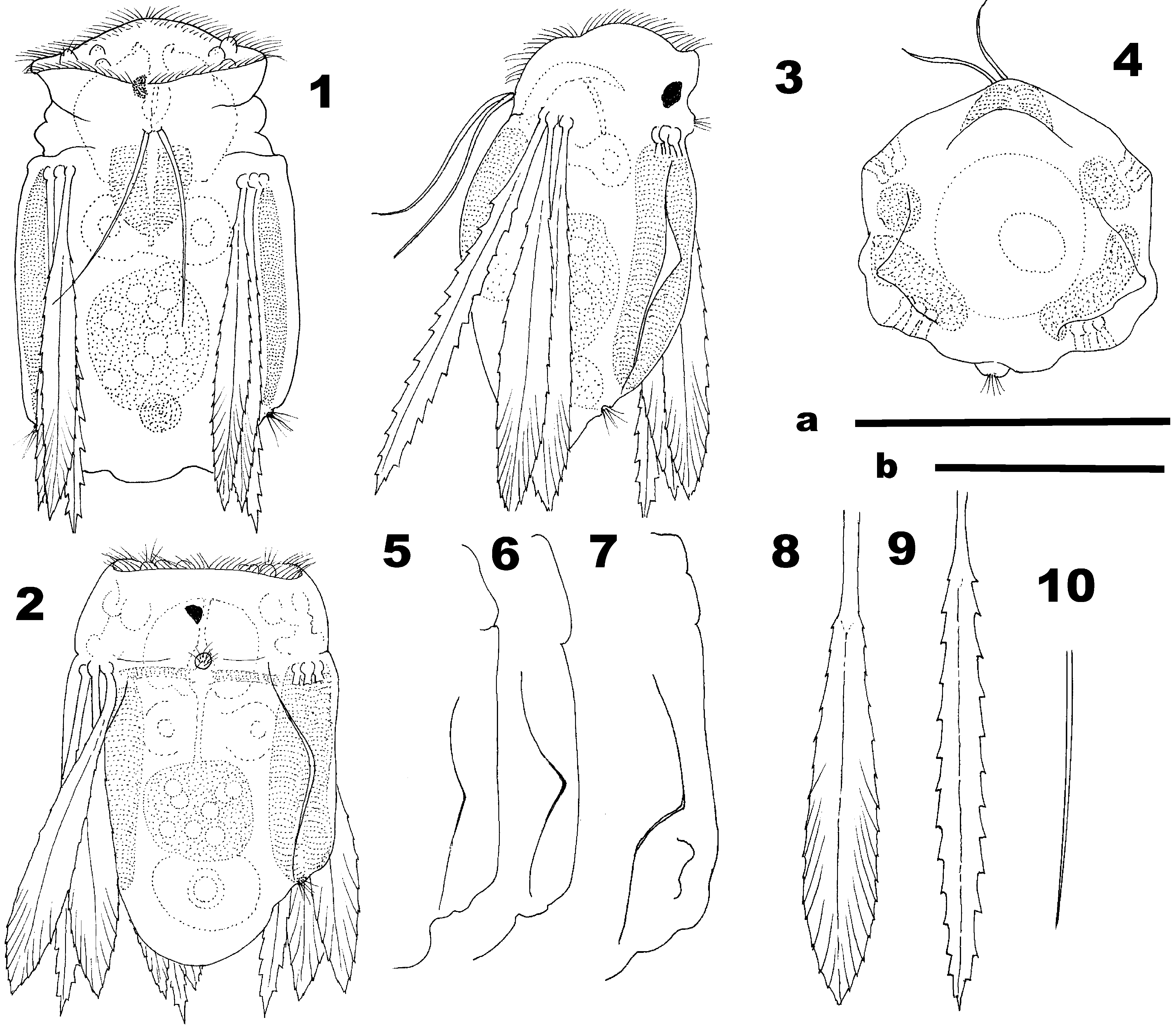
S hort diagnosis. Polyarthra with four bundles of well developed paddles, all similar in length, but shorter than body. Each bundle with two different types of paddles. Ventral accessory paddles present. Vitellarium with 8 nuclei. Unci with single tooth. Manubria gently curved in anterior half, with no projection or lamella present on its surface. Rami with single large tooth on inner margin. Distal part of rami, anterior to large tooth, round with one apical lobe.
Description of parthenogenetic female. Shape of body, in dorsoventral view, subrectangular or roughly subpentagonal ( Figs. 1–2 View FIGURES 1 – 10 ), more rarely, with lateral flanks progressively widening posteriorly. Maximum width about 60-70 % of total body length. Posterior end of body frequently produced into a wide, convex depressed process. Along the dorsal wall of the body, on both sides, and posterior to each bundle of paddles, there is an integumental expansion or ridge. This ridge is somewhat variable in shape, but could generally be described as being at an obtuse angle with the apex, located midway between the insertion of paddles and lateral antenna ( Figs 2, 3, 5–7 View FIGURES 1 – 10 ). On occasion this ridge is accompanied by a small expansions near end of body ( Fig. 7 View FIGURES 1 – 10 ). In lateral view, body progressively tapers to posteriorly, dorsal expansions do not reach outline of maximum thickness.
None of the studied specimens exhibited a “ proloba- like” ventral protuberance. Vitellarium with 8 nuclei. However, an additional nucleus was observed in just two specimens collected from two different localities.
Corona with relatively long cilia, one wide and low ciliate protuberance in central part, a pair of short, inconspicuous apical antennae close to outer limit of corona and two short protuberances positioned close to each antenna ( Figs. 1–3 View FIGURES 1 – 10 ). Eyespot conspicuous and relatively large (6.5–7% of body length), dark-red in colour and hemispherical in shape, with a posterolateral inclination of its plane ( Figs. 1–2 View FIGURES 1 – 10 ).
Dorsal antenna, easily visible due to a thickening that partially surrounds the pore, located midway between dorsal bundles of paddles ( Figs. 2–3 View FIGURES 1 – 10 ). Lateral antennae, not so conspicuous, located close to distal insertion of paddle muscles ( Fig. 2 View FIGURES 1 – 10 ).
Four bundles of major paddles located close to limit between head and rest of body. Each bundle with three paddles similar in length extending beyond the posterior end of the body by about 20–25 % of paddles length. Paddle bundles comprise two lanceolate and one ensiform paddle, all 10–20% shorter than body length, with a welldefined central longitudinal vein. Lanceolate paddles with maximum width, 13–15 % of length, at distal fourth fifth; with 14–17 short teeth on each side, those on distal third as end of a sort pattern of oblique veins or pleats ( Fig. 8 View FIGURES 1 – 10 ). Ensiform paddle, slightly longer than lanceolate paddles, with maximum width, 7–9 % of length, at middle; margins coarsely serrated, with 11–14 well defined larger teeth and surface of blade smooth ( Fig. 9 View FIGURES 1 – 10 ). Accessory pair of ventral paddles, ligulate and extremely slender, basal width 2.5–3.0 % of length, with smooth margins and as long as or slightly shorter than half of body length, inserted midway between ventral bundles ( Figs. 1, 3, 4 and 10 View FIGURES 1 – 10 ).
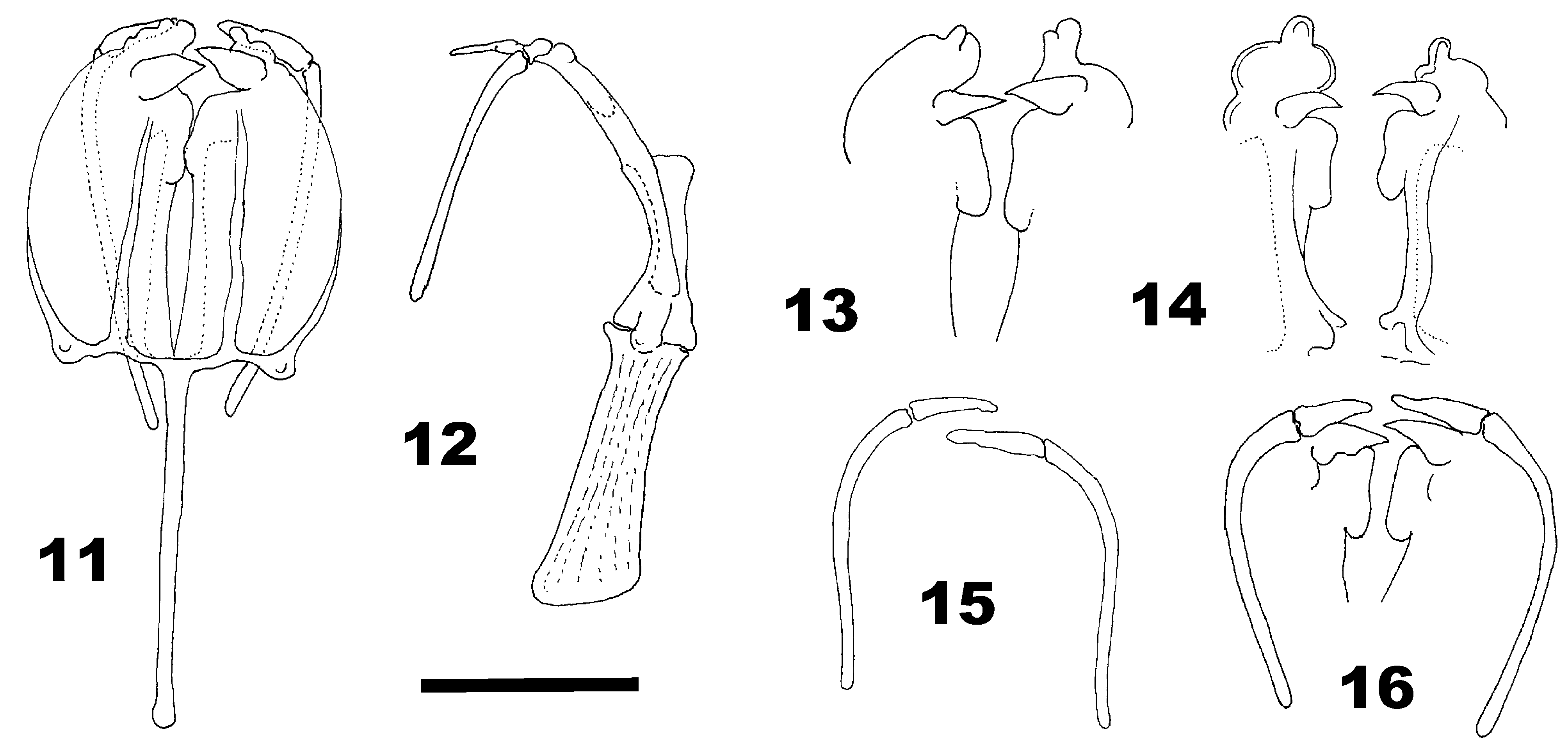
Throphi virgate, about half as long as body, somewhat poorly sclerotized ( Fig. 11 View FIGURES 11 – 16 ). In ventral view, fulcrum seems to be as long as rami, bacilliform with a small rounded expansion at distal end; in lateral view, shorter than rami, subrectangular, with dorsal margin straight and ventral margin concave, expanded at distal end (middle height = 16–17 % of length and distal height = 28–29 % of length) ( Figs. 11–12 View FIGURES 11 – 16 ).
In lateral view, it can be seen that rami are longer than fulcrum and curved from coronal plane to transverse plane ( Fig. 12 View FIGURES 11 – 16 ). Basal apophyses half as long as fulcrum and perpendicular to longitudinal axis of trophi, with distal part ending in a rounded, posteriorly expanded protuberance. Outer half of each ramus subhemicircular, with thin lamella whose anterior limit is barely visible. Inner part of each ramus with one conspicuously stout conical tooth at beginning of distal fourth; anterior to this tooth there is a rounded structure with one lobule at tip; posterior to stout conical tooth there is a sort of smooth flap followed by a smooth, gently concave margin ( Figs. 13–14 View FIGURES 11 – 16 ). Manubria rod-like, slender and curved, with distal part straight or slightly recurved in dorsoventral view; approximately as long as fulcrum; smooth surface without lamella ( Figs. 15–16 View FIGURES 11 – 16 ). Unci approximately as long as one third of manubria, tapered into one relatively blunt tip ( Figs. 15–16 View FIGURES 11 – 16 ).
Dimensions (in µm, mean ± standard deviation; n = 20). Body: length = 86.4 ± 7.0, width = 58.8 ± 4.7, height = 53.0 ± 2.4. Paddles: major lanceolate, length = 75.7± 5.3, width = 10.7 ±1.0; major enciform, length = 77.2 ± 3.4, width = 6.7 ± 1.1; accessory ventral, length = 38.2 ± 10, width = 1.1 ± 0.1. Trophi: total length = 41.3 ± 2.4 [48.5 ± 2.7]; fulcrum length = 20.9 ± 0.8, rami: length = 19.7 ± 9.8 [26.0 ± 0.9], width 10.8 ± 1.5; manubria length = 23.4 ± 1.5; unci length = 6.7 ± 0.5.
Due to the rami being curved in a sagittal plane two measurements are given for total length and rami length: 1) in ventral view, with coverslip supported on plasticine so as not to compress the trophi, 2) between square brackets, in lateral view or in ventral view but with rami straightened by crushing with a coverslip.
Etymology. The specific name refers to the origin of the studied material, which is water bodies located within the La Plata River Basin. La Plata Basin or Cuenca del Plata is the largest water system in South America.
Differential diagnosis and discussion. Polyarthra platensis sp. nov. should be included in the “ P. vulgaris group” due to the presence of ventral accessory paddles and taking into account certain features of the trophi. The species may be considered phylogenetically close to P. dolichoptera Idelson, 1925 . However, there are several consistent differences between these two species. First, there are two characters that seem to be autapomorphic to P. platensis ; specifically 1) the presence of a pair of lateral, normally angled expansions of dorsal integument and 2) the peculiar morphological heterogeneity of the main paddles.
The unusual presence of the lateral expansion of dorsal integument has not been reported before in any of the other species in the genus and may be interpreted as a hydrodynamic adaptation associated to the rapid skipping motion used to avoid predators, which is typical of the species of this genus.
Jersabek et al. (2003) show a photograph of a specimen, in lateral view, questionably identified as “ P. l u m i - nosa? Kutikova, 1962 ” (Catalog Number: ANSP 749), with a morphological feature that resembles this integumental expansion. However, it is not discernable in the dorsal view photographs of the same specimen. In comparison, the lateral expansions of integument are readily visible in dorsal view in P. platensis , even at low magnification.
With respect to the morphological differences between paddles within a bundle, at least two other cases are known, in P. luminosa Kutikova, 1962 and P. minor Voigt, 1904 . However, in both species this morphological heterogeneity is constrained to either the dorsal bundles, and only to the left dorsal bundle in P. minor . Of additional note, P. minor does not belong to “ P. vulgaris- group” because it lacks the accessory ventral paddles. In P. platensis sp. nov., all bundles have the same pattern of heterogeneity, while the distinct paddle in P. platensis sp. nov. is ensiform, in P. luminosa and P minor are dagger-shaped (i.e. leaf-shaped but widest in the proximal part).
Apart from these autapomorphies, P. platensis sp. nov. and P. dolichoptera may be differentiated by the following features: 1) in P. dolichoptera the general body shape tapers to posteriorly, while in P. p l a t e n s i s sp. nov. the posterior part is not tapered; 2) the main paddles in P. dolichoptera are as long as, or longer than the body, while in P. platensis sp. nov. the paddles are shorter than the body; 3) the ventral accessory paddles of P. dolichoptera have serrated edges and are relatively shorter: ca. one fourth of the main paddle length, while in P. p l a t e n s i s sp. nov. these paddles have smooth margins and are somewhat more than half as long as the major paddles; 4) in P. dolichoptera the manubria have well-developed lamellae, while in P. platensis sp. nov. lamellae are absent; and 5) in P. dolichoptera the basal apophyses of rami have a knob-like recurved (anteriorly directed) termination, while in P. platensis sp. nov. the protuberance is directed to posteriorly.
Other differences also exist, which are not easy to circumscribe in terms of shape or size, but are clearly identifiable when specimens are compared. Examples include, the shape of the tips and inner margin of the rami, the size of the teeth, and the basal structure of the rami.
P. platensis sp. nov. also shares several features with P. l u m i n o s a. However, these two species are clearly distinguished by the following features: 1) in P. l u m i n o s a the shape of the accessory paddles is broad, lanceolate and with serrated edges, while in P. platensis sp. nov. these paddles are narrow, ligulate, and with smooth edges; 2) in P. luminosa the manubria have a lamella, while in P. platensis sp. nov. the manubria are subcyclindrical and bare; 3) in P. l u m i n o s a the unci are almost as long as the manubria, while in P. platensis sp. nov. the unci are definitely shorter, measuring about one third of the length of the manubria; 4) in P. luminosa the outer side of rami are bare, while they have a wide lamella in P. platensis sp. nov.; and 5) in P. l u m i n o s a the basal apophyses are bifurcated terminally, while in P. p l a t e n s i s sp. nov. they end in a knob.
In P. platensis sp. nov., the anterior end of the rami (after the major View in CoL tooth) resembles that of P. i n d i c a, described by Segers and Babu (1999). However, P. indica View in CoL has four alternating teeth that precede the major View in CoL tooth, while the manubria have a well-developed lamella, and the paddles are homogeneous in shape. Specimens of Polyarthra View in CoL from the Broa reservoir in Brazil were named ‘ Polyarthra View in CoL sp. near vulgaris View in CoL Carlin’ by Segers and Dumont (1995). Because this species is also a member of the vulgaris View in CoL -group, similarities exist with P. platensis sp. nov. However, the former species has a number of traits that differ from P. platensis sp. nov., such as, the shape of accessory ventral paddles, the absence of a pair of dorso-lateral expansions of the integument, the homogenous shape of the main paddles, and several details of trophi structure.
The discovery of this new species, which, at first glance, is easily assignable to the P. vulgaris- group, suggests that a revision of the genus in the Neotropical region is pressingly needed.
| INALI |
Instituto Limnologia |
No known copyright restrictions apply. See Agosti, D., Egloff, W., 2009. Taxonomic information exchange and copyright: the Plazi approach. BMC Research Notes 2009, 2:53 for further explanation.