
Pseudrotasfer microincubator, Bohn, Jens Michael, 2007
|
publication ID |
https://doi.org/ 10.5281/zenodo.179963 |
|
DOI |
https://doi.org/10.5281/zenodo.5672536 |
|
persistent identifier |
https://treatment.plazi.org/id/335287D9-E471-D36C-FD9B-FB94FD591DF2 |
|
treatment provided by |
Plazi |
|
scientific name |
Pseudrotasfer microincubator |
| status |
sp. nov. |
Pseudrotasfer microincubator View in CoL spec. nov.
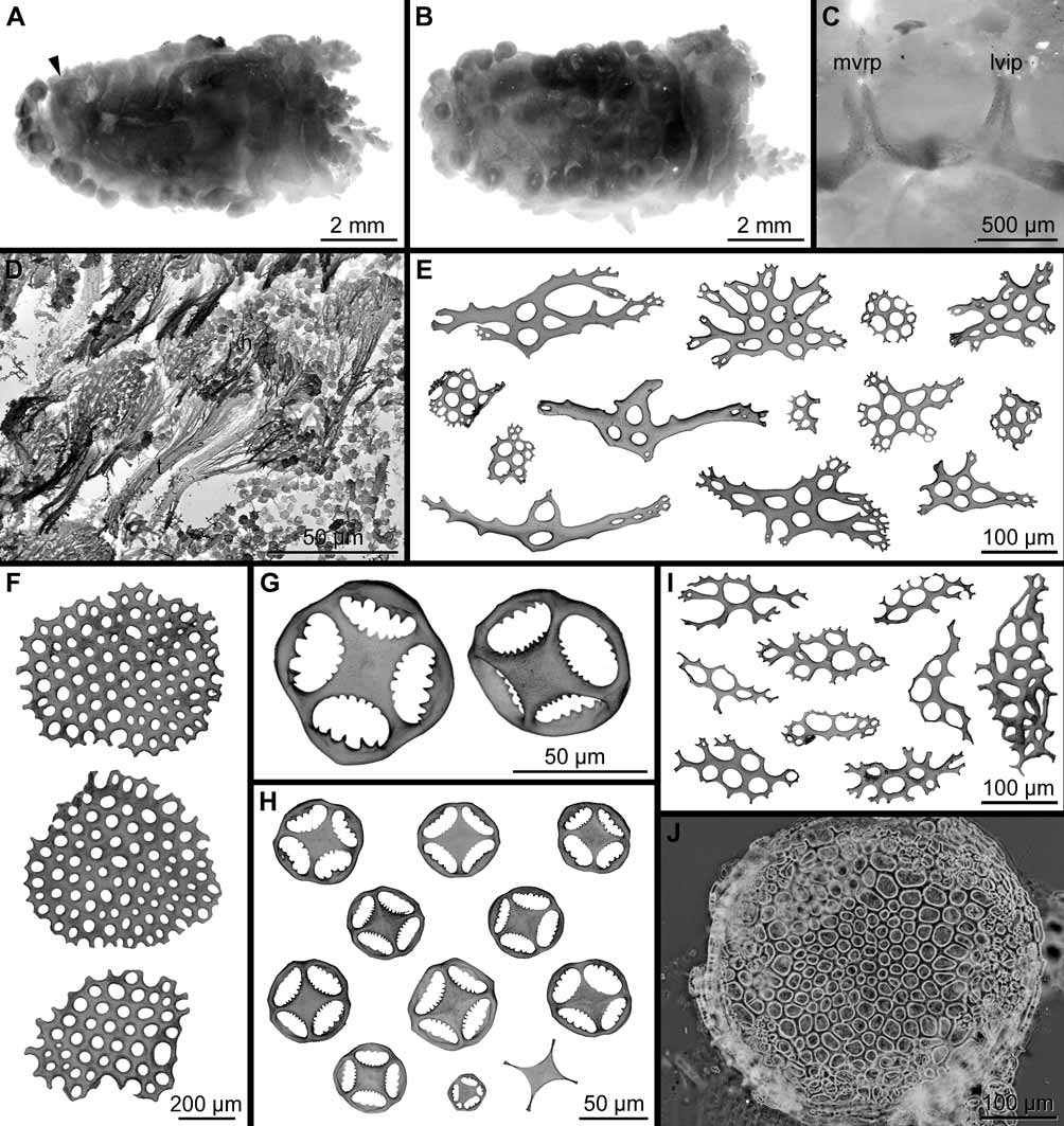
( Fig. 2 View FIGURE 2 A–J)
Material examined. Holotype. FS “Polarstern”, LAMPOS, station PS 61/150-1, 54 °30.22' S, 56°08.20' W, 286 m to 54°29.64' S, 56°08.13' W, 290 m, Agassiz trawl, 0 6 Apr. 2002 ( ZSM 20070012, 1 ɗ). Paratypes. FS “Polarstern”, LAMPOS, station PS 61/145-1, 54 °01.58' S, 62°01.03' W, 271 m to 54°01.11' S, 62°01.63' W, 272 m, Agassiz trawl, 0 5 Apr. 2002 ( ZSM 20070011, 1 Ψ); station PS 61/150-1 [for details see holotype] ( ZSM 20070013, 1 ɗ).
Description. Although three specimens are available (holotype: ɗ, body length 7 mm; paratypes: 1 ɗ, body length 6 mm; 1 Ψ, body length 5 mm), the description of the new species is mainly based on the holotype and the male paratype, due to the fact that the female paratype is in a defective state of preservation. Although the specimens are small, all of them are mature. Preserved, the specimens are of a whitish colour. The body is subcylindrical ( Fig. 2 View FIGURE 2 A–B), but with a flattened ventral sole, and rounded posterior end. Mouth terminal, anus subdorsal above ventral sole ( Fig. 2 View FIGURE 2 A: arrowhead). Tentacles 10, dendritic, two ventral considerably smaller than others.
The dorsal radii are almost devoid of tube feet, each radius with single radial tube foot present close to anterior end of body, and next to anus. These are cylindrical and considerably smaller than those on the sole. Likewise, a single tube foot is also present in each of the ventrolateral radii close to anterior end of body. Somewhat distanced from anterior end (1.6–1.7 mm in the current specimens), ventral sole extends to posterior end of body ( Fig. 2 View FIGURE 2 B). Tube feet defining sole conspicuous, cylindrical, with distinct terminal discs. Feet restricted to radii, a single row in each ventrolateral radius (ɗ paratype: 8 tube feet, holotype: 11 tube feet), and a double row in a zig-zag arrangement in mid-ventral radius (ɗ paratype: 8 tube feet, holotype: 13 tube feet).
Calcareous ring simple ( Fig. 2 View FIGURE 2 C), with no posterior processes. Anterior processes of all 10 plates about same height. They are oblong rectangular, incised anteriorly in radial plates. Interradial plates oblong triangular, except for middorsal interradial plate, which has a deeply incised V-shaped anterior process. Posterior margin of plates emarginated, more pronounced in radial plates than in interradials.
Retractor muscles arise from longitudinal muscles about one third body length from anterior end. A single tubular polian vesicle present in left lateral interradius (stone canal and madreporite not investigated due to delicate state of specimens). Intestinal tract consists of a short oesophagus, followed by an intestine with a long loop, and terminates in a short cloaca. Anterior descending intestine suspended on midventral mesentery, ascending anterior intestine on a mesentery fixed to left lateral interradius, and descending posterior intestine on a mesentery attached to right side of midventral longitudinal muscle. Right and left respiratory trees arise with a short common trunk from anterior dorsal side of cloaca. Both trees simple short tubules, which may have few short side branches.
Sexes are separate. Due to delicate state of specimens, position of gonopore could not be ascertained. Gonad consists of left and right bunch of few simple unbranched tubules attached to middorsal mesentery immediately posterior to middorsal interradial plate of calcareous ring. In males, each bunch is composed of 3–6 long tubules, as well as few short and probably developing tubules. Long tubules are densely filled with so-called spermatozeugmata ( Fig. 2 View FIGURE 2 D), bunch-like structures composed of numerous spermatozoa with agglutinated tails ( Fig. 2 View FIGURE 2 D: t). Female paratype with a bunch of simple balloon-shaped tubules on both sides of middorsal mesentery (two on left and about four on right side), filled with embryos (all of about same developmental stage). In addition, single small tubules filled with eggs present.
Tentacles supported by very variable rod- to plate-like ossicles ( Fig. 2 View FIGURE 2 E), up to 350 µm long, in outline elongated to rounded. Ossicles smooth, with holes of variable size, central holes usually larger than peripheral, often with irregular branching marginal outgrowths.
Body wall ossicles of two types, a deeper layer of scattered large plates and an upper layer of densely distributed wheel-like baskets. Plates of deeper body wall ( Fig. 2 View FIGURE 2 F) large (up to 700 µm in diameter), smooth, irregularly circular in outline, with holes of varying sizes. Baskets of upper body wall ( Fig. 2 View FIGURE 2 G–H) small (30– 75 µm in diameter), shallow, resembling four-spoked wheels, with hub-like broadened central primary cross and undulating rim connecting its four arms (“spokes”). While outer margin of hub is armed with several small outward-pointing teeth, inner surface of rim between spokes is equipped with fewer, usually larger, inward-pointing teeth.
Tube feet covered by a dense outer layer of wheel-like baskets, absent only from terminal disc. Baskets overlie a layer of smooth, usually slightly curved plates ( Fig. 2 View FIGURE 2 I), similar to those in tentacles, with irregular elongated outline, perforated by larger and smaller holes, and often with various marginal outgrowths. These plates restricted to distal ends of tube feet, adjacent to terminal plates. Terminal disc supported by single terminal plate (exceptionally by few smaller plates), up to 400 µm in diameter, smooth, roundish in outline, with irregular marginal outgrowths; central holes of plates smaller than peripheral ( Fig. 2 View FIGURE 2 J).
Reproduction and development. Brooding period includes at least the beginning of April. The only known female has its gonad tubules filled with juveniles, which are all at about the same developmental stage. The juveniles are about 1.2 mm long. The body is cylindrical to deformed due to packing within the gonad tubules. There are at least eight tentacles of about the same size, all retracted. No tube feet were detected and the body wall is covered by a layer of wheel-like baskets, which are also present in the adults.
Distribution. ( Fig. 1 View FIGURE 1 ) So far, Pseudrotasfer microincubator is only known from the Burdwood Bank in the south-western Atlantic Ocean, depth range 271 to 290 m.
Etymology. A small breeder ( microincubator ).

| ZSM |
Bavarian State Collection of Zoology |
No known copyright restrictions apply. See Agosti, D., Egloff, W., 2009. Taxonomic information exchange and copyright: the Plazi approach. BMC Research Notes 2009, 2:53 for further explanation.