
Melanoblossia ansie, Bird & Wharton, 2015
|
publication ID |
https://doi.org/ 10.5733/afin.056.0218 |
|
persistent identifier |
https://treatment.plazi.org/id/335B8795-3046-612F-FE4F-FD4DFCA72D27 |
|
treatment provided by |
Felipe |
|
scientific name |
Melanoblossia ansie |
| status |
sp. nov. |
Melanoblossia ansie View in CoL sp. n.
Figs 1–5 View Fig View Fig View Fig View Fig View Fig
Melanoblossia sp. Bird et al. 2015: 56, 81, 103, 122, 139, 188, figs 6B, 10B, 26, plates 36I, 154G, H, 157. Etymology:The species is named in honour of distinguished South African arachnologist, Dr Ansie Dippenaar-Schoeman , for her contribution to arachnology in Africa, and mentor to varioUs stUdents in this field, inclUding to the first aUthor in the present stUdy. NoUn (name) in apposition.
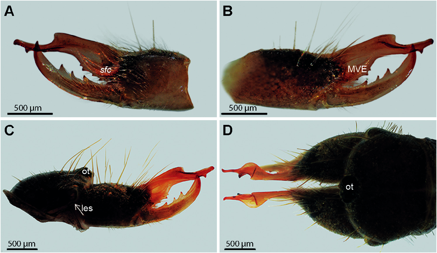
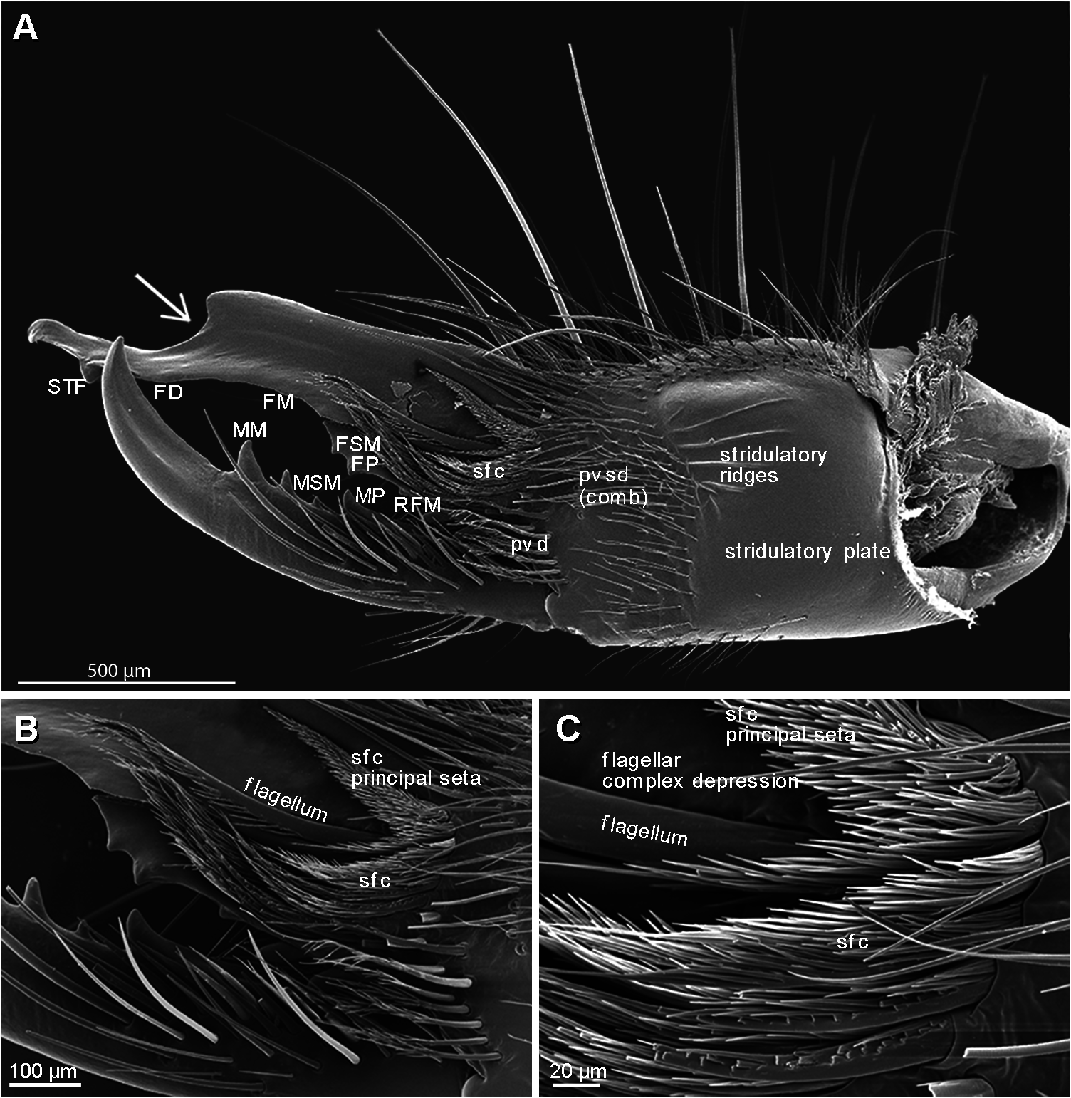
Affinities:This species shows closest affinities with Melanoblossia . It is separated from Daesiella Hewitt, 1934 , Unguiblossia Roewer, 1941 , and most Lawrencega by the presence of two tarsomeres on leg IV. Males are distinguished from Microblossia by the presence of a distinct type C setiform flagellar complex (type C sfc) ( Bird et al. 2015), and from Lawrencega by the presence of a prominent sfc principal seta ( Fig. 4 View Fig ), i.e. the dorsalmost seta in the flagellar complex, which is long, strongly differentiated and dorsodistally directed, similar to other species of Melanoblossia ( Fig. 4 View Fig ), compared to the short, weakly differentiated and more distally directed sfc principal seta in Lawrencega . Diagnosis: Males differ from all other known melanoblossiine species by the elaborate modifications distally on the fixed (dorsal) finger, comprising a prominent, cUrved retrolateral flange and a strong ventral dip in the dorsal margin of the finger ( Figs 3 View Fig , 4A View Fig ). The flagellar homolog (as defined by Bird et al. 2015) is distinct and clearly visible in prolateral view, similar to most Lawrencega species but different from other known Melanoblossia species, which have this modified seta more hidden behind the proximal row of setae in the sfc. Conversely, the sfc principal seta is prominent in this species, similar to other known Melanoblossia species but different from that of Lawrencega species ( Fig. 4 View Fig ).
Description:
Male.
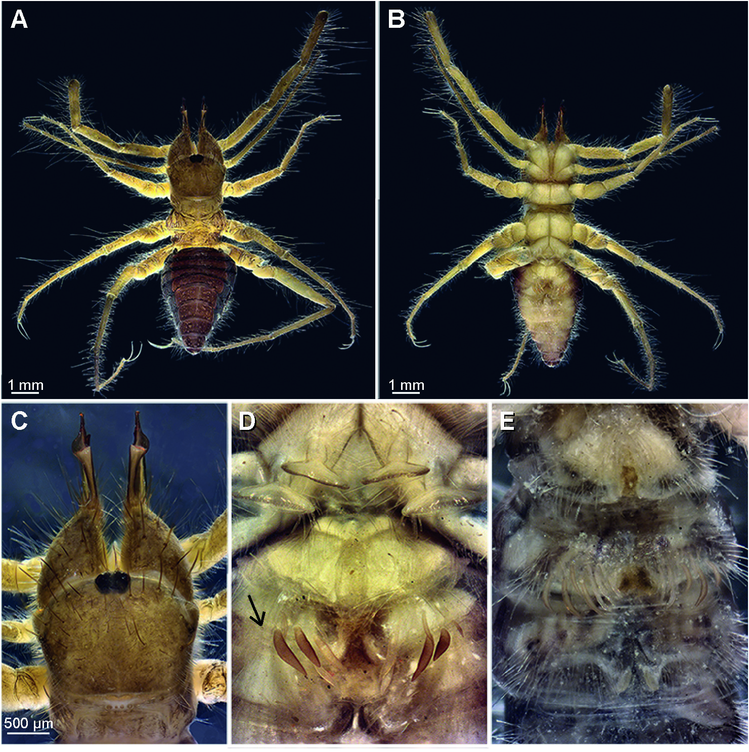
Habitus. Division between prosoma and opisthosoma broad; opisthosoma tapering posteriorly ( Fig. 1A, B View Fig ).
Coloration. Propeltidium and chelicerae dark yellowish brown. Ocular tubercle black. Opisthosomal tergites reddish brown with dark purplish bands on posterior margins, which are wider in anterior four segments that are visible in dorsal view. Lateral opisthosoma purple with whitish longitudinal folds in the intersegmental membrane. Opisthosomal sternites light cream brown, changing to purplish posteriorly. Anal segment completely reddish brown. Legs and pedipalps yellowish brown dorsally, similar to propeltidium, cream brown ventrally, similar to opisthosomal sternites; coxae, trochanters and genital sternite lighter cream brown ( Fig. 1A, B View Fig ).
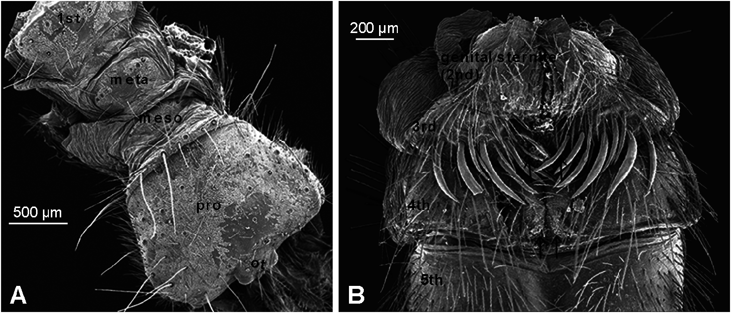
Prosoma. Median longitudinal furrow of propeltidium incomplete ( Figs 1C View Fig , 3D View Fig ) and sUperficial ( Fig. 2A View Fig ); anterolateral propeltidial lobes partially fUsed to propeltidiUm; lateral eyespots long and narrow, hidden from lateral view on ventral side of anterolateral propeltidial lobe along margin of lobe ( Fig. 3C View Fig , arrow indicates position); median plagula narrower than ocular tubercle ( Fig. 1C View Fig ); mesopeltidium and metapeltidium wider than long, mesopeltidium boatshaped, metapeltidium rectangular ( Fig. 2A View Fig ); coxae densely covered with bifid setae. Widely spaced reddish brown, long, bifid setae on propeltidiUm and retrodorsally on the chelicera, on propeltidium more numerous in anterior third ( Fig. 3B, C View Fig ), interspersed with shorter, less pigmented setae extending across the surface of chelicera and propeltidium.
Opisthosoma. Tergites and pleUrites sparsely covered in relatively long, bifid, transparent setae. Similar setae on sternites, but slightly longer and slightly more dense. Fleshy ctenidia on first post-genital sternite (third opisthosomal segment), 3–4 largest ones on each side reddish brown, interspersed with 4–5 slightly smaller cream-coloured ctenidia. Dense patch of slightly thickened setae on fourth post-genital sternite (sixth opisthosomal segment); less distinct in some specimens.
Chelicera. Shape: Fixed finger scUlptUred distally ( Fig. 3 View Fig ), with prominent retrolateral flange and strong ventral dip in dorsal margin of finger ( Fig. 4A View Fig , arrow); fixed finger medioventral excrescence ( MVE) pronounced ( Fig. 3B View Fig ); gnathal edge (=cutting edge) strongly retrolaterally compressed; sUbterminal flange ( STF) present on fixed finger mUcron, sitUated directly distal to fixed finger distal ( FD) tooth, if present; STF is a prominent toothlike flange originating distally on the gnathal edge and proximally merging into the retrolateral side of the finger; movable finger gradUally cUrving dorsally, mUcron approximately one third length of finger. Dentition ( Fig. 4A View Fig ): Movable finger dentition with well developed proximal tooth ( MP) and slightly larger medial tooth ( MM; most distal tooth on finger) separated by one ( Fig. 3B View Fig ), sometimes two ( Fig. 4A View Fig ) secondary teeth (movable finger sUbmedial teeth, MSM) increasing in siZe proximal to distal if two MSM. Fixed finger median series dentition redUced, with small bUt distinct proximal tooth ( FP), and smaller than FP bUt distinct secondary tooth (fixed finger sUbmedial tooth, FSM) directly distal to FP; other primary teeth (fixed finger medial, FM; fixed finger distal, FD) absent, to present in highly vestigial form; if present, barely visible as a slight, often darkened short ( FM) or elongated ( FD) protuberances. Fixed finger fondal teeth well developed in prolateral row with distinct, tall and narrow profondal medial ( PFM) and profondal proximal ( PFP) tooth, both larger than FP; not possible to examine PF row in holotype for risk of damage, but profondal subdistal ( PFSD) tooth present in another specimen examined. Retrofondal ( RF) row with four teeth, distalmost tooth in fixed finger retrofondal ( RF) row (pUtative retrofondal medial tooth, RFM) distinct, approximately half the size of FP; rest of RF row teeth small and insignificant, barely visible, with second most proximal tooth (retrofondal proximal tooth, RFP) slightly larger than the one distal (profondal submedial, PFSM) and proximal (profondal subproximal, PFSP) to it .
Flagellar complex. Setiform flagellar complex (sfc), type C (see Bird et al. 2015). Well defined clUster of modified pvd setae ( Fig. 4 View Fig ). Two rows of setae emerge from slight linear elevation into a broad flagellar complex depression ( Fig. 4C View Fig ). Distal row comprises three setae with limited plumosity; distalmost seta in row is the hypothesised setiform flagellUm homolog, which is laterally compressed and smooth with limited plUmosity ventrally and distally. Second (proximal) row of setae in sfc uniformly and strongly plumose throughout, curve dorsally along margin of MVE, except for sfc principal seta (dorsalmost setae in the proximal row of sfc), which is strongly plumose, especially at base, resulting in a general triangular-shaped seta.
Rostrum. Approximately 1.5× longer than wide; dorsal margin of epistosomo-labral plate weakly sinuous; interlocked anastomosed setae form rigid sieve; median setae in sieve shortest, and dorsal setae longer than ventral setae. Lateral lip flagella distinctly but not densely plumose, one situated on each lip dorsally at base of lip.
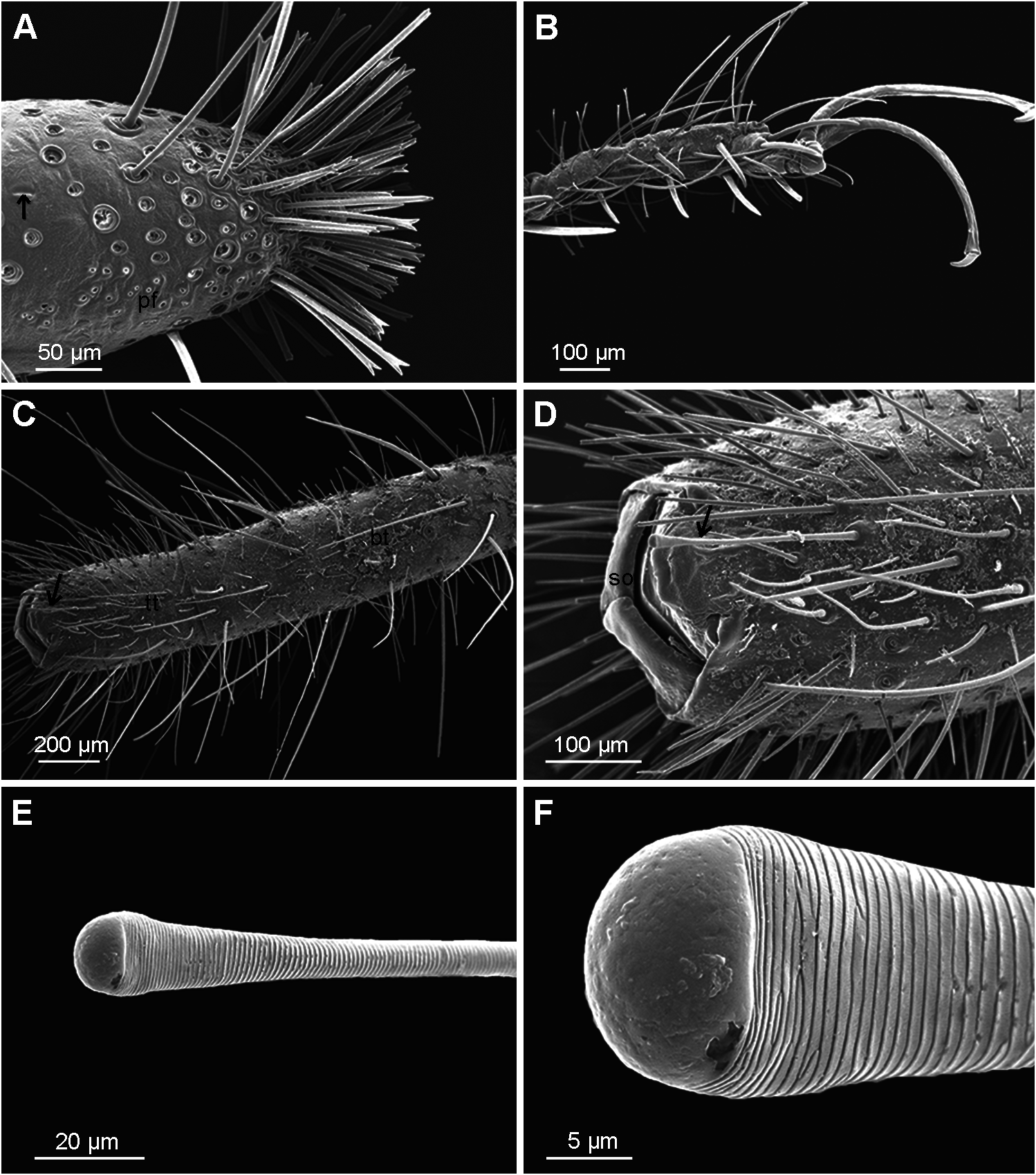
Pedipalp. TelotarsUs immovably fixed to basitarsUs, together Uniformly cylindrical throughout ( Fig. 5C View Fig ); tibia curved proventrally; femur more or less cylindrical ( Fig.1A, B View Fig ). Spiniform setae absent. Flexible apparent bifid setae of varioUs lengths on all segments, those situated proventrally longer and more ridged; few very long hairlike setae, longest ones medially on dorsal side of tarsus (basitarsus + telotarsus) and tibia. Dorsal to retrolateral femur with dense cover of short, transparent setae. Clubbed seta medioventrally on telotarsus ( Fig. 5E, F View Fig ).
Leg I. Patella, tibia and basitarsus cylindrical throughout, telotarsus slightly broadening towards apex. Concentration of bifid setae at apex. Narrow prodorsal field of tarsal pores ( Fig. 5A View Fig ). Slit sensilla at least on telotarsus ( Fig. 5A View Fig , arrow). Trochanter, femur and patella covered with short, transparent setae.
Walking legs. Claws long in all walking legs, longer than telotarsus in legs II and III. Second (distal) segment of claw approximately eighth the length of first (proximal) claw segment.Trochanter, femur and patella covered with short, transparent setae ( Fig. 1A, B View Fig ). Legs II and III: Telotarsus not divided into tarsomeres; spiniform pattern of telotarsus 2.2.2.1, with four setae on proventral and three on retroventral side ( Fig. 5B View Fig ). Leg IV: Telotarsus divided into two tarsomeres; spiniform pattern of tarsomeres 2.2 /2.2.2.
Measurements (from holotype; in millimetres). Total length 9.27; propeltidium length 1.47, width 1.96; ocular tubercle length 0.20, width 0.39; eye (ocellus) diameter 0.14; CL 2.42; CH 0.75, CW 0.75. Appendages (segment length): Palp total length 5.84, femur 1.95, tibia 1.79, basitarsus + telotarsus 2.10; leg I total length 5.84, patella 1.75, tibia 1.60, basitarsus 1.00, telotarsus 0.74; leg II total length 4.92, patella 1.44, tibia 1.17, basitarsus 1.10, telotarsus 0.61, claw 0.73 (distal claw segment 0.10); leg IV total length 9.95, patella 2.73, tibia 2.38, basitarsus 2.06, telotarsus 1.39, claw 0.79 (distal claw segment 0.09).
Female. Unknown.
Variation : Twelve males (24 chelicerae) were examined for variation in dentition. On the movable finger MP and MM present in all specimens, single MSM present in 14 chelicerae, second MSM, smaller than and sitUated proximal to the first MSM, present in 10 chelicerae. Large variation in size in the second (proximal) MSM, from denticlesized to only slightly smaller than distal MSM. The FP and FSM were present with little variation in size and shape, in all chelicerae, FM was absent in 14 chelicerae and present as a slight, generally darkened protuberance in all other specimens. Except for the FD, which was difficUlt to evalUate in chelicerae where the jaws were closed, bilateral asymmetry in absence/presence of teeth was found only in three specimens. In one a retrofondal tooth was absent on the left chelicera, and in the other two a second movable finger sUbmedial ( MSM) tooth was present on the left, bUt not on the right chelicera. One fixed finger was deformed towards the apex, resUlting in the loss of the STF. Ctenidia variable in nUmber, bUt difficUlt to evalUate as these seem to easily break off .
Holotype ♂: NAMIBIA: Lüderitz District : Diamond Area : Sperrgebiet National Park : Tsaukhaib Mountain , old transport route (26°42'58.0"S 15°40'02.6"E), 906 m, 24–30.viii.2006, EduVentures 9 Expedition. Deposited in the National Museum of Namibia, Windhoek ( NMNW 14227 ). GoogleMaps
Paratypes: Same collecting data as holotype: 1♂ ( MRAC 244095 ); 1♂ ( NCA 2015 /3479); 1♂ ( NMNW 13394 ); 1♂ ( NMNW 13395 ); 3♂ ( NMNW 13396 ); 3♂ ( NMNW 14179 ); 1♂ (SAM-ENW-C006956) GoogleMaps ; 1♂ ( SMFD) .
Other material examined: Same collecting data as types: 1♂ ( AMNH [ LP 9857 ]) .
Distribution: Known only from the type locality at the foot of the Tsaukhaib Mountain in the Sperrgebiet National Park, situated in the southern Namib Desert in the southwestern corner of Namibia.
Biology: The type locality of Melanoblossia ansie sp. n. is classified as a sUccUlent steppe vegetation zone, which falls within the Succulent Karoo biome ( Irish 1994; Mendelsohn et al. 2002). Melanoblossia ansie sp. n. was only collected during one pittrap cycle at the beginning of a three-year long pittrap survey of the Sperrgebiet National Park, during a year of particularly high rainfall. Soon after this pittrap cycle, pittraps moved approximately 50 metres further, and M. ansie sp. n. was not collected again. Targeted collection attempts later for this species did not deliver any specimens. Melanoblossia ansie sp. n. seems to have a restricted range of microhabitat and seasonal preference. These species are probably restricted to sandy habitats, as indicated by their long claws, and are likely diurnal, similar to other melanoblossiids, as indicated by their dark pigmentation.
| MP |
Mohonk Preserve, Inc. |
| MM |
University of Montpellier |
| MSM |
Marine Science Museum, Tokai Univ. |
| FM |
Department of Nature, Fujian Province Museum |
| AMNH |
American Museum of Natural History |
No known copyright restrictions apply. See Agosti, D., Egloff, W., 2009. Taxonomic information exchange and copyright: the Plazi approach. BMC Research Notes 2009, 2:53 for further explanation.
|
Kingdom |
|
|
Phylum |
|
|
Class |
|
|
Order |
|
|
Family |
|
|
Genus |