
Leucandra spinifera, Klautau, Imesek, Azevedo, Plese, Nikolic & Cetkovi, 2016
|
publication ID |
https://doi.org/ 10.5852/ejt.2016.178 |
|
DOI |
https://doi.org/10.5281/zenodo.3850369 |
|
persistent identifier |
https://treatment.plazi.org/id/3406810E-DD3F-B21F-FD4E-FEFA098A3C26 |
|
treatment provided by |
Valdenar |
|
scientific name |
Leucandra spinifera |
| status |
sp. nov. |
Leucandra spinifera View in CoL sp. nov.
urn:lsid:zoobank.org:act:280369B2-48FF-4F3D-88E3-73317D5919A5
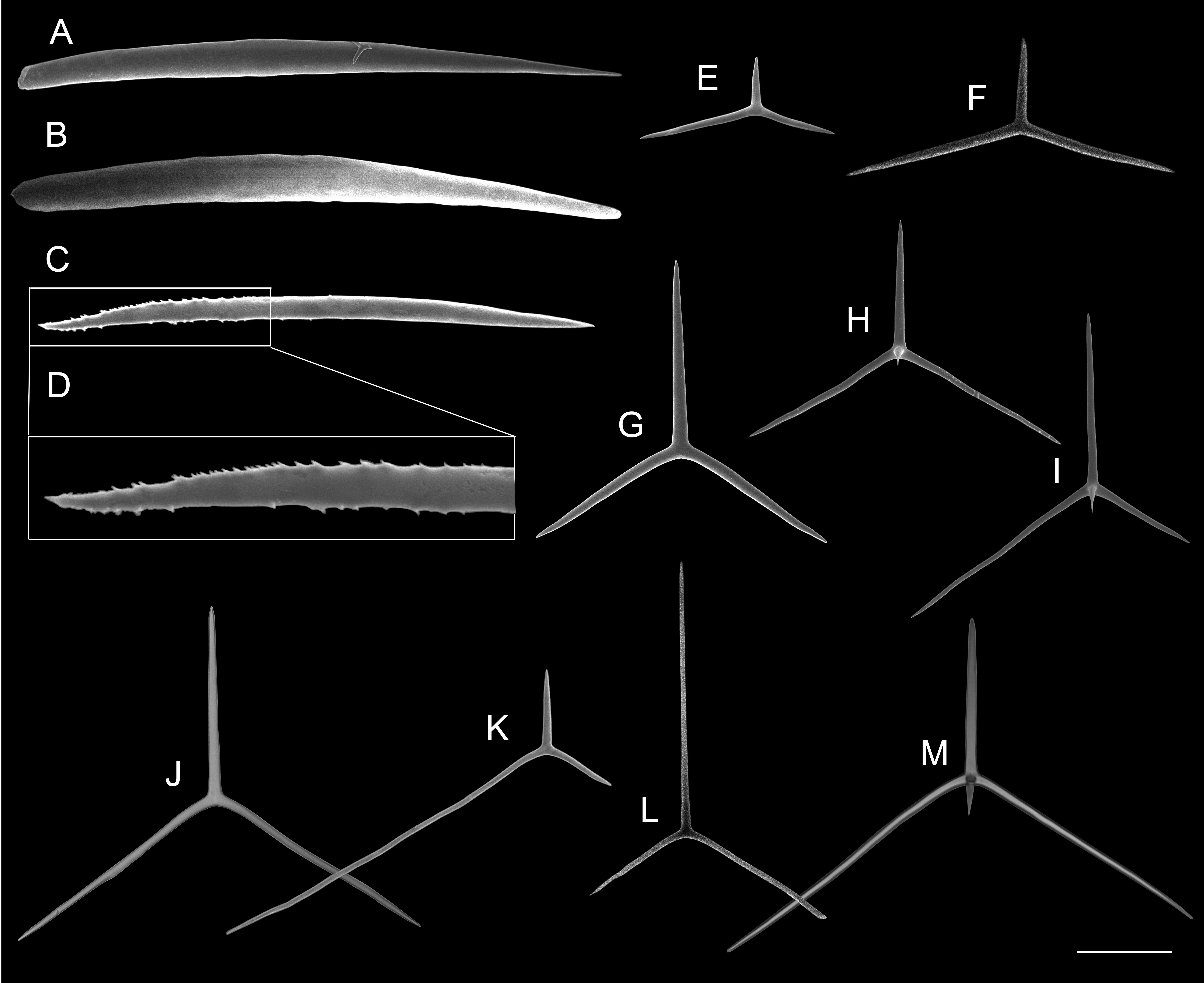
Figs 10–11 View Fig View Fig ; Table 9
Etymology
From the Latin spinifer, meaning prickly, for the presence of numerous diactines.
Material examined
Holotype
ADRIATIC SEA: Vrulja Cove, 43°24'01.3" N, 16°53'10.9" E, 10 m, collected by Vedran Nikolić, 24 Aug. 2011 ( IRB-SG3 = UFRJPOR 8348, in ethanol).
GoogleMapsParatype
ADRIATIC SEA: Island of Čiovo, 43°28'58.5" N, 16°21'25.6" E, 5 m, collected by B. Pleše and V. Nikolić, 6 Nov. 2010 (PMR-13742 = UFRJPOR 6861, in ethanol).
Colour
White in life and in ethanol.
Description
The body has the shape of a vase (0.8 × 0.4 cm), with a single apical osculum surrounded by a membrane and a crown of a few, or even no trichoxeas ( Fig. 10A View Fig ). The osculum is supported by sagittal tetractines, but a few triactines are also present. They are organised in parallel and point their apical actines to the osculum. They become disorganized, smaller, thinner and less sagittal farther from the osculum. They are also substituted by triactines. Numerous diactines on the surface make it very hispid. The aquiferous system is leuconoid and the atrium is large ( Fig. 10A View Fig ). The cortical skeleton is composed of tangential triactines, perpendicular giant diactines, microdiactines and rare trichoxeas ( Fig. 10 View Fig B–E). The giant diactines frequently cross the entire choanosome ( Fig. 10B View Fig ). The choanosomal skeleton has no organisation. It is composed mainly of subregular triactines, with curved paired actines. Tetractines are also present, but only surrounding canals. The atrial skeleton has triactines and a few tetractines that project their apical actines into the atrium ( Fig. 10F View Fig ). Microdiactines are also present in the atrium.
Spicules ( Table 9)
OSCULAR TRIACTINES (very few) AND TETRACTINES (abundant). Sagittal. Actines are cylindrical and blunt to sharp. The unpaired actine is thinner than the paired ones. The apical actine of the tetractines is conical, sharp, smooth and strongly curved towards the osculum aperture.
TRICHOXEAS. Very thin, long and straight. They are frequently broken. These spicules are rare, but can be found in the cortex and atrium.
DIACTINES. Almost fusiform. The tip that penetrates the choanosome is a little larger and more rounded ( Fig. 11 View Fig A–B). Size: 866.5/ 54.4 µm.
MICRODIACTINES. Fusiform ( Fig. 11C View Fig ). They are present in the cortex and atrium. They frequently have microspines ( Fig. 11D View Fig ), but smooth spicules are also present. Size: 100.4/ 4.2 µm.
CORTICAL TRIACTINES. Sagittal. Actines are slightly conical, with blunt tips. The unpaired actine is shorter than the paired ones, which are curved. One of the paired actines is frequently shorter than the other ( Fig. 11 View Fig E–F). Size: 189.5/ 12.9 µm (paired actine); 150.8/ 13.5 µm (unpaired actine).
CHOANOSOMAL TRIACTINES. Subregular to sagittal. The paired actines are curved, consequently the unpaired angle is smaller than the paired angles. Actines are slightly conical with blunt tips. They are almost the same length ( Fig. 11G View Fig ). These spicules are spread in the choanosome and surrounding the canals. Size: 192.8/ 12.8 µm (paired actine); 188.8/ 14.4 µm (unpaired actine).
CHOANOSOMAL TETRACTINES. Sagittal. The paired actines are curved, consequently the unpaired angle is smaller than the paired angles. Actines are slightly conical with blunt tips. The apical actine is straight or curved, conical, smooth and sharp ( Fig. 11 View Fig H–I). These spicules are present only surrounding the canals.
ATRIAL TRIACTINES AND TETRACTINES. Triactines are much more abundant. These spicules are strongly sagittal. The paired actines are curved and much longer than the unpaired one.Actines are slightly conical and blunt ( Fig. 11 View Fig J–L). The apical actine of the tetractines is straight or slightly curved near the end, conical, smooth and sharp ( Fig. 11M View Fig ). These tetractines are very similar to those of the choanosome. Size (triactine): 305.3/ 7.9 µm (paired actine); 211.4/ 9.8 µm (unpaired actine). Size (tetractine): 276.8/ 8.4 µm (paired actine); 222.0/ 9.7 µm (unpaired actine); 42.5/ 6.9 µm (apical actine).
Ecology
Specimens were collected on a cliff in a shaded area.
Remarks
This species differs from all other species of Leucandra mainly by the composition of the skeleton, particularly by the presence of mainly triactines in the atrial skeleton, with very long and slender paired actines and few spiny microdiactines in the cortex. The most similar species is the Californian L. heathi Urban, 1906 . However, this species has no tetractines, while L. spinifera sp. nov. has a few tetractines. Besides, microdiactines are not abundant in L. spinifera sp. nov., while in L. heathi they form a continuous palisade in the cortex.
We found 10 species of Leucandra recorded from the Mediterranean until now, and L. spinifera sp. nov. can be differentiated from all of them: L. aspera ( Schmidt, 1862) has no microdiactines; L. balearica ( Lackschewitz, 1886) has only tetractines in the atrium and its microdiactines are much smaller (12- 24/ 1 µm); L. globosa (Sarà, 1951) has different microdiactines; L. bolivari Ferrer-Hernandez, 1916 has no diactines; L. crambessa Haeckel, 1872 has no microdiactines and has tetractines only in the atrium; L. nausicaae (Schuffner, 1877) has no diactines and the atrial skeleton comprises only tetractines; L. riojai Ferrez-Hernandez, 1918 has only tetractines in the atrium; L. rodriguezii ( Lackschewitz, 1886) has shorter microdiactines (12-14/ 1 µm) which occur only in the atrium and the atrium is also composed of only tetractines; L. sulcata Ferrer-Hernandez, 1918 has microdiactines of a different shape, which are present abundantly only in the cortex, while the atrium is composed mainly of tetractines.
Abbreviations: at = atrium; cx = cortex.

| V |
Royal British Columbia Museum - Herbarium |
No known copyright restrictions apply. See Agosti, D., Egloff, W., 2009. Taxonomic information exchange and copyright: the Plazi approach. BMC Research Notes 2009, 2:53 for further explanation.
|
Kingdom |
|
|
Phylum |
|
|
Class |
|
|
SubClass |
Calcinea |
|
Order |
|
|
Family |
|
|
Genus |