
Palaeoginkgoxylon sp.
|
publication ID |
https://doi.org/10.35463/j.apr.2022.01.04 |
|
DOI |
https://doi.org/10.5281/zenodo.10973991 |
|
persistent identifier |
https://treatment.plazi.org/id/34188785-1D46-FFB5-9794-E225FC2D5EB7 |
|
treatment provided by |
Felipe |
|
scientific name |
Palaeoginkgoxylon sp. |
| status |
|
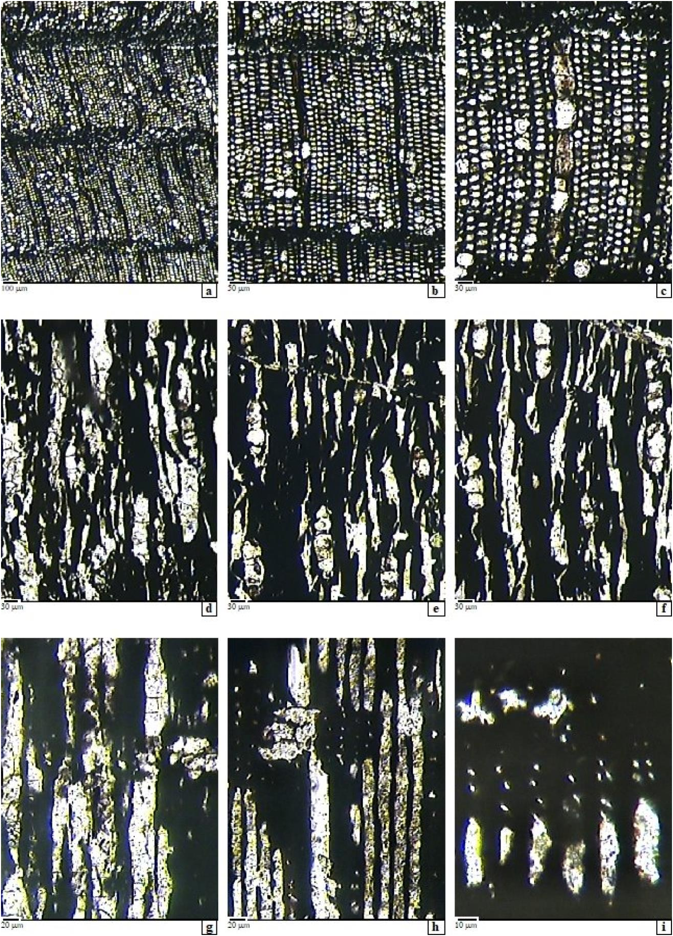
Fig. 5 View Fig , a-i.
Material
The studied material is represented by two samples of silicified wood, of decimetric dimensions, found in the Holbav locality area, on the Maiului brook. The studied samples are trunk or thick-branch fragments, dark to black color and by hand lens or even by naked eye a regular fibrous structure is visible. These two samples have the field-numbers 1032, 1036 in "Grădinaru Collection" and are stored under the inventory numbers 27674 and 27675, at the National Museum of Geology , in Bucharest .
Microscopic description
Growth rings – in cross sections show a tracheidoxylic structure with rather indistinct boundaries, with few parenchyma and sometimes with axial canals, most likely mucilaginous. And, the structure is marked by the presence of some swollen mucilaginous cells among the tracheids and also, inside the rays.
Tracheids – with polygonal cross-section, unequal in size and with rounded corners, with quite abrupt transition from the early- to the late-wood; thus, they have 25-35 / 35-40 μm r/tg diameters in the early- wood, smaller in the late-wood, and with relatively thick walls, of 4-7 μm the double wall. Between two successive rays there are 2-12 radial regular rows of tracheids, and the density is 1360- 1728 tracheids per mm 2. The tangential walls of the tracheids are usually unpitted. On the radial walls the pitting is of mixed type, with pits polygonal to slightly rounded, spaced or contiguous, in a single vertical row or, rarely, biseriate, alternate or slightly irregular to opposite. The bordered pits are slightly flattened, relatively small, of 8- 10(15) μm in diameter. Often, only the pit chamber is visible, the aperture is small round to short elliptic, of 1.5-2(3.5) μm. Crassulae or helical thickenings were not observed. In radial view sometimes the endings of the tracheids appear slightly bended, overlapping each other and bear some dark content inside.
Axial parenchyma - is visible in cross-section, sometimes as swollen cells dispersed among the axial tracheids, most probably with mucilages, commonly close to rays, or isolated, or in short vertical rows, sometimes difficult to observe due to poor preservation.
Rays – are thin, linear in tranversal view, with inflated secretory cells inside, and have simple pits. Tangentially, the rays are uniseriate and have 1-7(-16) cells, (i.e. 20- 180(-350) μm high). Sometimes, the taller rays have some few biseriate storeys. The ray-cells have polygonal to rounded shape or seem inflated and full of white substance, most probably mucilaginous. In some cases, on the tangential walls, simple pits are present. Ray density is 7-12 rays per horizontal tangential millimeter. In radial section the rays appear heterogeneous, because, beside the normal parenchymal cells, secretory cells occur, the cells are all procumbent; 19-20(-28) μm high and with moderately thick-walled: 3-6.5 μm the double wall. The cells of marginal rows are slightly taller, of 28-30(-40) μm. The cross- fields are of cupressoid type, having 1-4(- 6) pits or more, as rounded to oval cupressoid pits of 4- 8(-13) μm in diameter, with circular or short elliptic inclined apertures of 1-3.5 μm. Their arrangement is alternate or slightly irregular, on 1-2(-3) rows. In some cases, the walls of ray cells seem to be slightly wrinkled, but few details can be observed, due to poor preservation.
Axial canals - rarely were found, and only partially, probably at the limit of the vascular cylinder with the pith ( Fig.5 View Fig , photo a, arrow).
Affinities and discussion
Some xylotomical features, such as presence of inflated axial parenchyma cells, idioblasts, tracheids with curved tips and with opposite pit pairs separated by crassulae, cupressoid cross-fields, with irregular aspect, which are evidence of a wood structure similar to the current Ginkgo , a living fossil naturally surviving in China.
There are several fossil stem genera described by the study of remains of fossil ginkgophyte trunks found in different Mesozoic sites in the world: Ginkgoxylon (Saporta) Süss 2003 ; Proginkgoxylon (Khudajberdyev) Zheng and Zhang (in Zheng et al., 2008); Szeioxylon Wang, Jiang et Qin, 1994 ; Sinopalaeospiroxylon Zhang et Zheng, 2006 (in Zhang et al., 2006); Primoginkgoxylon Süss, Rössler, Boppré and Fischer, 2009 ; Palaeoginkgoxylon Feng, Wang and Rössler, 2010 ; and Baieroxylon Greguss, 1961 (see in Martínez and Lutz, 2007).
Taking into account the observed xylotomical details of our specimens which are very similar to Palaeoginkgoxylon of Feng, Wang and Rössler (2010), we compared the description of our specimens with the diagnosis and description of Palaeoginkgoxylon , which has primary structure and pith (absent in our material), and also a well-developed pycnoxylic secondary xylem, with tracheids slightly irregularly arranged. In radial section, the tracheids show bent ends overlapping each other, close to the intersection with rays, and have radial pitting usually uniseriate; the ray cells are irregularly pitted on the horizontal walls and the cross-field pits are cupressoid; also, some axial parenchyma is present.
We have studied two fragmentary specimens that represent only the secondary wood, and showing similar features, even though they are rather badly preserved. Thus, we observed the radial pitting usually 1-2 seriate of mixed type on tracheids, the cross fields with numerous cupressoid pits, irregularly or alternately arranged and the presence of swollen parenchyma cells. The central part of the stem is absent. In the first part of our study (see Iamandei et al., 2018) we described some other wood remains found in Hobav area, having similar anatomical features, and were attributed to Palaeoginkgoxylon sp.
The studied two new specimens were collected from the same area and show a very similar xylotomy; for this reason, we assign them to the same form, Palaeoginkgoxylon sp. , hoping for a better-preserved material in order to be described at species level.
In this context, we mention that Givulescu and Czier (1990) gave firstly a short macroscopic description and figuration of a well preserved Ginkgoites leaf from the Șuncuiuș area (Apuseni Mountains). However, it is worth to mention, that some years after, Czier (1994, 1998, 2000, 2005) has restudied the material, has revised the entire Ginkgo foliage literature of the Carpathian Basin, described and figured both macro- and microscopically numerous new specimens, new species and combinations, and has elaborated for the ginkgoalean leaves a new determination method based on mathematics. Finally, Czier (1998) proposed the transfer of Ginkgoites and Baiera to the genus Ginkgo . Czier's proposal seems to be correct, and we support it as acceptable, because in addition to the similar foliage structure of these genera, our Jurassic material is very similar in its xylotomic features to the current Ginkgo wood.
No known copyright restrictions apply. See Agosti, D., Egloff, W., 2009. Taxonomic information exchange and copyright: the Plazi approach. BMC Research Notes 2009, 2:53 for further explanation.
|
Kingdom |
|
|
Phylum |
|
|
Class |
|
|
SubClass |
Ginkgoidae |
|
Order |
|
|
Family |
|
|
Genus |