
Mesovelia andamana, Jehamalar & Chandra & Polhemus, 2019
|
publication ID |
https://doi.org/10.11646/zootaxa.4651.3.4 |
|
publication LSID |
lsid:zoobank.org:pub:D1C1327C-D098-499D-9A2F-81504ED52C0D |
|
DOI |
https://doi.org/10.5281/zenodo.5930889 |
|
persistent identifier |
https://treatment.plazi.org/id/34238792-FFF7-7E53-FF3A-90C3FE56FCB4 |
|
treatment provided by |
Plazi |
|
scientific name |
Mesovelia andamana |
| status |
sp. nov. |
Mesovelia andamana sp. nov.
( Figs. 5 View FIGURES 5 A–G)
Material examined. Holotype (apterous ♂): INDIA, ANDAMAN & NICOBAR ISLANDS, Andaman District , South Andaman, Mongulton, 27 m a.s.l., 11.59673 0 N, 92.66133 0 E, 6.iii.2012, Coll. E.E. Jehamalar. Paratypes: 1 ♂ apt., 1 ♀ apt., same data as for holotype. 1 ♀ apt., South Andaman , Wimberly Gunj, 69 m a.s.l., 11.74037 0 N, 92.7102 0 E, 2.iii.2012, Coll. E.E. Jehamalar ; 1 ♀ apt., South Andaman , Guptapara, 22 m a.s.l., 11.5816 0 N, 92.6823 0 E, 6.iii.2012, Coll. E.E. Jehamalar.
Repository. The specimens are deposited in National Zoological Collection, Hemiptera Section, ZSI, New Alipore, Kolkata, West Bengal, India. Holotype Reg. No. 8331/H15 and Paratypes Reg. No. 8332/H15 to 8335/H15.
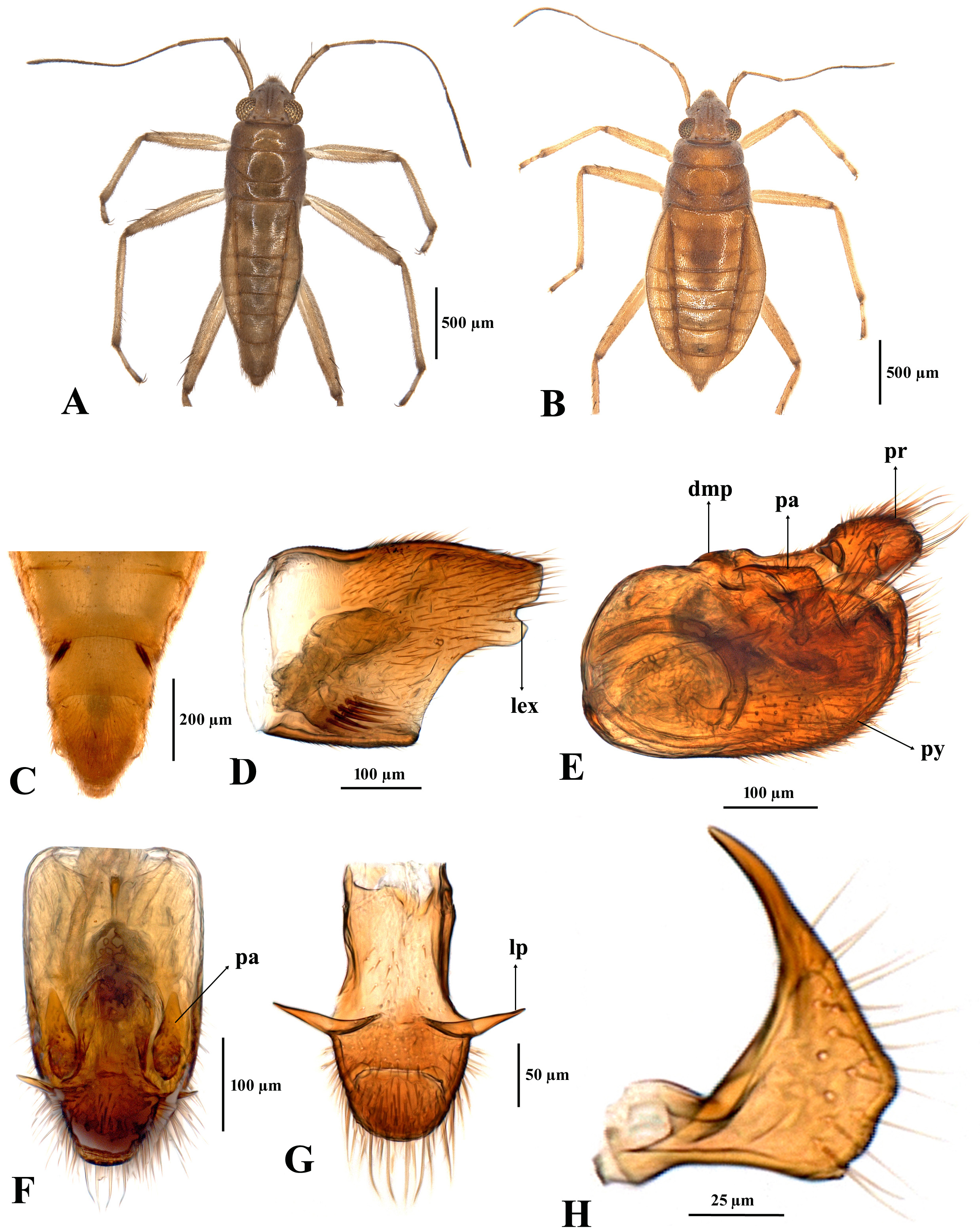
Diagnosis. Mesovelia andamana sp. nov. can be identified by the presence of mostly a single spine on the flexor region of the fore and mid femora subapically; the slightly broad male paramere with a constriction near the dorsolateral subapex ( Fig. 5G View FIGURES 5 ); and the setal tufts with 4–6 spiniform setae on male abdominal sternum VIII ( Fig. 5C View FIGURES 5 ).
Description. Apterous male ( holotype): ( Figs. 5A View FIGURES 5 , C–G). Body length 2.05 (1.94–2.05, n=2), body width at metanotum 0.56 (0.56, n=2), body width at tergum IV 0.49 (0.49–0.54, n=2).
Colour. Yellowish brown; dorsum of body covered with minute brown setae, meso and metanota and terga I–IV with indistinct brown marks; antenna dark brown; legs yellowish brown; femora of all legs subapically with brown hue; apex of tibia and first and last tarsomeres of all legs brown; claws, setiform spines on appendages and spiniform setal tufts on abdominal sternum VIII black.
Structural characters. Frontoclypeal region with 7–8 thick setae. Head length 0.22, head width across eyes 0.46; synthlipsis 0.19; eye length 0.18, eye width 0.10. Lengths of antennomeres I–IV 0.31, 0.30, 0.61, 0.59.
Pronotal length 0.16, width 0.51; mesonotal length 0.18, width 0.55; metanotal length 0.14, width 0.56. Lengths of leg segments: foreleg: femur 0.63, tibia 0.57, tarsomeres I–III 0.03, 0.06, 0.08; mid leg: femur 0.77, tibia 0.78, tarsomeres I–III 0.04, 0.15, 0.12; hind leg: femur 0.91, tibia 1.30, tarsomeres I–III 0.04, 0.21, 0.15. Widths of fore, mid, hind femora 0.11, 0.12, 0.12. Flexor region of fore and mid femora subapically with single spine; flexor region of second hind tarsomere with two widely spaced thin, short setae.
Dorsal abdominal length 1.25; intersegmental suture between abdominal terga I–III visible, terga I–VIII 0.13, 0.14, 0.11, 0.12, 0.12, 0.14, 0.20, 0.23; sterna IV–VIII 0.10, 0.10, 0.14, 0.15, 0.12. Combined length of abdominal sterna V–VII 0.40. Basal region of sternum VIII sublaterally with pair of dark brown spiniform setal tufts, each setal tuft with 4–6 spiniform setae arranged irregularly ( Fig. 5C View FIGURES 5 ); sternum VIII midlaterally at the level of posterior margin with very small tubercle (more evident in alcohol after dissection), posterolaterally excavated; inner length of spiniform setal tuft on sternum VIII 0.07, basal width 0.04, width between two tufts 0.15; without any space between posterior margin of abdominal sternum VII and anterior margin of spiniform setal tuft. Terminalia: length of pygophore 0.24, anterior part of proctiger subequal to length of bowl-shaped posterior part, clothed with long setae posteriorly, posterior part ventromedially convex, not excavated, median lateral process long with acute tip ( Fig. 5E View FIGURES 5 ); paramere slightly broad basally, medially not twisted, apical part, slender, medium sized and slightly curved ( Fig. 5G View FIGURES 5 ), paramere, when attached to pygophore, with apical part slightly curved and directed laterad in lateral view ( Fig. 5F View FIGURES 5 ) and in dorsal view tip directed anterad ( Fig. 5D View FIGURES 5 ).
Apterous female ( paratype): ( Fig. 5B View FIGURES 5 ). Colour: similar to apterous male. Body length 2.38 (2.38–2.74, n=3), width across metanotum 0.68 (0.68–0.81, n=3), width across tergum IV 0.80 (0.80–1.00, n=3), head length 0.32, head width 0.52, eye length 0.19, eye width 0.12, synthlipsis 0.25; lengths of antennomeres I–IV 0.37, 0.30, 0.61, 0.63; pronotal length 0.18, width 0.57, mesonotal length 0.20, width 0.64, metanotal length 0.13, width 0.68. Lengths of leg segments: foreleg: femur 0.62, tibia 0.55, tarsomeres I–III 0.03, 0.06, 0.10; mid leg: femur 0.80, tibia 0.74, tarsomeres I–III 0.03, 0.15, 0.13; hind leg: femur 1.07, tibia 1.43, tarsomeres I–III 0.05, 0.23, 0.16. Widths of fore, mid, hind femora 0.10, 0.11, 0.14. Flexor region of fore and mid femora subapically with single spine, rarely with one of the mid femora with two spines.
Dorsal abdominal length 1.48, lengths of abdominal terga III–VIII, 0.16, 0.17, 0.16, 0.18, 0.20, 0.17; proctiger 0.12, dorsal gonoplac length 0.07, abdominal sterna V–VII, 0.13, 0.11, 0.15, abdominal length from sternum VIII to abdominal tip 0.63; combined length of abdominal sterna V–VII 0.39; maximum connexivum width at tergum V 0.16, ventral abdominal length from sternum V to abdominal tip 1.02.
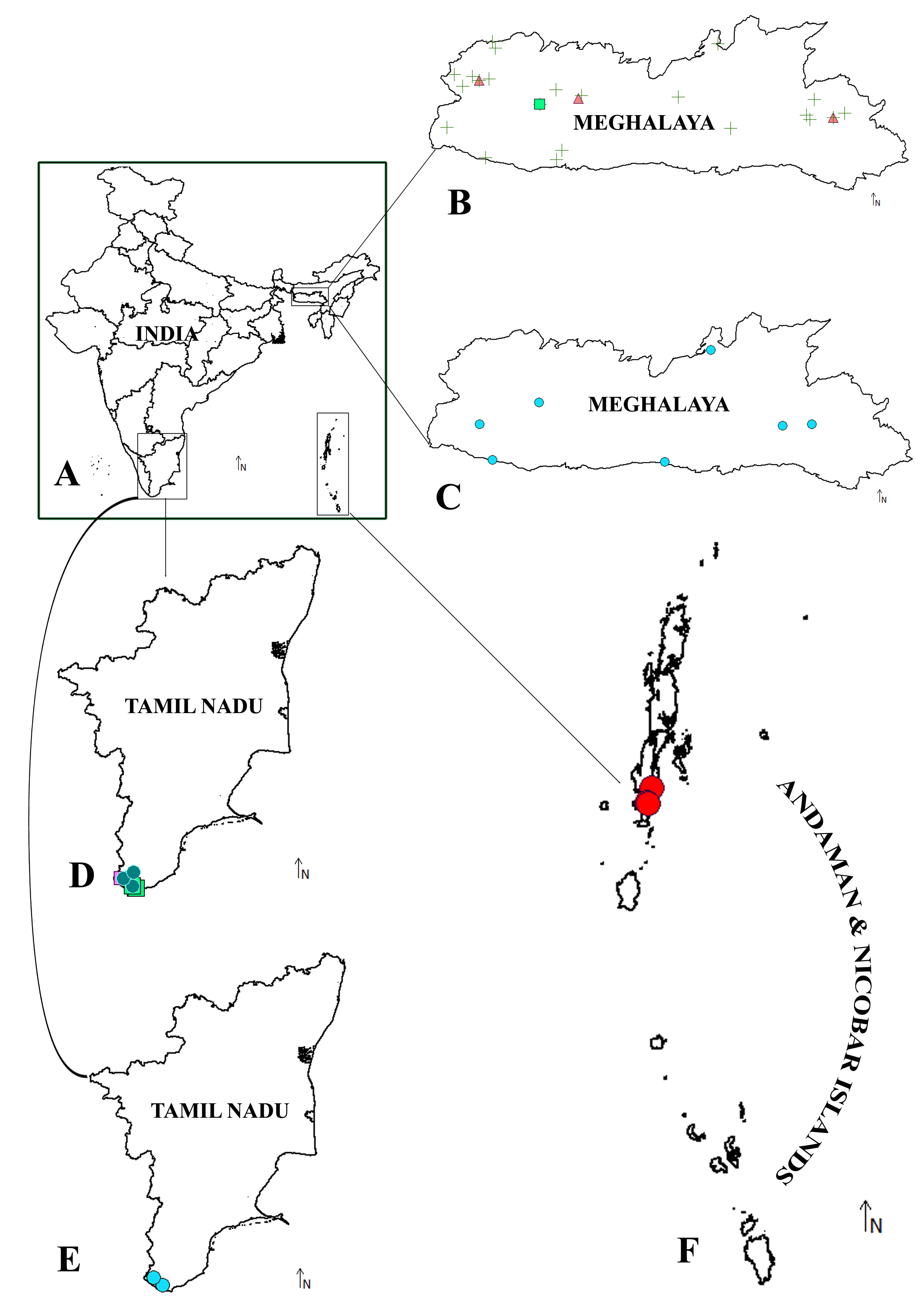
Distribution. Presently known only from South Andaman Island, Andaman and Nicobar Islands, India ( Fig. 9F View FIGURES 9 ).
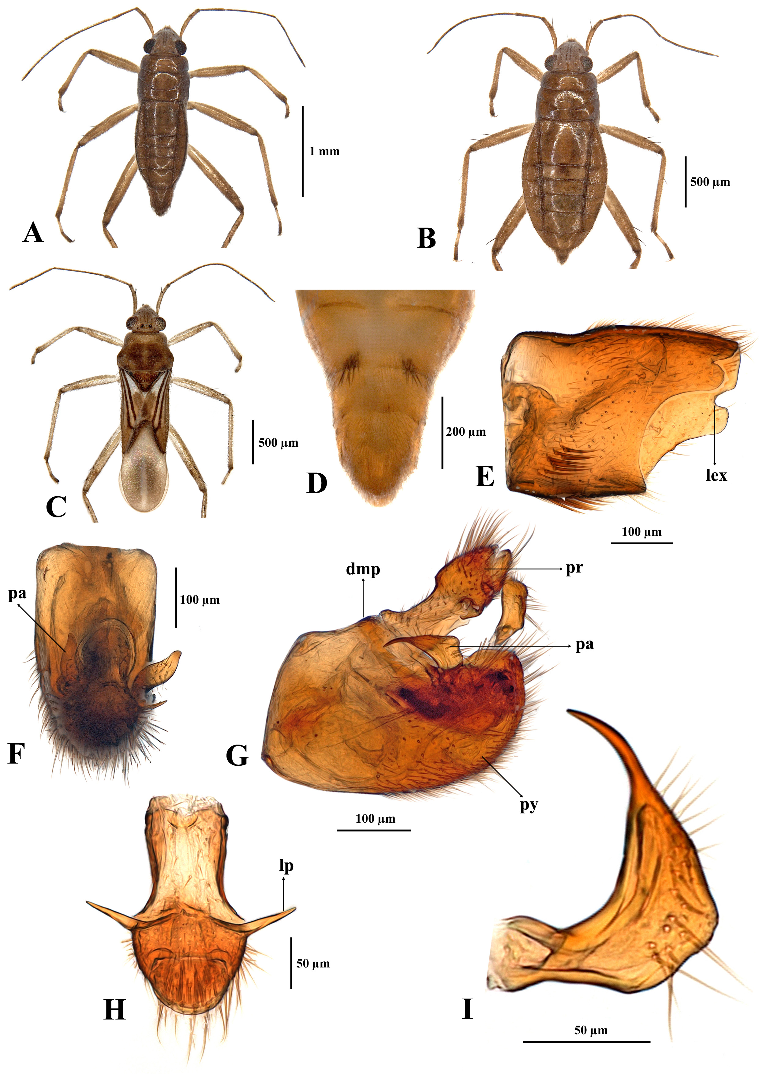
Comparative notes. The spiniform setal tufts on the eighth abdominal sternum of the male are similar in appearance in both M. tenuia ( Fig. 8C View FIGURES 8 ) and M. andamana ( Fig. 5C View FIGURES 5 ); however, M. tenuia can easily be separated from M. andamana by its slender male paramere and the presence of a single spine on the flexor regions of fore and mid femora ( Fig. 8H View FIGURES 8 ). Mesovelia melanesica and M. tenuia share a common characteristic of a slender paramere, but both species can be easily separated from each other by the number of spines on the flexor region of the fore and mid femora. Mesovelia bispinosa and M. isiasi can easily be distinguished from M. tenuia and M. andamana by the shape of the male paramere and the presence of 8–13 spiniform setae in each of the setal tufts on the eighth abdominal sternum of the male ( Fig. 6D View FIGURES 6 , Fig. 7E View FIGURES 7 ). Mesovelia isiasi can be separated from M. bispinosa by the presence of a single spine on the flexor region of the fore femur, and the mid femur usually has a single spine and rarely two spines on one side; however, the flexor region of the fore femur of M. bispinosa has one or two spines and mid femur has two spines.


No known copyright restrictions apply. See Agosti, D., Egloff, W., 2009. Taxonomic information exchange and copyright: the Plazi approach. BMC Research Notes 2009, 2:53 for further explanation.
|
Kingdom |
|
|
Phylum |
|
|
Class |
|
|
Order |
|
|
InfraOrder |
Gerromorpha |
|
Family |
|
|
Genus |