
Cycloneuroterus abei Melika & Tang
|
publication ID |
https://doi.org/10.11646/zootaxa.4088.4.1 |
|
publication LSID |
lsid:zoobank.org:pub:3A08C8E6-0516-40D8-B96F-5C6311B9B775 |
|
DOI |
https://doi.org/10.5281/zenodo.5672115 |
|
persistent identifier |
https://treatment.plazi.org/id/347B594E-FFD1-4A2C-FF43-FAF9C9CCF876 |
|
treatment provided by |
Plazi |
|
scientific name |
Cycloneuroterus abei Melika & Tang |
| status |
sp. nov. |
Cycloneuroterus abei Melika & Tang , new species
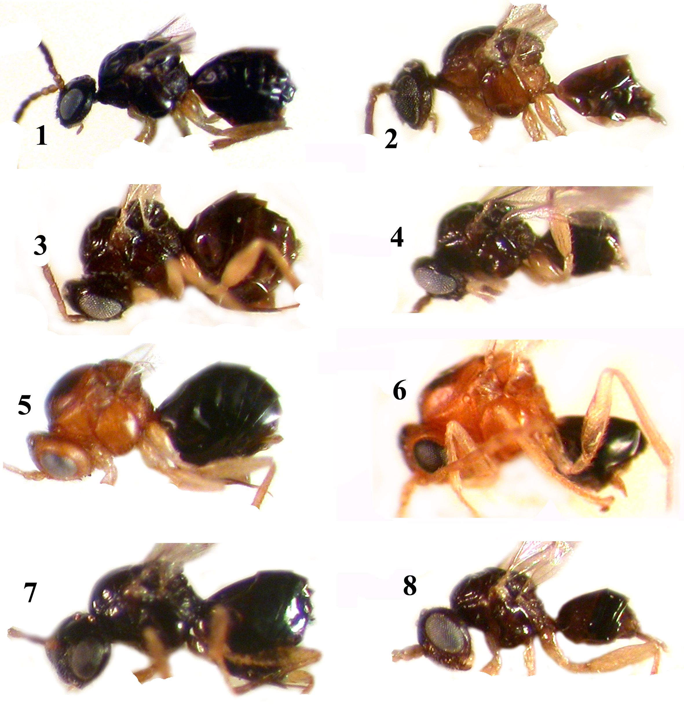
Figs 1–2 View FIGURES 1 – 8 , 25–42 View FIGURES 25 – 32 View FIGURES 33 – 37 View FIGURES 38 – 42
Type material: HOLOTYPE female: TAIWAN, Taichung City, Lineng logging road, Heping Dist., ex Quercus globosa , 24.III.2011 ( TAI 126), 24.164114ºN, 120.957922ºE, 740m, ex integrated round leaf gall (spJPl6), adult em. 30.III.2011, leg. C. T. Tang, F. Sinclair, J. Hearn, K. Lohse. Nineteen female and 12 male PARATYPES: 6 males and 3 females with the same labels as the holotype; 4 males: TAIWAN, Taichung City, Lineng logging road, Heping Dist., ex. Quercus globosa , 24.III.2011 ( TAI 126), 24.164114ºN, 120.957922ºE, 740m, ex integrated round leaf gall (spJPl6), adult em. 29.III.2011, leg. C. T. Tang, F. Sinclair, J. Hearn, K. Lohse; 2 males and 1 female: TAIWAN, Taichung City, Lineng logging road, Heping Dist., ex Quercus globosa , 24.III.2011 ( TAI 126), 24.164114ºN, 120.957922ºE, 740m, ex integrated round leaf gall (spJPl6), adult em. 1.IV.2011, leg. C. T. Tang, F. Sinclair, J. Hearn, K. Lohse; 7 females: TAIWAN: New Taipei City, Mt. Erge, Shihding Dist., ex Quercus glauca , 25.III.2011 ( TAI 118), 24.967203ºN, 121.619744ºE, 678m, ex swollen roundish leaf gall projects on both sides of leaf, adult em. 27.III.2011, leg. C. T. Tang, F. Sinclair, J. Hearn, K. Lohse; 5 females: TAIWAN: New Tiapei City, Mt. Erge, Shihding Dist., ex Quercus glauca , 25.III.2011 ( TAI 118), 24.967203ºN, 121.619744ºE, 678m, ex swollen roundish leaf gall projects on both sides of leaf, adult em. 30.III.2011, leg. C. T. Tang, F. Sinclair, J. Hearn, K. Lohse; 1 female paratype: TAIWAN: New Taipei City, Mt. Erge, Shihding Dist., ex Quercus glauca , 25.III.2011 ( TAI 118), 24.967203ºN, 121.619744ºE, 678m, ex swollen roundish leaf gall projects on both sides of leaf, adult em. 31.III.2011, leg. C. T. Tang, F. Sinclair, J. Hearn, K. Lohse; 2 females: TAIWAN: New Tiapei City, Mt. Erge, Shihding Dist., ex Quercus glauca , 25.III.2011 ( TAI 118), 24.967203ºN, 121.619744ºE, 678m, ex swollen roundish leaf gall projects on both sides of leaf, adult em. 1.IV.2011, leg. C. T. Tang, F. Sinclair, J. Hearn, K. Lohse.
The female holotype, 5 female and 3 male paratypes are deposited in NMNS, 6 female and 4 male paratypes in PHMB, 2 female and 1 male paratypes in USNM, 6 female and 4 male paratypes in NCHU.
Etymology. Named in honour of the Japanese cynipidologist, Prof. Yoshihisa Abe (Biosystematics Laboratory, Graduate School of Social and Cultural Studies, Kyushu University, Fukuoka, Japan).
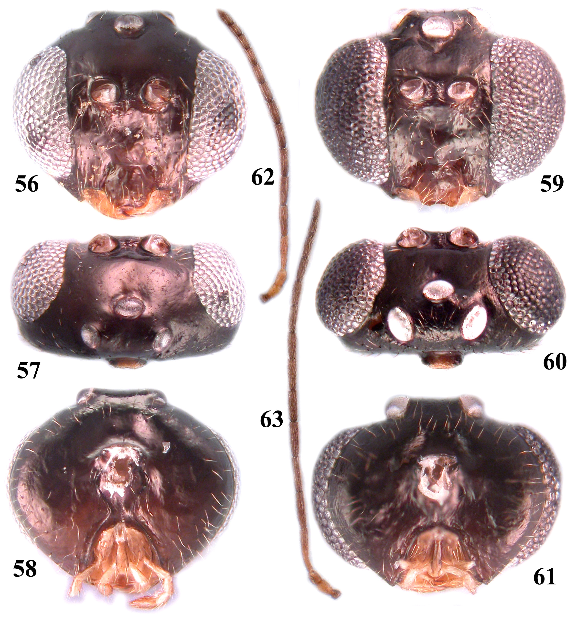
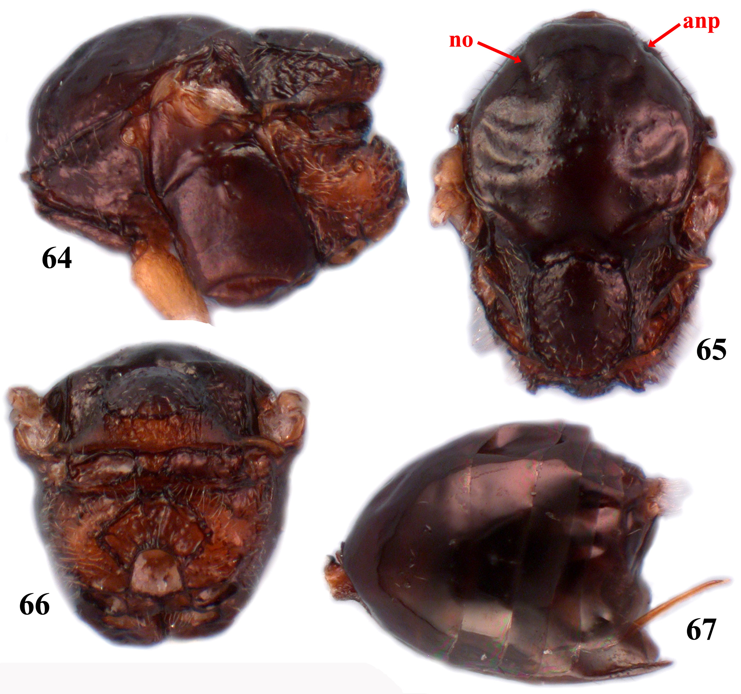
Diagnosis. Cycloneuroterus abei most closely resembles C. gilvus and C. fortuitusus in the body coloration (black to dark brown) and the body length (female usually> 2.0 mm) but can be distinguished by characters as below. In C. abei inner margins of eyes slightly converge ventrally in females ( Fig. 25 View FIGURES 25 – 32 ), parallel in males ( Fig. 28 View FIGURES 25 – 32 ); the diameter of male lateral ocellus 1.2× as long as in female ( Figs 26, 29 View FIGURES 25 – 32 ); the lower face, vertex and occiput delicately alutaceous, frons smooth and glabrous ( Figs 25–27, 28–30 View FIGURES 25 – 32 ); F1–F4 nearly equal in length ( Fig. 31 View FIGURES 25 – 32 ); the mesopleuron smooth ( Fig. 33 View FIGURES 33 – 37 ). In C. gilvus the eye of male is larger than in female ( Figs 56, 59 View FIGURES 56 – 63 ); the male eye distinctly broader than the gena in dorsal view ( Fig. 60 View FIGURES 56 – 63 ); diameter of the male lateral ocellus 1.4× as long as in the female ( Figs 57, 60 View FIGURES 56 – 63 ); the head of male and female smooth, glabrous; F 1 1.2 × as long as F2 ( Fig. 62 View FIGURES 56 – 63 ); the mesopleuron smooth ( Fig. 64 View FIGURES 64 – 67 ). In C. fortuitusus inner margins of eyes parallel in the male and female ( Fig. 16 View FIGURES 14 – 20 ); the diameter of male lateral ocellus 1.27× as long as in female; the lower face, frons, vertex and occiput are coriaceous, matt ( Fig. 16 View FIGURES 14 – 20 ); F2 slightly shorter than F1; the mesopleuron with delicate sculpture medially ( Fig. 17 View FIGURES 14 – 20 ). The postocciput and postgena of C. abei and C. gilvus are smooth, glabrous ( Figs 27 View FIGURES 25 – 32 , 58 View FIGURES 56 – 63 ), while in C. fortuitusus coriaceous and matt. Radial cell of the fore wing in C. abei and C. gilvus 4.0× as long as wide ( Figs 38 View FIGURES 38 – 42 , 68 View FIGURES 68 – 70 ), while C. fortuitusus 4.5× as long as wide.
Description. SEXUAL FEMALE. Head dark brown to black, except for light brown clypeus; mandibles, maxillary and labial palps yellowish; scape and pedicel light brown, flagellomeres progressively darker till last one; mesosoma and metasoma black to dark brown, except lighter tegula; legs yellow, base of coxae slightly darker.
Head 2.28× as broad as long in dorsal view, 1.3× as long as broad in frontal view, slightly narrower than mesosoma. Gena delicately alutaceous, not broadened behind eye, 0.5× as long as cross diameter of eye. Malar area alutaceous, without striae and malar sulcus, 0.2× as high as height of eye. Eyes converging ventrally. POL nearly 1.4× as long as OOL; OOL 1.6× as long as lateral ocellus and 1.5× as long as LOL; ocelli ovate, elongated, central one thinner. Transfacial distance 1.2× as long as height of eye; diameter of torulus 1.3× as long as distance between toruli, distance between torulus and eye 1.5× as long as diameter of torulus. Lower face alutaceous, setose; median elevated area narrow, delicately coriaceous. Clypeus elevated above lower face, quadrangular, flat, alutaceous, ventrally emarginate, with slight median ventral incision. Anterior tentorial pit small, distinct; epistomal sulcus and clypeo-pleurostomal line distinct, deep. Frons and interocellar area uniformly alutaceous, interocellar area with few white setae. Vertex and occiput alutaceous. Postocciput and postgena smooth, without setae. Posterior tentorial pit large, ovate, deep, area below not impressed. Postgenal bridge higher than height of occipital foramen, shorter than oral foramen. Antenna with 12 flagellomeres, shorter than length of body; pedicel subglobose, longer than wide, nearly equal in length to broadened part of scape; F1 nearly as long as F2–F4, 1.8× as long as pedicel; F5 to F11 progressively shorter; F 12 1.6 × as long as F11; placoid sensilla on F1–F12.
Mesosoma 1.15× as long as high in lateral view. Pronotum smooth, short dorsally, without parallel striae laterally; strongly impressed along anterior rim; propleuron alutaceous, glabrous, with few setae and smooth area centrally. Mesoscutum smooth, with few white setae, 1.2× as broad as long (largest width measured across mesoscutum on the level of the base of tegulae). Notaulus, anterior parallel line, parapsidal line and median mesoscutal line absent, rows of setae do not indicate them; parascutal carina broad. Mesoscutellum trapezoid, longer than wide, broadest in posterior 1/3, smooth, with few setae, foveolate laterally and posteriorly, slightly overhanging metanotum. Scutellar foveae absent, semilunar transverse depression present anteriorly, with smooth and glabrous bottom. Mesopleuron and speculum smooth, glabrous, without setae, impressed along acetabular carina; mesopleural triangle alutaceous, glabrous, without setae. Dorsoaxillar area reticulate, with few white setae; lateroaxillar area alutaceous, without setae. Subaxillular bar smooth, glabrous, posteriorly as high as height of metanotal trough. Postalar process with parallel delicate striae. Metapleural sulcus reaches posterior margin of mesopectus in 1/2 of its height. Metascutellum uniformly coriaceous, metanotal trough smooth, glabrous; ventral impressed area smooth, without striae, nearly 2.0× as high as height of metascutellum. Central propodeal area broad, glabrous, with few delicate, longitudinal rugae; lateral propodeal carina strong, high, strongly curved outwards in middle height; lateral propodeal area with rugae, dense setae. Nucha without irregular rugae.
Radial cell of fore wing 4.0× as long as wide. Rs+M distinct, lighter at apex, reaches basalis in lower half of its height. Areolet large, triangular, distinct. Wing margin with long cilia. Rs and R1 reach wing margin.
Metasoma shorter than length of head+mesosoma, longer than high in lateral view; 2nd metasomal tergite occupy 0.44 length of metasoma in dorsal view, with few white setae laterally; all subsequent tergites without setae, smooth, glabrous. Ventral spine of hypopygium short, prominent part 2.04× as long as wide in ventral view, with sparse white setae, extending beyond apex of spine.
Body length 2.3–2.4 mm (n=5).
MALE. Similar to female. Head and mesosoma light brown to dark brown, with mesoscutum and mesoscutellum much darker; metasoma black to dark brown; antenna light brown. Eye larger. Diameter of lateral ocellus 1.2× as long as in female. Antenna with 13 flagellomeres, nearly equal to body length; F1 curved and swollen apically, 1.1× as long as F2; F1–F12 progressively shorter; F 13 1.3 × as long as F12; placoid sensilla on all flagellomeres. Body length 1.8–2.1 mm (n=4).
Gall ( Figs 39–42 View FIGURES 38 – 42 ). The gall is an integral young leaf swelling, protruding on both sides of the leaf blade (on Q. globosa galls were found to grow on the leaf petiole; Fig. 42 View FIGURES 38 – 42 ); 2.5–6.0 mm in diameter, with a single or multiple larval chambers. Sometimes the gall growth causes distortion of leaves. Tissues outside of larval chamber are succulent during larval development.
Biology. Gall growth coincides with the point of host sprouting in mid-February. Galls develop to largest size when the host leaves wholly expanded, and remain on the host until defoliation. Wasps emerged from galls at room temperature immediately after collection (late March till mid-April). Asexual generation is unknown.
Distribution. Taiwan: Shihding District, New Taipei City; Fuhsing Township, Taoyuan County; Heping District, Taichung City.


No known copyright restrictions apply. See Agosti, D., Egloff, W., 2009. Taxonomic information exchange and copyright: the Plazi approach. BMC Research Notes 2009, 2:53 for further explanation.
|
Kingdom |
|
|
Phylum |
|
|
Class |
|
|
Order |
|
|
Family |
|
|
Genus |