
Mycale (Aegogropila) cavernosa Bergquist, 1965
|
publication ID |
https://doi.org/ 10.11646/zootaxa.4912.1.1 |
|
publication LSID |
lsid:zoobank.org:pub:9536C1CF-4AEF-47F8-959B-48CD7A5392D8 |
|
DOI |
https://doi.org/10.5281/zenodo.4450922 |
|
persistent identifier |
https://treatment.plazi.org/id/361087A7-FFDE-FFBB-55AB-FF3250A7C967 |
|
treatment provided by |
Plazi |
|
scientific name |
Mycale (Aegogropila) cavernosa Bergquist, 1965 |
| status |
|
Mycale (Aegogropila) cavernosa Bergquist, 1965 View in CoL
Figs 15 View FIGURE 15 a–h
Mycale cavernosa Bergquist, 1965: 170 View in CoL , figs 23a–c.
Mycale (Aegogropila) cavernosa View in CoL ; Hajdu et al. 1995: 7.
Material examined. USNM 23703 View Materials , small fragment of the holotype, Palau Islands , 1.75 miles NE of Ngabadangel, depth 30.6 m, coll. Coral Fish Project Expeditions, stat. 125, 24 August 1955 .
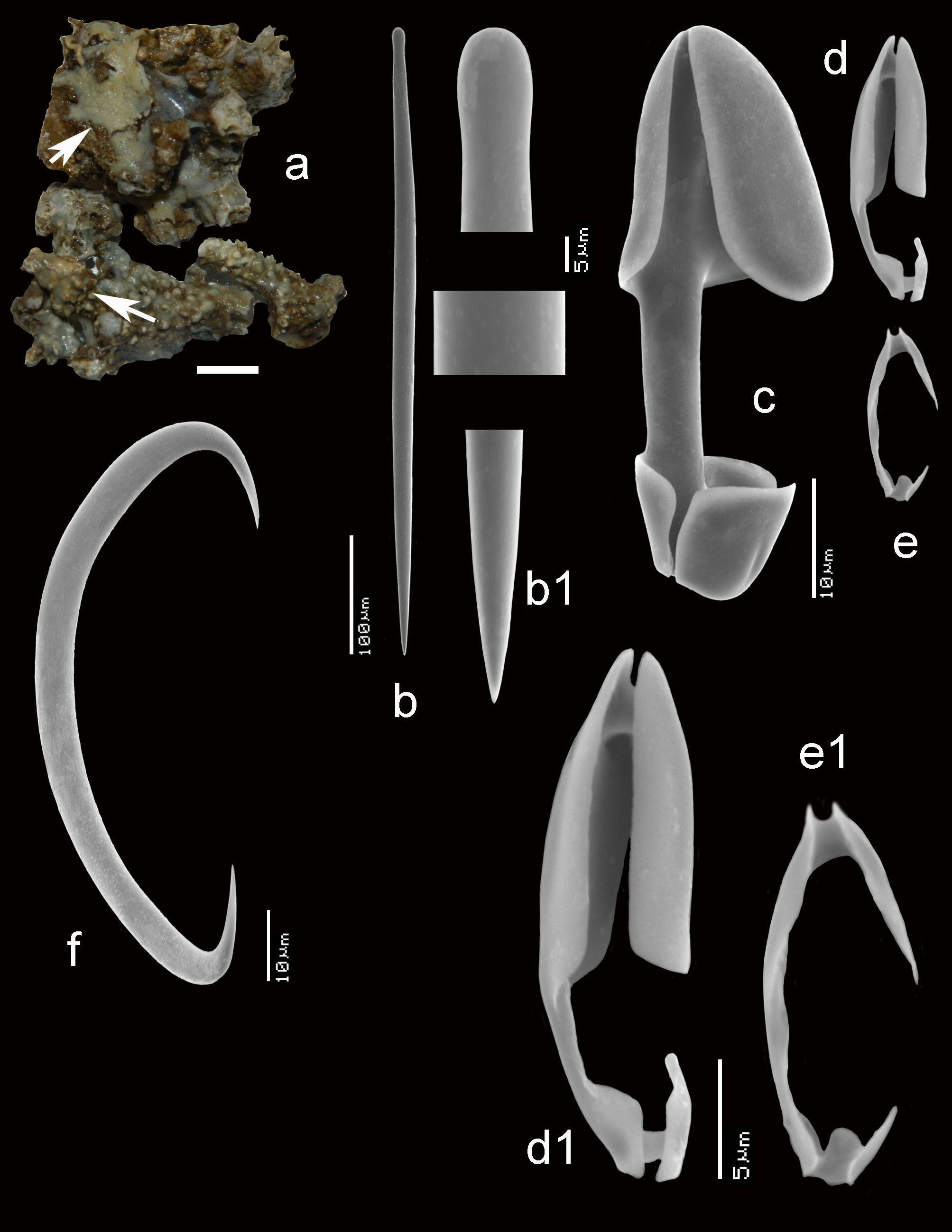
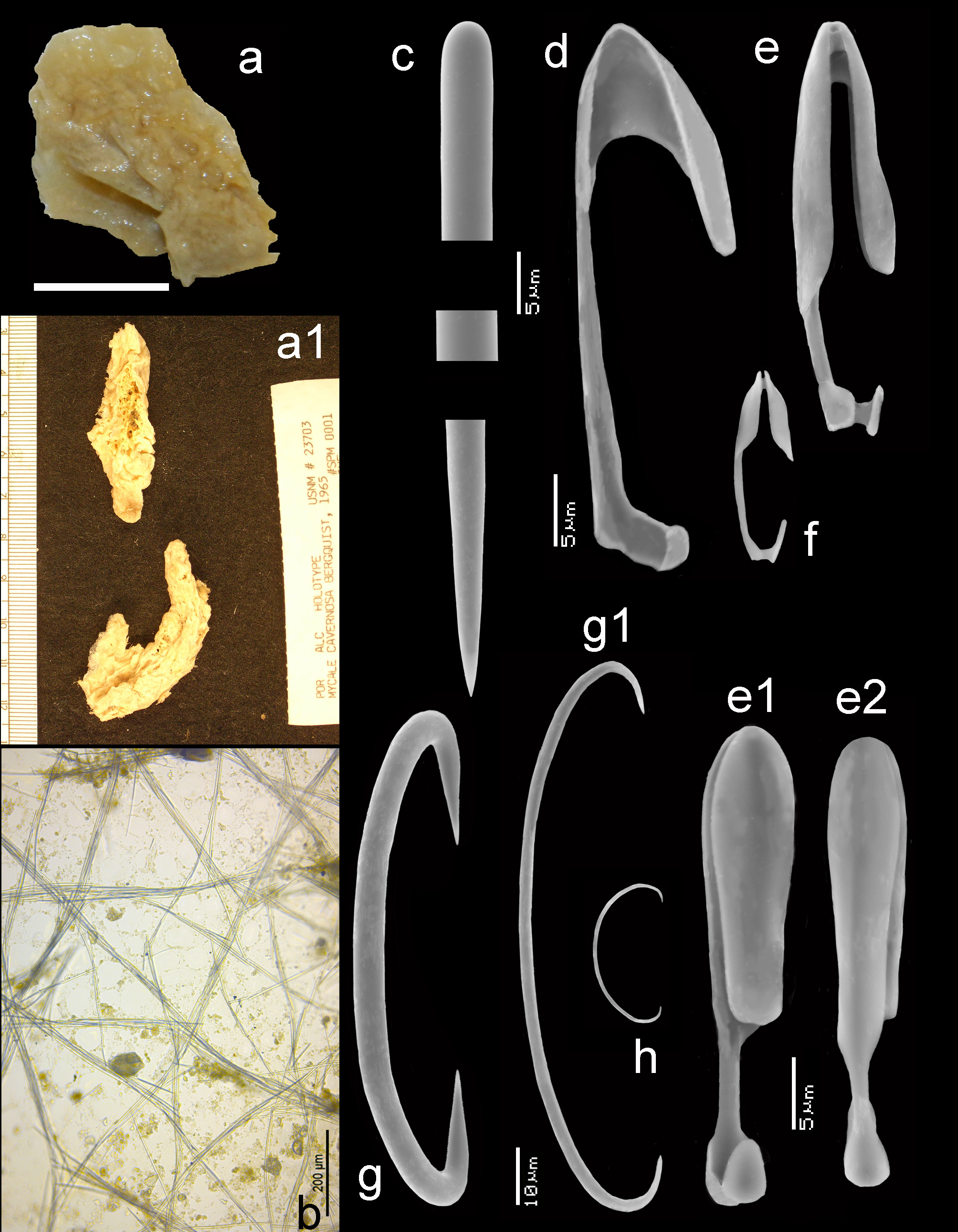
Summary description. The lobate single specimen of 20 x 5 x 1.5 cm is broken in two finger-shaped fragments ( Figs 15a,a View FIGURE 15 1 View FIGURE 1 ). The fragments are greyish white, no life color is known. The surface is characteristically folded, probably due to shrinking. Surface skeleton is of the aegogropila-type ( Fig. 15b View FIGURE 15 ), with thin tracts 4–38 µm in diameter (1–8 spicules in cross section) delimiting triangular meshes of up to 400 µm in widest dimension. The choanosomal skeleton is plumoreticulate with notable spongin cementing the tracts, with in the interior stout spicule tracts (90–185 µm in diameter), thinning out towards the surface (20–50 µm in diameter), where there are spicule brushes carrying the tangential surface skeleton. Spicules ( Figs 15 View FIGURE 15 c–h) mycalostyles ( Fig. 15c View FIGURE 15 ) (Bergquist: 262– 306 –351 x 4– 4.2 – 8 µm), remeasured 273– 330.7 –391 x 4– 5.9 – 7.5 µm, anisochelae I ( Fig. 15d View FIGURE 15 ) (if proper, not mentioned by Bergquist) 39–60 µm, anisochelae II ( Figs 13e,e View FIGURE 13 1 View FIGURE 1 ,e 2 View FIGURE 2 ) (Bergquist 29–33– 40 µm), remeasured 28– 33.4 – 36 µm, anisochelae III ( Fig. 15f View FIGURE 15 ) (Bergquist 11– 13.5 – 15 µm), remeasured 11– 13.7 – 16 µm, sigmas I ( Figs 15g,g View FIGURE 15 1 View FIGURE 1 ) (Bergquist 92– 97.5 – 105 µm), remeasured 96– 99.4 – 104 µm, sigmas II ( Fig. 13h View FIGURE 13 ) (Bergquist 19– 23 – 26.5 µm), remeasured 21– 24.2 – 26 µm. No additional microscleres, and no rosettes of anisochelae were observed.
Distribution. Known only from the Palau Islands.
Comments. We were able to make some slides and a SEM stub, and confirmed Hajdu et al. ’s (1995: 7) discovery of anisochelae I in low quantity and with reduced shape. These were not mentioned by Bergquist, so it remains somewhat uncertain whether they are proper. The anisochelae II are similar to those of M. (Ae.) sulevoidea . We consider the present species as doubtfully valid, because toxas were sometimes rare in some specimens of M. (Ae.) sulevoidea . If the anisochelae I mentioned above are proper to the sponge, there is an outside possibility that M. (Ae.) cavernosa will prove to be a junior synonym of M. (Ae.) sulevoidea , but lack of toxas precludes this conclusion.


No known copyright restrictions apply. See Agosti, D., Egloff, W., 2009. Taxonomic information exchange and copyright: the Plazi approach. BMC Research Notes 2009, 2:53 for further explanation.
|
Kingdom |
|
|
Phylum |
|
|
Class |
|
|
Order |
|
|
Family |
|
|
Genus |
Mycale (Aegogropila) cavernosa Bergquist, 1965
| Van, Rob W. M., Aryasari, Ratih & De, Nicole J. 2021 |
Mycale (Aegogropila) cavernosa
| Hajdu, E. & Zea, S. & Kielman, M. & Peixinho, S. 1995: 7 |
Mycale cavernosa
| Bergquist, P. R. 1965: 170 |