
Maraenobiotus slovenicus, Brancelj & Karanovic, 2015
|
publication ID |
https://doi.org/ 10.1080/00222933.2015.1022620 |
|
publication LSID |
lsid:zoobank.org:pub:92302CF9-21BA-4454-A3DD-337BB7152DCE |
|
DOI |
https://doi.org/10.5281/zenodo.4328310 |
|
persistent identifier |
https://treatment.plazi.org/id/09F4D0BE-3D4A-41F8-A0AF-F9DE7C47B6C3 |
|
taxon LSID |
lsid:zoobank.org:act:09F4D0BE-3D4A-41F8-A0AF-F9DE7C47B6C3 |
|
treatment provided by |
Carolina |
|
scientific name |
Maraenobiotus slovenicus |
| status |
sp. nov. |
Maraenobiotus slovenicus sp. nov.
( Figures 1–8 View Figure 1 View Figure 2 View Figure 3 View Figure 4 View Figure 5 View Figure 6 View Figure 7 View Figure 8 )
Material examined
Holotype. adult female (length 658 µm), completely dissected and mounted on a slide in glycerol and sealed with nail polish; collected on 20 January 2014 in the temporary spring Močilo near village Gornji Ig (Ljubljana, Slovenia); deposited in the Slovenian Museum of Natural History (Ljubljana), registration no. PMS 2014.1352 View Materials .
Allotype. adult male (length 551 µm), completely dissected and mounted on a slide in glycerol and sealed with nail polish; collected on same date at same location as holotype; deposited in the Slovenian Museum of Natural History (Ljubljana), registration no. PMS 2014.1353 View Materials .
Paratypes. 10 females, 3 males (stored in 70% alcohol); collected on 8 February 2014 at the same location as holotype and allotype; deposited in the Slovenian Museum of Natural History (Ljubljana), registration no. PMS 2014.1354 View Materials . Five females, 2 males (partly damaged) on a single SEM stub; deposited in the Slovenian Museum of Natural History (Ljubljana), registration no. PMS 2014.1355 View Materials .
The remaining specimens (20 females, one male) collected on 22 February 2014 at the same location as the holotype remain in authors’ collections .
Description
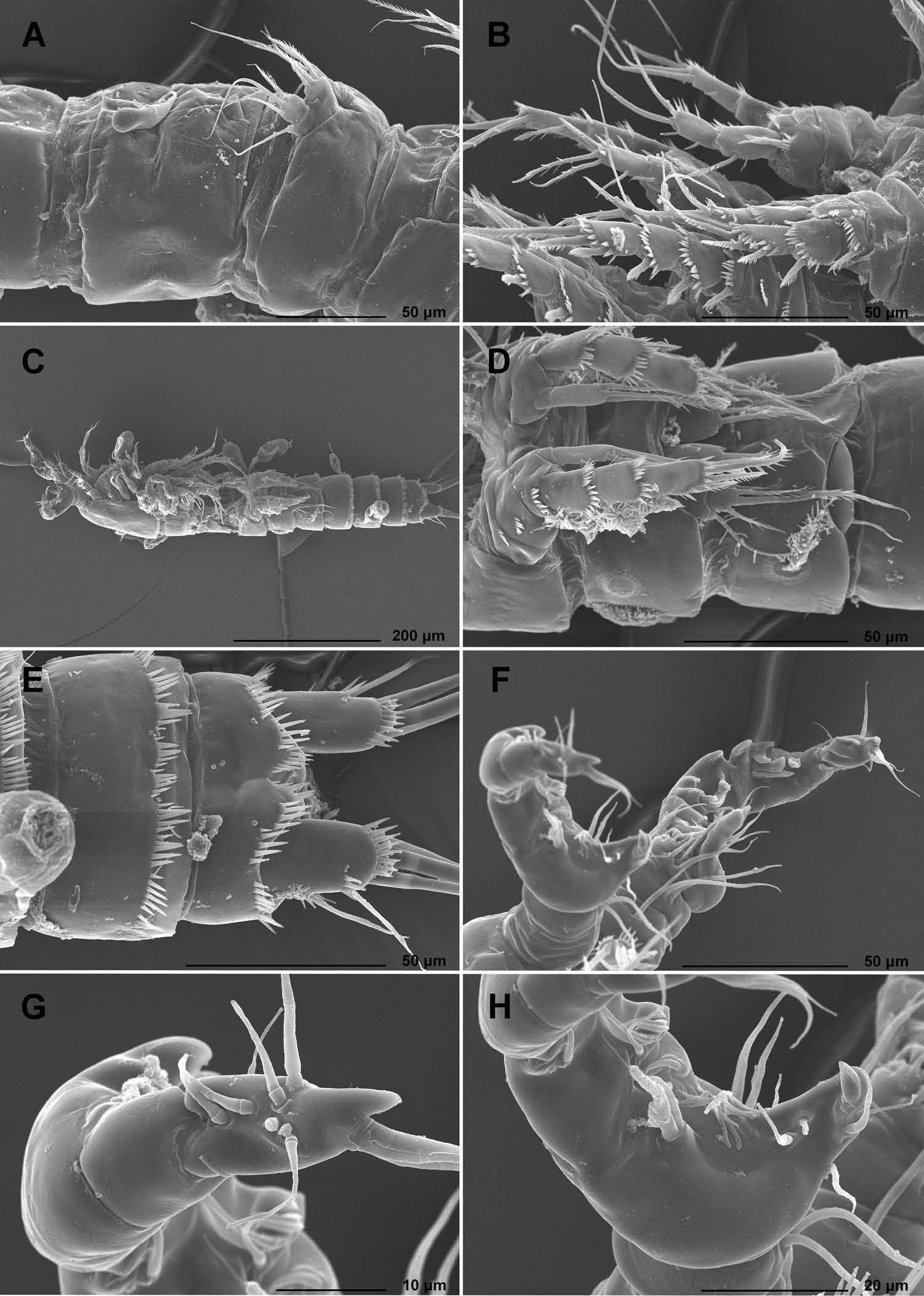
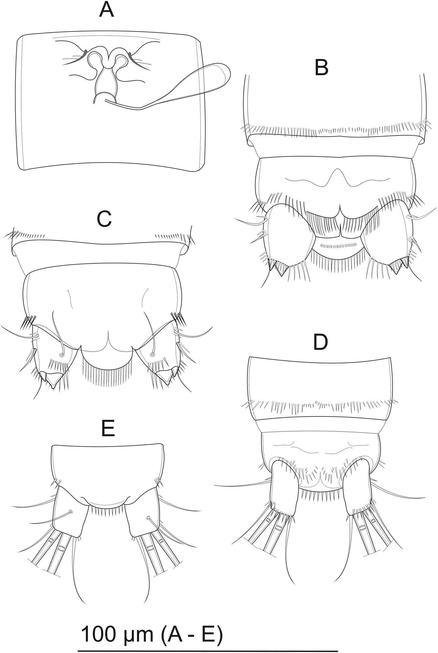
Female, body length, measured from tip of rostrum to posterior margin of caudal rami, 609–651 µm (mean: 636 µm; n = 10), elongated, width constant to end of double genital somite, last three urosomal somites slightly narrower, evenly tapering toward anal somite, colourless ( Figures 1A View Figure 1 , 2A View Figure 2 ). Body length/width ratio about 4.5; prosome/urosome ratio 1.1; length/width ratio of cephalothorax in dorsal view about 1; cephalothorax about 22% of entire body length. Naupliar eye not discernible. Rostrum small, rounded ( Figure 1B, C View Figure 1 ). Integumental windows not discernible. Posterior margins of thoracic and abdominal somites dorsally smooth ( Figure 1A, E, F, G View Figure 1 ). Genital double-somite with no spines laterally or ventrally ( Figures 1A, F View Figure 1 , 2C, D View Figure 2 , 7A View Figure 7 ). Genital complex with single large copulatory pore, sclerotized, bellshaped; seminal receptacles small, simple and heavily sclerotized. Fused plate with reduced sixth legs positioned well above seminal receptacles ( Figure 7A View Figure 7 ).
Short row of spinules laterally on fourth urosomal somite; fifth urosomal somite dorsolaterally and ventrally with continuous row of spinules, smallest in ventromedial part ( Figures 1A, G View Figure 1 , 2D View Figure 2 , 7B, C View Figure 7 ). Anal somite laterally with short row of strong spinules; ventrally four groups of long and slightly curved spinules ( Figures 1A, G View Figure 1 , 7B, C View Figure 7 ).
Anal operculum large, rounded, overreaching distal end of anal somite, with some 20 well developed spinules along distal margin ( Figures 1A, G View Figure 1 , 7B, C View Figure 7 ). Ventral side with additional row of minute spinules. Anal operculum about 58% of total anal somite width.
Caudal rami conical, slightly diverging, with inner and outer margins slightly convex; ramus as long as wide, with no dorsal keel ( Figures 1A, G, H View Figure 1 , 2E View Figure 2 , 7B, C View Figure 7 ). Inner distal corner dorsally with 6–10 strong spines, ventrally inner corner with row of long and slightly curved spinules followed by smaller spinules toward outer corner. Anterolateral external accessory seta (I) slightly shorter than furcal ramus, inserted close to base; anterolateral external accessory seta (II) shorter than seta I and thin. Posterolateral seta (III) positioned below insertion of outer terminal seta (IV), shorter than seta II, very thin. Outer terminal seta (IV) reduced to minute knob, heavily chitinized. Inner terminal seta (V) reduced to minute knob, bigger then seta IV, heavily chitinized. Inner accessory seta (VI) reduced, not discernible from nearby spines. Dorsal seta (VII) slightly longer than caudal ramus, inserted on internal side, at about mid length.
Antennule ( Figures 1B, C, D View Figure 1 , 2F View Figure 2 , 5A View Figure 5 ) relatively short, eight-segmented. Aesthetasc on fourth segment cylindrical, with rounded tip, reaching base of 8th segment. Second aesthetasc on terminal segment as long as first, slim. Both aesthetascs combined as acrotheck (common base of spine and aesthetasc). Setal formula: 1.7.5.2.1.2.2.8. Length ratio of antennular segments from proximal to distal end and along caudal margin 1:2:1.4:1:1:1.4:1:2.2.
Antenna ( Figures 2B View Figure 2 , 5B View Figure 5 ) with allobasis, and robust, one-segmented Exp and Endp. Three strong spines on outer margin of Endp, increasing in length distally; terminal armature consisting of one short spine, one twice as long as short spine, and four geniculate setae. Exp with four spiniform setae, one ornamented with spinules on one margin.
Mandible ( Figure 5C View Figure 5 ) short and robust, with two strongly chitinized teeth on gnathobase. Dorsal seta near gnathobase. Mandibular palp very short, fused with coxa, with five setae, one of them twice as long as the other.
Maxillule ( Figure 5D View Figure 5 ) with strong and robust spines on praecoxal arthrite. Coxa with strong, chitinized spine and slim seta. Basis with strong, beak-like outgrowth, with long seta and long setules unilaterally. Exp and Endp reduced, fused with basis; each represented by two weak setae.
Maxilla ( Figure 5E View Figure 5 ) two-segmented; syncoxa with 2 endites with 3 and 2 elements. Basis with strong spine and long seta. Endp reduced to three setae.
Maxilliped ( Figure 5F View Figure 5 ) comprising syncoxa, basis, and one-segmented Endp. Syncoxa with seta distally. Basis three times as long as wide, with 8–10 spinules positioned near palmar margin dorsally, equal in length. Endp drawn out into strong, acutely curved claw; as long as basis and armed with several spinules in distal half; additional armature represented by a short seta.
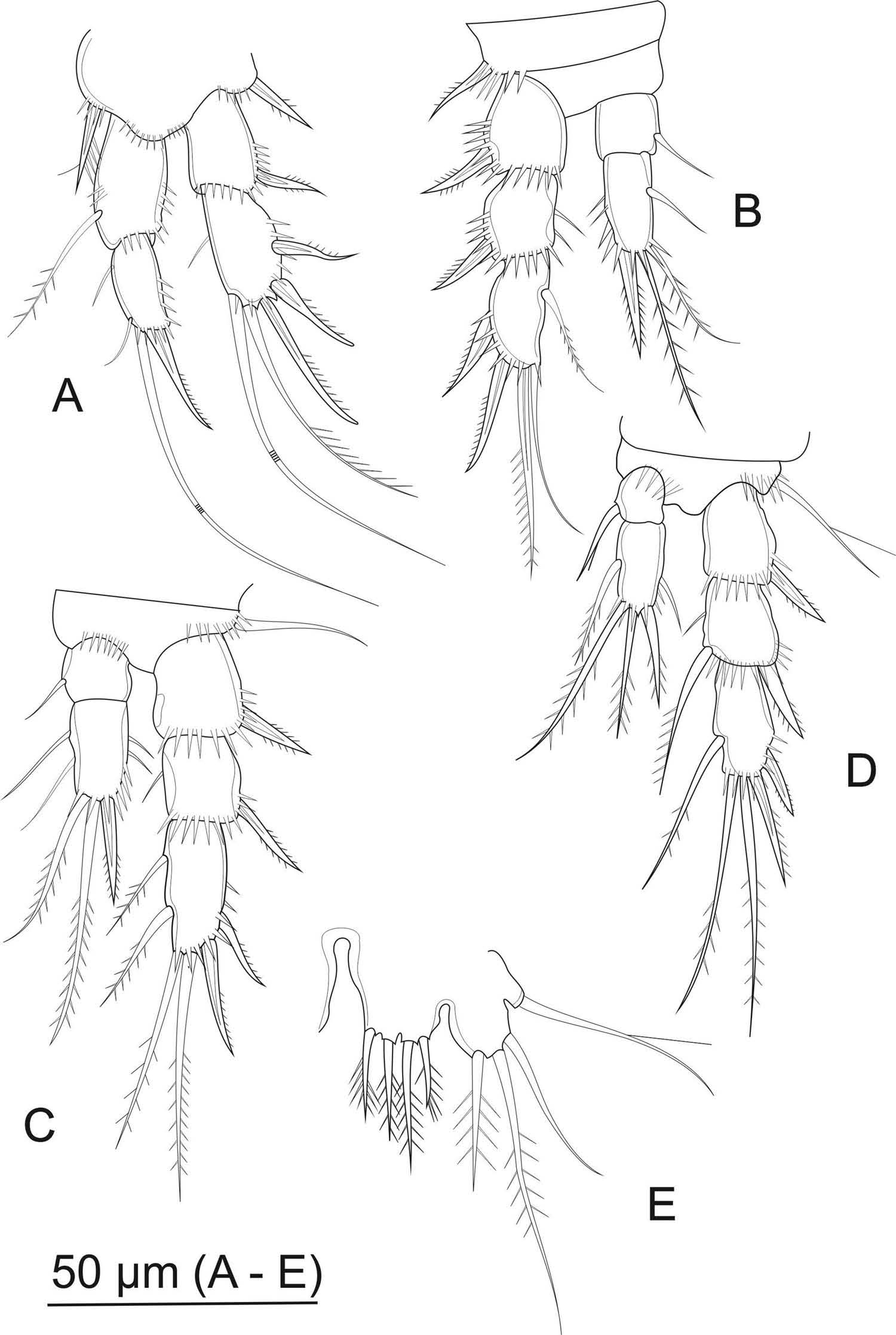
P1 ( Figures 3B View Figure 3 , 6A View Figure 6 ) with two-segmented Exp and Endp, equal in length. Basis with strong and robust inner spiniform seta and stout outer basal spine. Exp–1 with strong outer spine; Exp–2 with two strong spines laterally, spine and two long geniculate setae terminally; inner terminal seta longer than Exp. Endp–1 with long seta on inner margin at two thirds length. Endp–2 terminally with three setae; innermost short and soft, terminal seta long and geniculate, outer one spiniform, with spinules on outer margin.
P2 ( Figures 3B View Figure 3 , 6B View Figure 6 ) on basis with spiniform outer seta. Three-segmented Exp and two-segmented Endp. Endp as long as Exp–1 and Exp–2 combined. Exp–1 1.3 times as long as wide, with strong spine at distal corner. Exp–2 with strong outer spine. Exp–3 twice as long as wide, with two outer spines, terminal spiniform seta with strong spinules unilaterally and slim and bare seta, as long as spiniform seta. Endp–1 as long as wide, with seta on inner margin, at about two thirds of segment length. Endp–2 twice as long as wide, with seta along inner margin at half length of segment. Terminally two long soft setae, sub-equal in length; robust spiniform outer terminal seta, as long as Endp–2.
P3 ( Figures 2G View Figure 2 , 3B View Figure 3 , 6C View Figure 6 ) with long and thin outer basal seta. Three-segmented Exp and two-segmented Endp. Endp slightly shorter as Exp–1 and Exp–2 combined. Exp–1 and Exp–2 similar to that of P2. Exp–3 with relatively weak spine on 2/3 of outer margin, robust spine subterminally and two robust setae terminally. Additional two robust setae on inner margin at 1/3 and 2/3 length of segment, respectively. Endp two-segmented; Endp–1 as in P2. Endp–2 with a row of strong spines on outer margin. Terminally two long soft setae, sub-equal in length; robust spiniform outer terminal seta, as long as Endp–2; two long and weak setae on inner margin at 1/3 and 2/3 length of segment, respectively.
P4 ( Figures 2G View Figure 2 , 6D View Figure 6 ) with outer basal seta long and thin, with additional very long and thin spinules. Three-segmented Exp and two-segmented Endp. Endp reaching about to middle of Exp–2. Exp–1 and Exp–2 similar to those of Pa and P3 but with additional robust seta on inner distal corner of Exp–2. Exp–3 twice as long as wide, with relatively weak spine located subterminally on outer margin. Terminally two long soft setae, sub-equal in length; robust spiniform outer terminal seta, shorter than Exp–3; long seta on inner margin at half length of segment, respectively. Endp–1 as wide as long, smaller as in P2 or P3 respectively. Spine on inner margin with distinct long spinules. Endp–2 with short spine on outer margin, at 2/3 of segment length. Terminally robust spiniform seta on outer position and two setae unequal in length. Seta on inner margin at mid length of segment.
P5 ( Figures 2D, H View Figure 2 , 3A View Figure 3 , 6E View Figure 6 ) Exp and baseoendopod not distinctly separated; baseoendopodal lobe well developed, with four strong pinnate spines of unequal length; innermost three increasing in length toward outside; outermost shortest, Exp lobe longer than baseoendopodal, as long as wide, with three apical setae of unequal length; innermost shortest, with strong spinules; medial longest, with strong spinules; outer one bare. Outer lateral seta on baseoendopodal lobe long and bare, but one very long setula.
P6 ( Figure 7A View Figure 7 ) fused, small, forming simple plate; each with thin seta with several long setulae.
Male, body length, measured from tip of rostrum to posterior margin of caudal rami, 524–554 µm; average: 543 µm (n = 4), elongated, widest at distal part of cephalosome, rest of body slightly narrower, evenly tapering toward anal somite, colourless ( Figure 3C, D, E View Figure 3 ). Naupliar eye not discernible. Rostrum small. Integumental window not discernible. Posterior margins of thoracic and abdominal somites dorsally and ventrally smooth ( Figures 3D, E View Figure 3 ). Short row of small spinules laterally on first urosomal somite; continuous row of small spinules dorsally on second urosomal somite ( Figure 4F View Figure 4 ); third urosomal somite dorsally with three groups of small spinules; fourth and fifth urosomal somite dorsally with four groups of small spinules, laterally with longer spinules; fourth urosomal somite ventrally with continuous row of spines equal in length; fifth urosomal somite ventrally with continuous row of spinules unequal in length ( Figures 3C, E View Figure 3 , 7D View Figure 7 ). Anal somite laterally with short row of strong spinules; ventrally row of spines between bases of furcal rami ( Figures 3E View Figure 3 , 4G View Figure 4 , 7D View Figure 7 ). First and second urosomite with cuticular window each; large pore next to cuticular window on first urosomite ( Figures 3D View Figure 3 , 4F, H View Figure 4 ).
Anal operculum well developed, rounded, overreaching distal end of anal somite, with about 10 well-developed spinules along distal margin ( Figure 7D, E View Figure 7 ).
Caudal rami almost rectangular, slightly longer than wide, diverging; outer margin slightly convex; with no dorsal keel ( Figures 3C, E View Figure 3 , 7D, E View Figure 7 ). Ventrally row of spinules at distal part of ramus ( Figures 3E View Figure 3 , 7D View Figure 7 ). Anterolateral external accessory seta (I) thin, slightly longer than ramus, inserted at 1/3 of ramus length ( Figures 4G View Figure 4 , 7D, E View Figure 7 ); anterolateral external accessory seta (II) as long as seta I, thin; positioned at distal corner of ramus. Posterolateral seta (III) absent. Outer terminal seta (IV) about 4 times as long as seta II, with fracture plane. Inner terminal seta (V) about 2.5 times as long as seta III, straight, robust, with fracture plane. Inner accessory (VI) slightly longer than seta I. Dorsal seta (VII) slightly longer than caudal ramus, inserted on internal side, at about mid length.
Antennule ( Figures 3C, F, G, H View Figure 3 , 4C, E View Figure 4 , 8A View Figure 8 ) relatively short, robust, ninesegmented, strongly chitinized. Geniculated articulation between sixth and seventh segments. Aesthetasc on fifth segment cylindrical, short, does not extend over fifth segment, aesthetasc on ninth segment spine-like. Both aesthetascs combined as acrotheck (common base of spine and aesthetasc). Proximal part of fifth segment extended medially, with strong chitinous tooth at inner corner ( Figures 3F, H View Figure 3 , 8A View Figure 8 ). Setal formula: 0.1.1.2.9.3.0.0.9.
Antenna, mandible, maxillule, maxilla, maxilliped, P1 similar to those in female.
P2 ( Figure 8B View Figure 8 ): Exp similar to female. Endp two segmented. Endp as long as Exp–1 and Exp–2 combined. Endp–1 0.75 times as long as wide, with seta on inner distal corner. Endp–2 three times as long as wide, with seta along inner margin at 1/3 length of segment. Terminally two soft setae, sub-equal in length, slightly shorter than segment; well-formed incision present on inner margin subterminally; at half length of segment on inner margin short spine, and at 1/4 two long spinules.
P3 ( Figure 8C View Figure 8 ) with long and thin outer basal seta. Three-segmented Exp and two-segmented Endp. Endp reaching middle point of Exp–2. Exp–1 and Exp–2 similar to that of female. Exp–3 with relatively weak spine on 2/3 of outer margin, robust spine subterminally, long robust outer seta and short inner spiniform seta terminally. Additional two robust bare spiniform setae on inner margin at 1/3 and 2/3 length of segment, respectively. Endp two-segmented; Endp–1 with long spear-like apophysis, reaching tip of Exp–3. Endp–2 with relatively short blunt spine curving outward and spear-shaped seta as long as spear-like apophysis of Endp–1.
P4 ( Figures 3D View Figure 3 , 8D View Figure 8 ) with outer basal seta long and thin, with additional two very long and thin spinules. Three-segmented Exp and two-segmented Endp. Endp extending just beyond Exp–1. Exp armature similar to female but less robust. Endp–1 half as wide as long, with no spine/seta on inner margin. Endp–2 with short spine on outer margin, at 2/3 of segment length. Terminally spiniform seta on outer position and two setae unequal in length. No seta on inner margin.
P5 ( Figures 3D View Figure 3 , 4F View Figure 4 , 8E View Figure 8 ) Exp and baseoendopod well separated; baseoendopod well developed, with two strong pinnate spines of unequal length; inner longer. Exp longer than baseoendopod, as long as wide, with four elements; inner three setae increasing in size toward outside; innermost seta bare; medial with unilateral spinules, outer with strong spinules bilaterally. Outermost seta as long as second innermost seta, with unilateral spinules. Outer lateral seta long with few long setules unilaterally.
P6 ( Figures 3D View Figure 3 , 4F View Figure 4 , 8E View Figure 8 ) fused, small, forming simple plate; each with thin bare seta.
Variability
No variability was observed other than minor variation in the number of spinules on the abdominal somites.
Etymology
The new species is named after Slovenia, where the specimens were collected for the first time.
Remarks
This Slovenian new species undoubtedly belongs to the Maraenobiotus vejdovskyi complex, which is mostly defined by the armature formula of its appendages ( Lang 1948). Its truncated female principal caudal setae would put it close to Maraenobiotus veydovskyi truncatus Gurney, 1932 , but a closer examination of this subspecies (which is elevated to the full specific rank below) and detailed comparison with our population reveals a number of significant differences. For example, M. vejdovskyi truncatus has a much shorter anal operculum than M. slovenicus sp. nov., which is ornamented with minute spinules (versus long spinules in M. slovenicus ). Also, M. vejdovskyi truncatus has no distal lateral caudal setae in female (versus well-developed setae in M. slovenicus ), and the transformed Endp of the male third leg has but a single apical element (versus two apical elements in M. slovenicus ). Unfortunately, many other characters could not be compared because of a limited set of illustrations provided by Gurney (1932) for M. vejdovskyi truncatus . There are three other reported and illustrated populations from this complex with truncated female principal caudal setae, one from Japan ( Ishida 1987) and two from Italy ( Pesce et al. 1994), which we describe below as new species. However, both M. ishidai sp. nov. from Japan and M. galassiae sp. nov. from Italy (see below) have much longer and cylindrical caudal rami. Maraenobiotus pescei sp. nov. from Italy (see below), however, has caudal rami very similar in shape to those of M. slovenicus , but the two differ in the lateral armature of the caudal rami (lateral setae very reduced or absent in M. pescei versus well developed in M. slovenicus ), number and size of spinules on the anal operculum (longer and fewer in M. slovenicus ), segmentation of the antennal Exp (two-segmented in M. pescei versus one-segmented in M. slovenicus ), number of apical elements on the second Endp segment of the male P3 (one in M. pescei versus two in M. slovenicus ), and relative length of Endp spine on the male P5 (the inner one almost twice as long as the outer one in M. pescei versus nearly equal in M. slovenicus ). Unfortunately, illustrations and descriptions of other four species are not as complete as those of M. slovenicus , so many morphological characters could not be compared.


No known copyright restrictions apply. See Agosti, D., Egloff, W., 2009. Taxonomic information exchange and copyright: the Plazi approach. BMC Research Notes 2009, 2:53 for further explanation.
|
Kingdom |
|
|
Phylum |
|
|
Class |
|
|
Order |
|
|
Family |
|
|
Genus |