
Neusticurus Duméril and Bibron, 1839
|
publication ID |
https://doi.org/10.1080/00222933.2018.1439541 |
|
publication LSID |
lsid:zoobank.org:pub:33DCF862-11CF-4FD0-B4D6-706E2C6A339E |
|
persistent identifier |
https://treatment.plazi.org/id/37447D69-FF9C-825E-FE4E-11511418FB10 |
|
treatment provided by |
Felipe |
|
scientific name |
Neusticurus Duméril and Bibron, 1839 |
| status |
|
Genus Neusticurus Duméril and Bibron, 1839 Neusticurus rudis Boulenger, 1900
( Figures 4 – 6 View Figure 4 View Figure 5 View Figure 6 ; Table 2)
Material examined
VENEZUELA. Bolívar state, Mount Roraima [± 1100 m elevation]: 1 adult ♂ ( BM 1946.8.31.64, holotype; high resolution digital photographs) . GUYANA. Cuyuni- Mazaruni district, Wayalayeng [ 678 m elevation]: 1 adult ♂ ( IRSNB18432 View Materials ) and 1 juvenile ( IRSNB18431 View Materials ); slopes of Maringma-tepui [ 1060 – 1376 m elevation]: 4 adult ♂♂ ( IRSNB18146 View Materials , IRSNB18433 View Materials , IRSNB18435 View Materials , IRSNB18437 View Materials ) and 4 adult ♀♀ ( IRSNB18147 View Materials , IRSNB18434 View Materials , IRSNB18436 View Materials , IRSNB18438 View Materials ) .
Referred material examined is listed in Table 2.
Note on the type locality of N. rudis
We follow the argument developed by Kok et al. (2018a) about specimens collected by F. V. McConnell and J .J. Quelch in 1898 at the foot of Mount Roraima , and consider the type locality of N . rudis to be on the southern slope of Mount Roraima in Bolívar state, Venezuela .
Diagnosis
Neusticurus rudis is characterised by the following combination of characters: (1) size moderate (maximum known SVL 80.5 mm); (2) tail 1.7 – 2.1 times SVL; (3) tympanum moderately recessed, auditory meatus short; (4) lower eyelid with semi-transparent disc of 5 – 8 palpebrals; (5) frontonasal single or paired; (6) occipital scales in contact with posterior edge of parietals and interparietal 7 – 11; (7) enlarged dorsal tubercles usually arranged in poorly defined, discontinuous longitudinal rows containing 41 – 45 scales; (8) flanks with enlarged trihedral scales, not forming distinct vertical rows, surrounded by smaller scales usually heterogeneous in size; (9) ventral scales in 25 – 30 transverse rows; (10) subdigital lamellae under 4th finger 16 – 22; (11) subdigital lamellae under 4th toe 27 – 35; (12) total number of femoral pores in males 40 – 55, 24 – 49 in females; (13) tail compressed with 4 – 5 transverse rows of lateral scales corresponding to two subcaudal scales and 2 – 3 dorsal tubercles (= verticils), the last dorsal tubercle of each verticil poorly overlapping the next verticil; (14) hemipenis non-capitate, lacking capitular groove on sides; (15) hemipenial lobes elongate with pointed tips, lacking lobular knobs.
Comparison with congeneric species
Neusticurus rudis is immediately distinguished from N. bicarinatus , N. medemi and N. racenisi in having the tympanum moderately recessed (deeply recessed in N. bicarinatus , N. medemi and N. racenisi ), and by a larger number of femoral pores in females ( 24 – 49 in N. rudis vs 4 – 15 in N. bicarinatus , 9 – 10 in N. medemi , and 10 – 15 in N. racenisi ). Neusticurus rudis is further distinguished from N. medemi and N. racenisi , and also from N. tatei , in having enlarged tubercles on the dorsum (absent in N. tatei , N. medemi and N. racenisi ), and from N. tatei in having fewer femoral pores in males ( 40 – 55 in N. rudis vs 60 – 61 in N. tatei ) and a larger number of femoral pores in females ( 24 – 49 in N. rudis vs eight in N. tatei ). Neusticurus rudis is principally distinguished from N. surinamensis in having enlarged dorsal tubercles usually arranged in poorly defined, discontinuous longitudinal rows containing 41 – 45 scales (dorsal tubercles arranged in well-defined, straight and continuous longitudinal rows containing 30 – 41 scales in N. surinamensis ), and by a larger number of femoral pores in females ( 24 – 49 in N. rudis vs 6 – 10 in N. surinamensis ). Neusticurus rudis is further distinguished from N. bicarinatus and N. surinamensis by non-capitate and nonenlarged lobes on the hemipenis (hemipenis with lobes distinctly enlarged, capitate and detached from the hemipenial body in N. bicarinatus and N. surinamensis ). Neusticurus rudis can be further distinguished from N. medemi , N. racenisi and N. tatei by having flounces of the hemipenial body divided by a nude area in the centre of the asulcate face (not interrupted in N. medemi , N. racenisi and N. tatei ).
Redescription
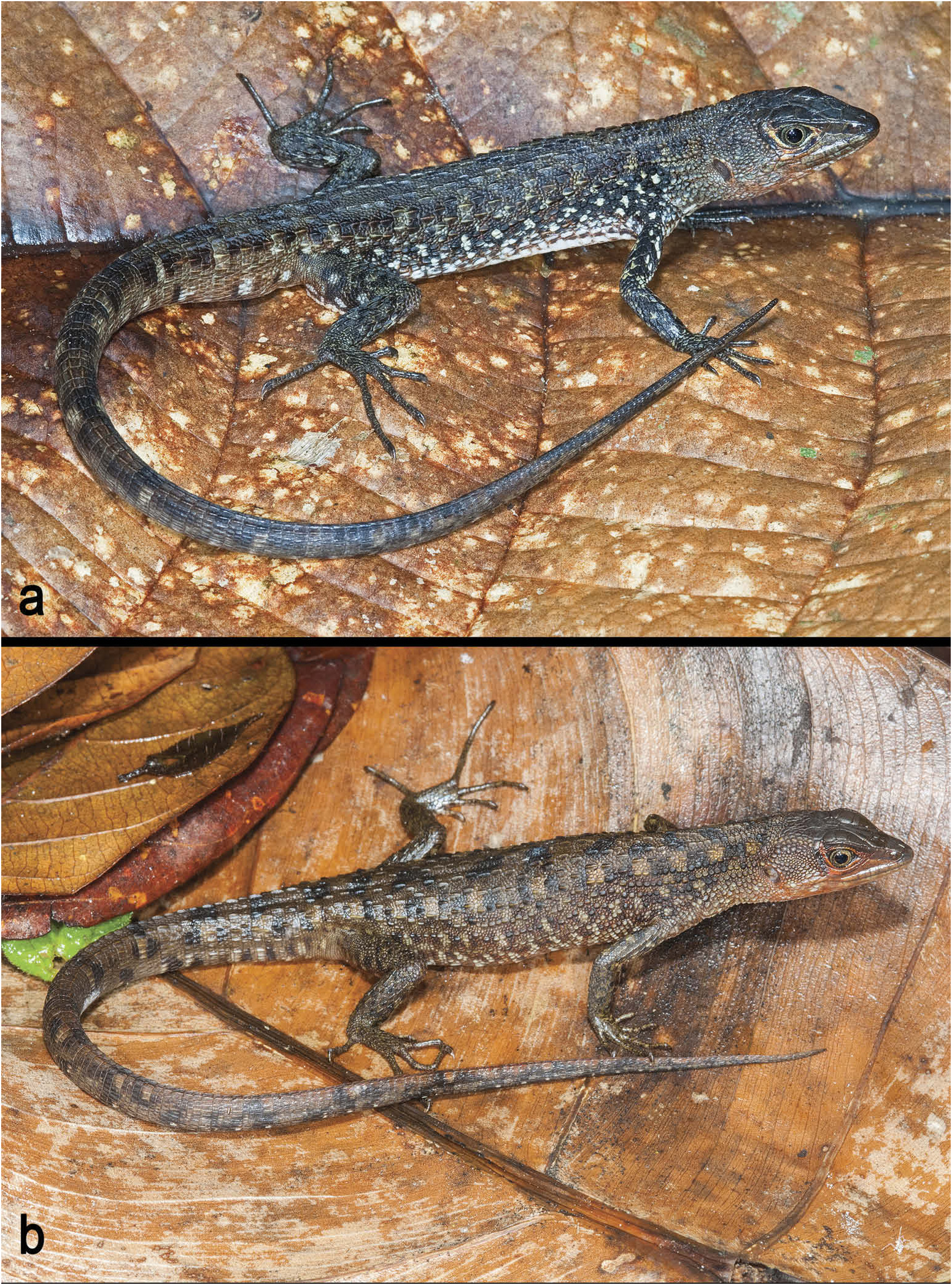
A Neusticurus of moderate size ( Figure 4 View Figure 4 ), maximum SVL 80.5 mm in males, 79.8 mm in females. Tympanum moderately recessed, external auditory meatus short. Rostral well visible from above, wider than long, rounded anteriorly, in contact with first supralabials, nasal and frontonasal. Frontonasal pentagonal, single or paired, laterally in contact with nasal and loreal, posteriorly in contact with prefrontals; if single, frontonasal may be partly divided anteriorly; when paired, the two frontonasals are similar in shape. An azygous scale is found between the frontonasal(s) and the prefrontals in six of the 11 specimens examined (55%). Prefrontals irregularly pentagonal or hexagonal, in broad contact medially, anteriorly in contact with frontonasal(s), laterally in contact with loreal, first supraciliary and first supraocular, posteriorly in contact with frontal; prefrontals with an azygous scale between them in three of the 11 specimens examined and two azygous scales between them in one specimen. Frontal longer than wide, bell-shaped, irregularly hexagonal, wider anteriorly, partially divided in three of the 11 specimens examined, anteriorly divided into several smaller scales in one specimen, in contact with prefrontals, frontoparietals and three anteriormost supraoculars. A pair of irregularly pentagonal, rarely rhomboidal ( one specimen), frontoparietals in broad contact medially, in contact with frontal, the two posteriormost supraoculars, parietals and interparietal; frontoparietals sometimes divided into multiple smaller scales; suture between frontoparietals indented by interparietal. Interparietal large, longer than wide, hexagonal to rhomboidal; short grooves in interparietal present in only two of the 11 specimens examined; interparietal projecting further posteriorly than parietals. Parietals variable in shape, irregularly pentagonal to rhomboidal, as wide as, or slightly wider than, the interparietal. Numerous occipitals highly variable in shape and size, 7 – 11 in contact with posterior edge of parietals and interparietal. Supraoculars four, the anteriormost smaller and often divided into two or more small scales or granules; occasionally one or two granules between 2nd supraocular and frontal; 1st supraocular contacting prefrontal, frontal and two anteriormost supraciliaries, rarely in point contact with loreal; 2nd supraocular in contact with 2nd and 3rd supraciliaries, and with frontal; 3rd supraocular in contact with frontal and frontoparietal, usually separated from supraciliaries by a row of small scales; 4th supraocular in contact with frontoparietal and parietal, rarely in contact with 5th and/or 6th supraciliaries. Supraciliaries 4 – 8, usually 5 – 6. Canthus rostralis well defined, nasal irregularly quadrangular or pentagonal, undivided, nostril near the centre, in contact with rostral (separated from rostral by two scales in one specimen), 1st supralabial (and 2nd supralabial in one specimen), loreal (or its lower segment when present), and frontonasal. Loreal irregularly quadrangular or pentagonal, usually divided in two scales by a horizontal suture (sometimes on one side only), the lower segment smaller; loreal – or its lower segment when present – in contact with 2nd and 3rd supralabials in one specimen, with 2nd supralabial in one specimen, with nasal, 1st supraciliary, frontonasal, prefrontal, frenocular and one or two preocular granules/scales. Frenocular pentagonal, usually longer than high (rarely higher than long), in contact with preocular granules/scales, loreal, 2nd and 3rd supralabials, followed by three – rarely two or four – usually subequal suboculars (second enlarged in a few specimens, first the smallest when four suboculars occur). Usually 5 – 6 postoculars, but up to eight in a few specimens (in these specimens several postoculars are highly reduced and granular), uppermost postocular the largest, in contact with temporals, parietal (although separated from parietal by one scale in one specimen), and separated from supraciliaries and supraoculars by a few granular scales. Translucent palpebral disc with 5 – 8 palpebrals. Supralabials seven or eight, rarely five or nine; posteriormost 2 – 3 supralabials usually rounded with a blunt keel. Temporal region with juxtaposed scales, heterogeneous in size and shape, some keeled, especially in males (keels occur in a few females). Cephalic scales smooth, with small sensory pits along their edges, except on most anterior and lateral scales on which pits are usually more homogeneously distributed over the scales. Ear opening large, anterior border ellipsoid, posterior border straight, auditory meatus short.
Mental wider than long, rounded anteriorly, straight posteriorly. Postmental pentagonal, usually pointed posteriorly, wider than long, posteriorly in contact with a pair of genials, laterally in contact with 1st and 2nd infralabials (with 1st infralabial only in two of the 11 specimens examined). Two pairs of genials (chinshields), 1st pair in broad contact medially (except in three of the 11 specimens examined in which the 1st pair is separated by an elongate medial scale), laterally in contact with infralabials; 2nd pair smaller, medially separated by 3 – 4 small scales; both pairs in broad contact with infralabials; second pair of genials followed by a row of 2 – 4 enlarged scales. Infralabials 3 – 4. Pre-gulars smooth, round to ovoid, juxtaposed, slightly increasing in size laterally. A distinct (rarely indistinct) line of small scales, 2 – 4 scales wide, separate gulars from pre-gulars. Gulars smooth, smaller anteriorly, increasing in size posteriorly, smaller scales juxtaposed, larger scales imbricate. One row of paired enlarged gulars. Collar distinct, containing 6 – 8 enlarged scales, collar fold covered with smooth juxtaposed granules.
Neck covered with trihedral tubercles, in contact longitudinally, arranged in continuous rows separated by a series of small, irregularly disposed and juxtaposed scales. Lateral neck scales strongly protruded, trihedral, usually arranged in rows as on dorsal surface of neck. Dorsum covered with two different types of scales: (1) small, juxtaposed smooth or weakly keeled scales, and (2) large, prominently keeled ovoid tubercles. Dorsal tubercles usually disposed very irregularly, occasionally arranged in semi – longitudinal rows of 41 – 45 scales (from occipital scales to posterior margin of hind limb). If so, tubercles often misaligned. Tubercles smaller and more rounded dorsolaterally, becoming conical, although still keeled, on flanks where they are rarely arranged in poorly defined oblique ( one specimen) or longitudinal ( three specimens) rows. Ventrals smooth, slightly imbricate, rounded posteriorly in 25 – 30 transverse and eight longitudinal rows (including one row of ‘ lateral scales ’ on each side). Scales on preanal plate equal to, or smaller than, ventral scales, mostly pentagonal, diamond-shaped or convex posteriorly. Preanal scale pattern highly variable, in most specimens median scale largest, overlapping a smaller posterior scale. The latter is part of a transverse row of 5 – 7 (mostly five) scales delineating the posterior margin of the preanal plate. Median scale overlaps with two larger, quadrangular scales on the posterior margin of the plate. Multiple smaller, more or less rounded scales on the sides of the preanal plate, surrounding the scales mentioned above. Total number of femoral pores in males 40 – 55, 24 – 49 in females. Pores usually in a continuous row, separated from the preanal plate by 1 – 3 scales. Each femoral pore is surrounded by 4 – 5 small scales, which gives it a flower-like appearance.
Tail compressed, 1.7 – 2.1 times SVL, with a – usually discontinuous – double row of dorsal tubercles. Each tail segment (verticil) corresponds to two subcaudal scales and contains 4 – 5 transverse rows of keeled lateral scales and 2 – 3 keeled dorsal tubercles, the last dorsal tubercle of each verticil poorly overlapping the next verticil. Subcaudals smooth, imbricate, quadrangular and with convex posterior margin.
Scales on dorsal surface of forelimbs weakly keeled, imbricate, with rhomboidal distal margin, arranged in longitudinal rows. Scales become smoother and smaller towards the ventral surface of the forelimbs. Scales on hand smooth, rhomboidal and strongly imbricate. Hind limbs with keeled rhomboidal and imbricate scales anteriorly; proximal and posterior region covered with granular scales. Subdigital lamellae on hand and foot smooth and single; subdigital lamellae on foot slightly tuberculate at the base. Number of lamellae on 4th finger 16 – 22, on 4th toe 27 – 35. Palms and soles covered with small, granular scales.
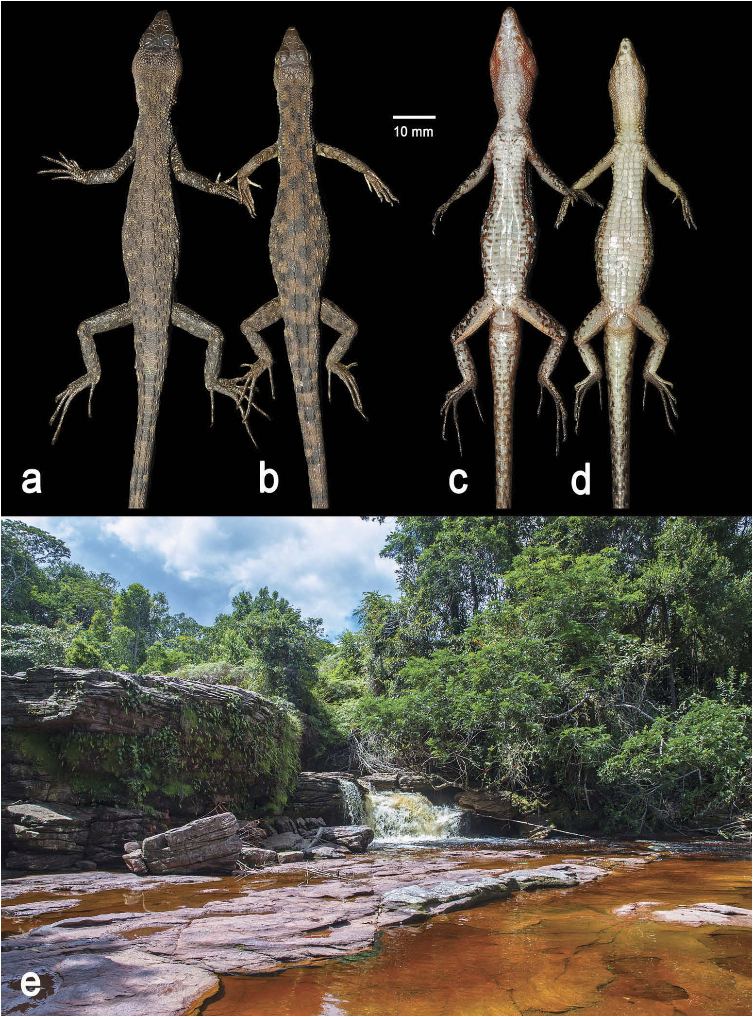
The hemipenis of IRSNB18432 ( Figure 5 View Figure 5 ) is bilobed, with the hemipenial body ornamented with eight transverse flounces with no vestige of spines or any other calcified structure. Hemipenial body roughly conical, with the base thinner than the distalmost portion. Lobes not distinctly detached from hemipenial body, non-capitate and with pointed tips, which gives an overall Y-shape to the organ. The sulcus spermaticus is deep and central in position, originating at the base of the organ, and proceeding in a straight line towards the lobular basis, bifurcating just before the lobular crotch. A small and V-shaped fleshy fold divides the sulcus, and the resulting branches extend across the lobes, ending on the ventral face of each lobular tip, between two conspicuous knobs. Two narrow areas with no ornamentation are parallel to the sulcus spermaticus. A narrow nude area separates the eight hemipenial body transversal flounces in two lateral sets on the asulcate face. Each of the body flounces runs downwards from the sulcate face, changing its direction upwards and returning to the downward direction on the sides towards the asulcate face, making a W shape.
Sexual dimorphism present, females slimmer than males, usually smaller, head of males larger and broader, neck much narrower in relation to head ( Figure 6 View Figure 6 ). Maximum number of femoral pores higher in males, variation in femoral pores greater in females (24 – 49) than in males (40 – 55). Colour pattern similar in both sexes, males sometimes darker than females and/or with brighter colour on lower sides of head ( Figures 4 View Figure 4 and 6 View Figure 6 ).
Colour in life ( Figure 4 View Figure 4 )
Ground colour variable, brown, olive green, or grey; top of head grey or black. Scales on sides of head usually grey or greyish brown. Labials and lateroventral scales reddish brown, orange, beige or yellowish. Iris grey or reddish brown. Area encircling the eyes usually orange or yellow, especially below the eye and in the preocular and postocular region. Chinshields, pregulars and gulars mottled grey and white, often with a hint of orange or yellow. Sides of neck dark brown, with yellowish-ochre spots. Dorsal pattern usually uniformly dark brown, occasionally greyish brown with numerous irregular black spots on the middorsal line. Middorsal line covered with quadrangular to circular markings bordered with black. Flanks often with numerous white ocelli. Flanks becoming more reddish brown towards the ventral side of the body. Upper surface of limbs similar to the dorsal region, with multiple ill-defined spots on the sides. Ventral surface of body bluish white or beige, often with brown markings laterally. Underside of limbs white or beige, without flecks. Palms and soles brown or grey. Tail often light brown proximally, becoming heavily spotted distally. In juveniles, the middorsal part of the tail is often reddish brown.
Colour in preservative
Males and females uniform light or dark brown, dorsal part of head dark grey. Dorsal and dorsolateral region of back and tail often with numerous irregular dark grey flecks. Flanks lighter, greyish white scales often occurring ventrolaterally. Ventral part of head white or beige, chinshields often mottled with brown or grey flecks. Ventral scales white or beige, ventrolaterals frequently with greyish spots. Underside of limbs white or beige. Tail white anteriorly, becoming brown or dark grey posteriorly.
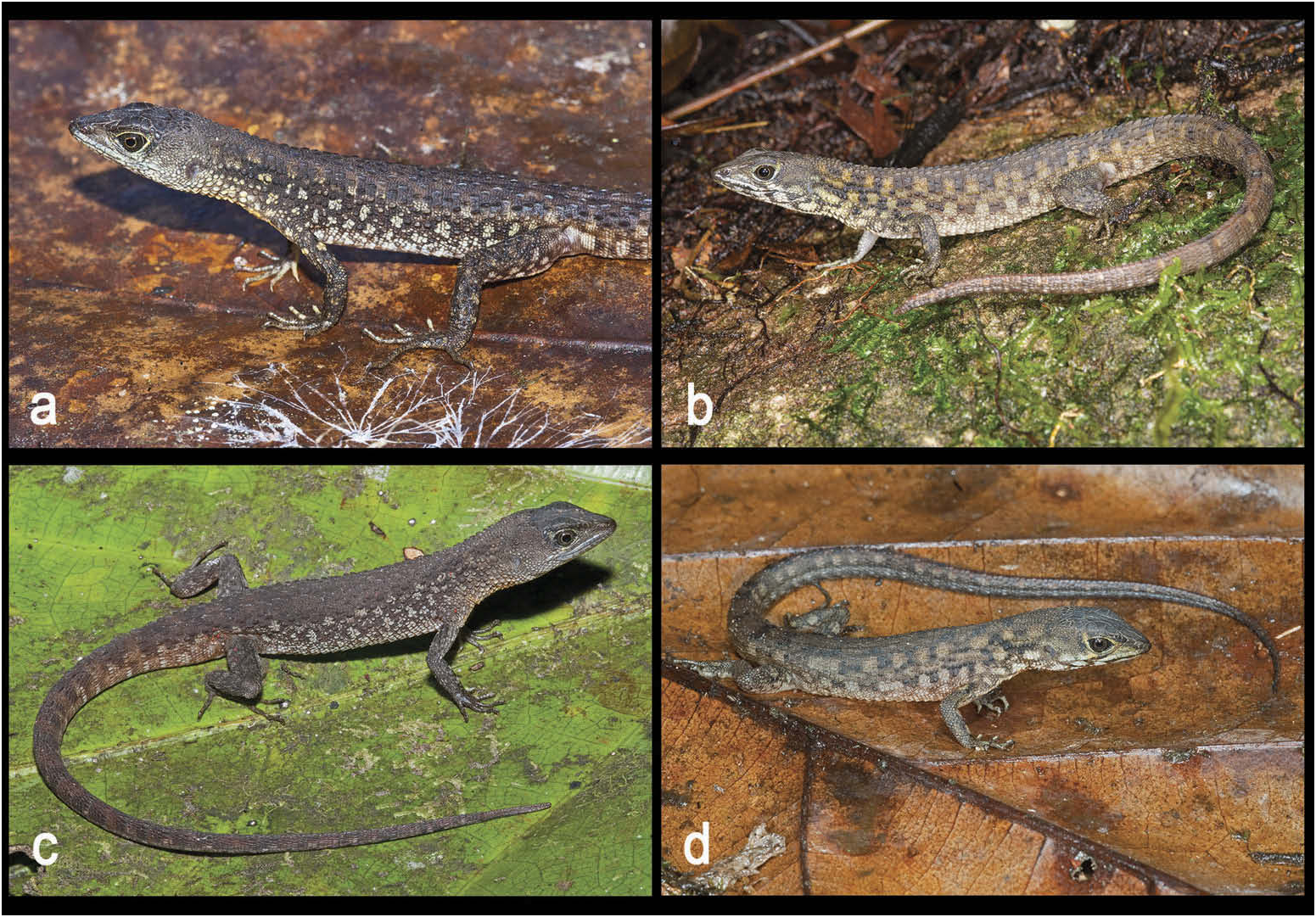
Geographic variation within the N. rudis clade ( Figures 7–8 View Figure 7 View Figure 8 ; Table 2)
There is some variation among the three localities under study in this clade (see also Discussion).
All specimens from Kaieteur National Park, Guyana ( Neusticurus rudis UCS 1, 200 – 580 m elevation, Figure 7 View Figure 7 ) have paired frontonasals and an azygous scale between frontonasals and prefrontals. Tubercles on the tail are smaller compared to those of specimens from other populations. Tubercles rarely imbricate at the base of the tail, mostly separated from the adjacent tubercle by 2 – 5 small, juxtaposed scales. Tubercles increase in number distally, frequently coming in contact with each other.
In specimens from the Iwokrama Mountains, Guyana ( N. rudis UCS 2, 70 – 200 m elevation, Figure 7 View Figure 7 ) the sexual dimorphism is much reduced ( Figure 8 View Figure 8 , compared to Figure 6 View Figure 6 ). Heads of males are distinctly narrower than those of males from Mount Roraima, Maringma-tepui and Kaieteur National Park (head width only slightly wider than neck vs distinctly wider in specimens from Mount Roraima, Maringma-tepui and Kaieteur National Park). Labials and region above supralabials are bluish white, with black or brown markings, extending onto the ventral side of the head. Sides of neck are beige or yellowish-ochre, with irregular pattern of dark brown markings or several rather large white spots. Ventral scales are creamy white to beige, nearly immaculate, occasionally with a few small flecks on the two lateralmost rows of scales. Occipital scales are fewer in specimens from the Iwokrama Mountains than in specimens from the other two localities.
Although hemipenes from the Kaieteur population are not available, there is substantial variation between the hemipenial organ of N. rudis sensu stricto and the organ of the Iwokrama population, mainly regarding the lobular shape and capitation. The hemipenis of the specimens from Iwokrama has distinctly capitate lobes, with the tips detached from the hemipenial body and ornamented with rounded knobs, whereas the hemipenis of N. rudis sensu stricto is non-capitate with no tip detached from the hemipenial body and no apical ornamentation ( Figure 5 View Figure 5 ).
Distribution and natural history
Neusticurus rudis sensu stricto as redefined here is currently restricted to east of the Venezuelan Gran Sabana, from the slopes of Mount Roraima in Venezuela through Wayalayeng and the slopes of Maringma-tepui to Mount Ayanganna in Guyana, between 678 – 1500 m elevation, a region drained by tributaries of the Essequibo and Mazaruni rivers ( Figure 1 View Figure 1 ). Other populations provisionally referred to that species (see above and Discussion) are currently found exclusively east of the Venezuelan Gran Sabana, from Mount Wokomung in the Pakaraima Mountains of Guyana to the Iwokrama Forest Reserve in Guyana between 159 and 1234 m elevation.
All specimens were found closely associated with streams in primary forest. The eight specimens collected on the slopes of Maringma-tepui were caught moving along the edges of cascading streams during the day, or sleeping on rocks along streams at night. Two specimens collected close to Wayalayeng were collected during the day on rocks in the Ataro River, at the top of Ataro Fall ( Figure 6 View Figure 6 ). Disturbed specimens quickly dove into the water.


| BM |
Bristol Museum |
| V |
Royal British Columbia Museum - Herbarium |
No known copyright restrictions apply. See Agosti, D., Egloff, W., 2009. Taxonomic information exchange and copyright: the Plazi approach. BMC Research Notes 2009, 2:53 for further explanation.