
Phyllium (Phyllium) geryon Gray, 1843
|
publication ID |
https://doi.org/ 10.11646/zootaxa.4365.2.1 |
|
publication LSID |
lsid:zoobank.org:pub:B166473D-1089-4DD2-866B-9339D152E616 |
|
DOI |
https://doi.org/10.5281/zenodo.5671937 |
|
persistent identifier |
https://treatment.plazi.org/id/39543029-FFE3-C31F-FF2C-71B1FEB63744 |
|
treatment provided by |
Plazi |
|
scientific name |
Phyllium (Phyllium) geryon Gray, 1843 |
| status |
|
Phyllium (Phyllium) geryon Gray, 1843 View in CoL
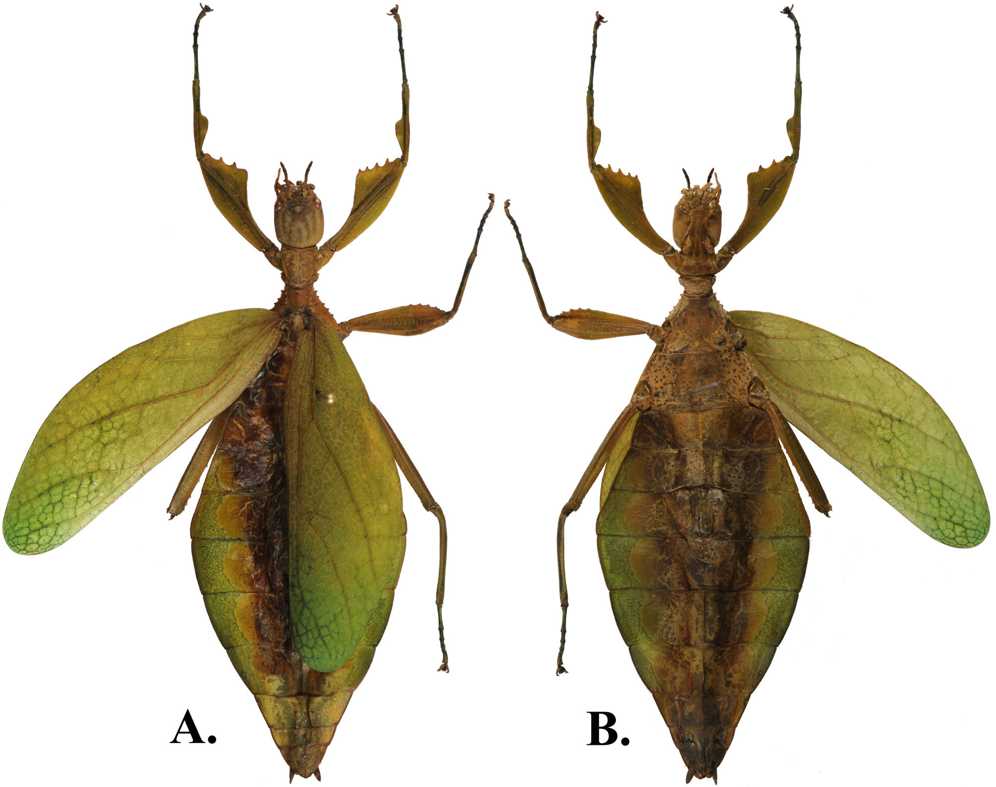
( Figs. 19A&B View FIGURE 19 & 20A,B,C&D View FIGURE 20 )
Material examined [ 2♀]: PHILIPPINES: 1♀: HOLOTYPE: 42. 72 Phil. Isl.; Philippine Islands; geryon, G.R.Gray, Cumings ; Phyllium geryon G.R.Gray , Philippine Islds.; BMNH (E) # 845232 [examined from detailed photos available on Phasmida Species File http:// phasmida .speciesfile.org (author: Paul D. Brock)]; 1♀: Philippines, Luzon, Aurora Province, Dingalan Municipality , August, 2017 [Coll. RC 17-256 ].
Discussion. Originally only known from the inexact type locality of “Inhabits the Philippine Islands ” from Gray’s 1843 description, Phyllium (Phyllium) geryon represents the tenth species of Phylliidae confirmed from Luzon. The small Phyllium specimen in the first author’s collection was originally expected to be difficult to compare morphologically to the damaged holotype, because many of the most frequently used features are missing. However, several unique features that still remain on the holotype allow us to confidently identify the punitive specimen. The granulation/spination of the head and thorax of the holotype were the most important features of the broken specimen to allow confident identification.
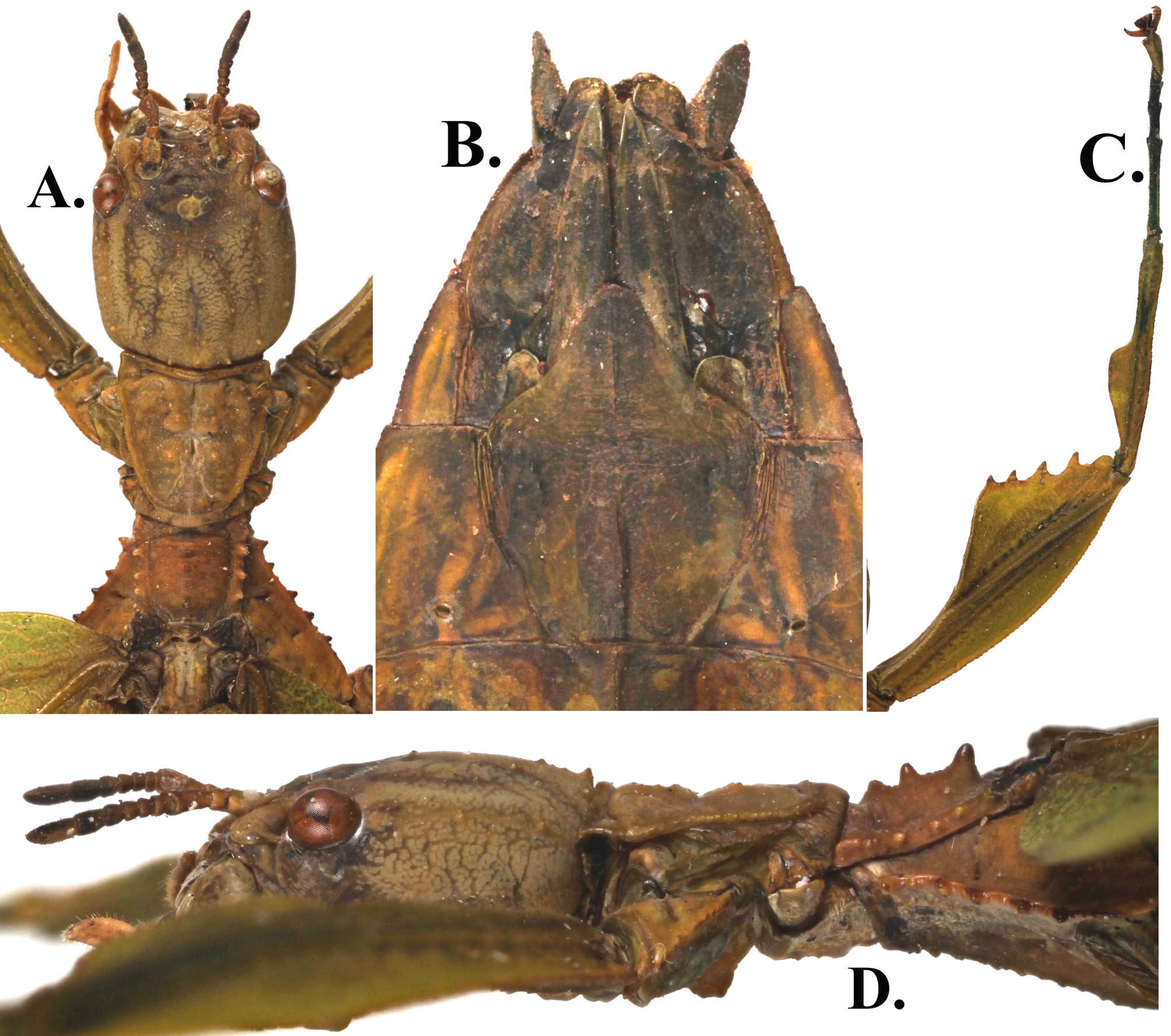
The most striking feature on the thorax that identifies Phyllium (Ph.) geryon is the spination along the crest of the mesopraescutum. The anterior rim lacks a spine, and is instead followed by a notable spine in the center and ends in a large prominent spine on the posterior that, in profile, is at least twice the size of the anterior rim ( Fig. 20D View FIGURE 20 ). Also the five well-defined tubercles of the mesopleurae with one to two small nodes placed between each were perfectly reflected in the specimen and is not something that we have seen in examination of dozens of other Phyllium species. The other features of the thorax also match perfectly but are not as unique as the posterior spine of the mesopraescutum crest.
The only feature that did not match perfectly between specimens was the apex shape of the subgenital plate. In the holotype the apex is sharply pointed and in the specimen from the first author’s collection the apex is rounded. The length of the subgenital plate however matches between the two individuals (both short, only reaching the posterior of segment IX) and the difference in terminal shape is assumed to be the result of being worn down, or a slight deformity resulting in a dulled point.
Many of the features described below are described for the first time, as it appears the holotype has been damaged for most of its existence. The profemora that was illustrated in the original description is the only feature that has drawn notable discussion over the years, as it is unclear if it belonged to the actual holotype or if it was a recreated illustration from deHaan’s work the year before (de Haan, 1842). The profemora illustration in Gray’s work certainly appears to be the same as the male nymph illustrated the year before which de Haan listed as belonging to a Phyllium (Phyllium) siccifolium male nymph. The determination by de Haan is likely erroneous as he lists the specimen as coming from “Timor, Nova Guinea ” a locality not known to have Phyllium (Phyllium) siccifolium and is instead more likely to belong to one of the many New Guinea native Phylliidae that have since been described.
We do find it odd however that while Gray appears to have illustrated a male nymphs profemora and assumed that it was equivalent to the Phyllium (Ph.) geryon holotype, he did place a great deal of importance on the shape of the profemora of Phylliidae , dividing his 1843 work into three divisions based solely on the exterior lobe of the profemora. From the specimen in the first author’s collection it is clear that the exterior lobe of the profemora, although not very wide (slightly thinner than the interior lobe) is complete and present, not absent as Gray describes. This leads us to believe that the holotype, even from the time of Gray’s original observation was missing its forelegs and Gray looking to use the profemora as a key feature in his work went looking for a specimen with a similar body shape to the Phyllium (Ph.) geryon holotype and found de Haan’s male nymph adequate. The first mention that the forelegs were missing from the holotype, and first to suggest that perhaps Gray only illustrated the nymph from de Haan, was Westwood only sixteen years latter when he observed the specimen and found the forelegs “wanting” ( Westwood, 1859). Rehn and Rehn in 1933 also discussed their suspicion of the missing exterior lobe of the profemora and surprisingly guessed that Phyllium (Ph.) geryon was native to Luzon, a guess incorrectly based on their assumption that Phyllium (Ph.) geryon was closely related to Microphyllium pusillulum which they based on the small size and thin exterior lobe of the profemora.
♀♀. Coloration. Pale to darker green throughout most of the body and tegimina, some areas discolored from drying. Compound eyes burnt orange. The granulation throughout is of a similar color to the surface upon which it is found or slightly lighter such as those found on the head capsule.
Morphology. Head capsule slightly longer than wide with a moderately detectable pattern of granules. Lateral to the prominent posteromedian tubercle there is a node about half the size, followed by two to three other similarly sized nodes following the margins of the head. Interior to those nodes there are several nodes on the posterior half of the head capsule pointing from the compound eyes back to the posteromedian tubercle forming a “V” pattern ( Fig. 20A View FIGURE 20 ). The small protuberance between the compound eye and antennal base is marked with a notable pit in the center. The frontal suture is also well furrowed. The frontal convexity has a slight covering in setae and is only slightly smaller than the size of the compound eyes, which are also smaller and less bulbous. Antennal fields only slightly wider than the base of the antennae. Antennae moderately slender and elongate (4.0 mm), approximately the same length as the postocular section of head capsule, and consisting of nine segments ( Fig. 20A View FIGURE 20 ). Antennae covered in setae of varying sizes, most sparse except for antennomeres VIII and IX with the most variety in size and the greatest quantity of setae. Apical antennomere cylindrical with rounded apex, about 2.5x longer than wide and about 1.5x as long as segment VIII which is notably longer than any of the other preceding four segments. Pronotum roughly trapezoidal, widest at the anterior, which is at least twice the length of the anterior rim. Lateral rims on the pronotum are roughly parallel for the first quarter of the length and then converging to the posterior margin. The anterior rim is distinct and slightly concave, lateral rims are moderate to weak and the posterior margin has no rim. Face of pronotum irregularly granulose with a distinct furrow on the median plane on the anterior half. Mesopraescutum almost a square with a length that equals the width and lateral margins that are only slightly converging. Lateral margins marked with three large tubercles and slight granulation on the anterior end. Mesopraescutum disk crest has an anterior rim slightly granulose and lacking a spine, followed by a notable spine in the center of the disk and ending in a large prominent spine on the posterior that, in profile, is at least twice the size of the anterior rim ( Fig. 20D View FIGURE 20 ). Mesopleurae evenly diverging with lateral margins armed with five evenly sized but slightly unevenly spaced tubercles. Between each tubercle there is one to two small nodes. Mesopleurae face smooth except for two clear pits, one on the anterior third and one on the posterior third. Prosternum irregularly granulous throughout, the anterior half is sloped into a point, not flat like the posterior half. Mesosternum irregularly granulous throughout, those along the sagittal plane slightly larger. Tegmina (length 39.2 mm, maximum width 12.6 mm) extending almost half way into abdominal segment VII. Alae rudimentary. Abdominal segments II–IV gradually widening with the posterior of segment IV the widest segment and V–X uniformly tapering towards the apex. Anal segment at its widest, wider than long (width-length ratio 1.67:1), with a relatively rounded apex. Subgenital plate short, only reaching the posterior of segment IX, sides slightly convex and ending in a rounded point ( Fig. 20B View FIGURE 20 ). Gonapophyses long and reaching the apex of the subgenital plate ( Fig. 20B View FIGURE 20 ). Profemora with narrow exterior lobe, slightly thinner than the interior lobe, margin appears smooth without magnification, but under low magnification, the entire margin is marked with small tight, evenly spaced dentition. Interior lobe wider than exterior lobe, obtuse in angle, and marked on this specimen with prominent teeth, two teeth on the left profemora and four teeth on the right profemora. Protibiae lacking an exterior lobe and the interior lobe is only present on the posterior half of the tibiae as a rounded triangle that drops sharply to the tibiae near the middle. Meso- and metafemora exterior and interior lobe gently rounded, interior lobe with notable serrate dentation and approximately twice the width of the exterior lobe. Meso- and metatibiae simple, lacking lobes or dentition.
Measurements [mm] of specimen [Coll. RC 17-256]: Length of body 65.7, length/width of head 6.5/5.6, length of pronotum 4.0, length of mesonotum 5.0, length of tegmina 39.2, greatest width of tegmina 12.6, length of alae - -, greatest width of abdomen 23.9, length of profemora 12.7, length of mesofemora 11.2, length of metafemora 13.8, length of protibiae 9.1, length of mesotibiae 8.0, length of metatibiae 12.2, length of protarsi 8.3, length of antennae 4.0.
| BMNH |
United Kingdom, London, The Natural History Museum [formerly British Museum (Natural History)] |
No known copyright restrictions apply. See Agosti, D., Egloff, W., 2009. Taxonomic information exchange and copyright: the Plazi approach. BMC Research Notes 2009, 2:53 for further explanation.
|
Kingdom |
|
|
Phylum |
|
|
Class |
|
|
Order |
|
|
Family |
|
|
Genus |