
Phyllium (Phyllium) bourquei Cumming & Le Tirant, 2017
|
publication ID |
https://doi.org/ 10.11646/zootaxa.4365.2.1 |
|
publication LSID |
lsid:zoobank.org:pub:B166473D-1089-4DD2-866B-9339D152E616 |
|
DOI |
https://doi.org/10.5281/zenodo.5671935 |
|
persistent identifier |
https://treatment.plazi.org/id/39543029-FFE6-C313-FF2C-700EFA123726 |
|
treatment provided by |
Plazi |
|
scientific name |
Phyllium (Phyllium) bourquei Cumming & Le Tirant |
| status |
sp. nov. |
Phyllium (Phyllium) bourquei Cumming & Le Tirant View in CoL sp. nov.
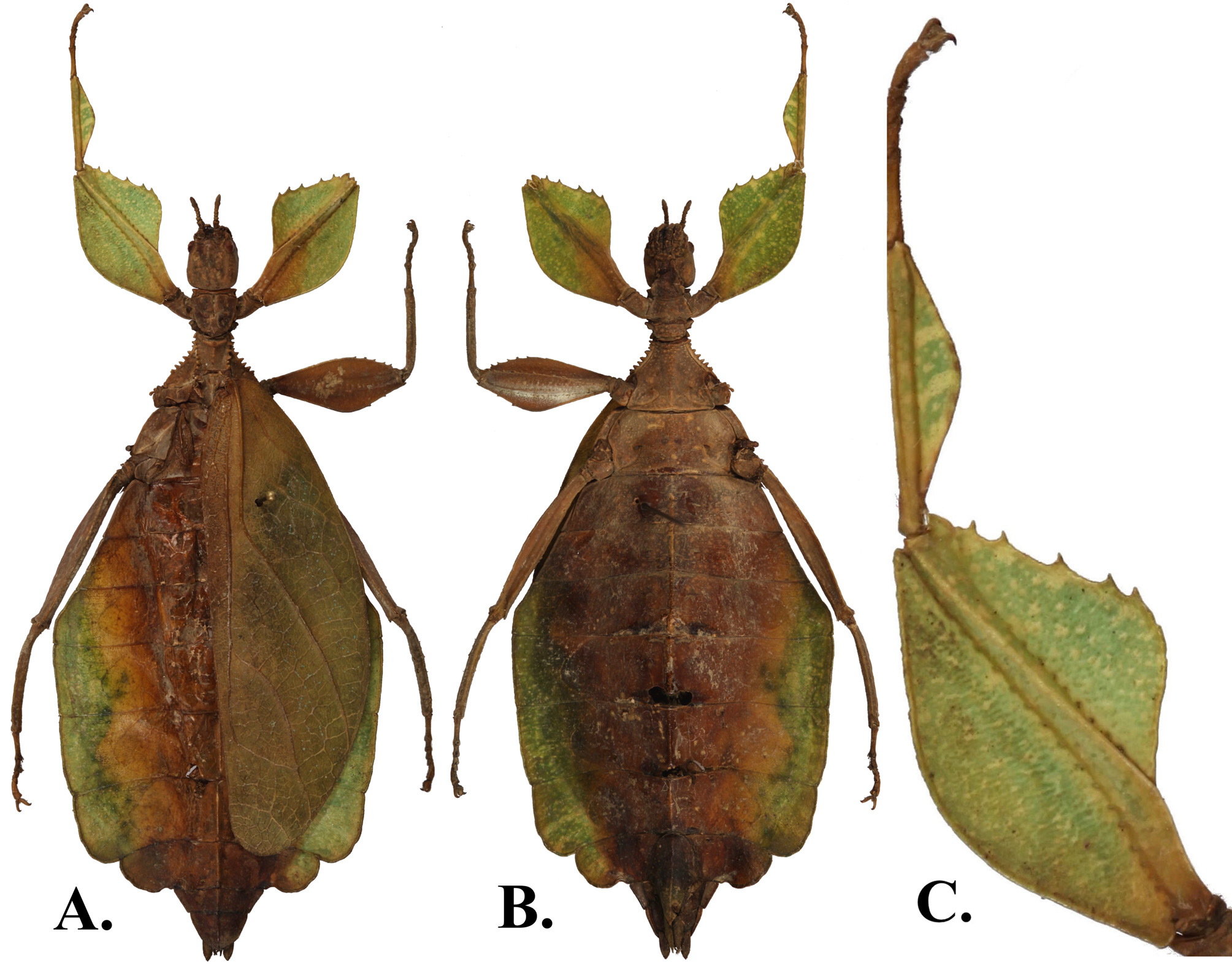
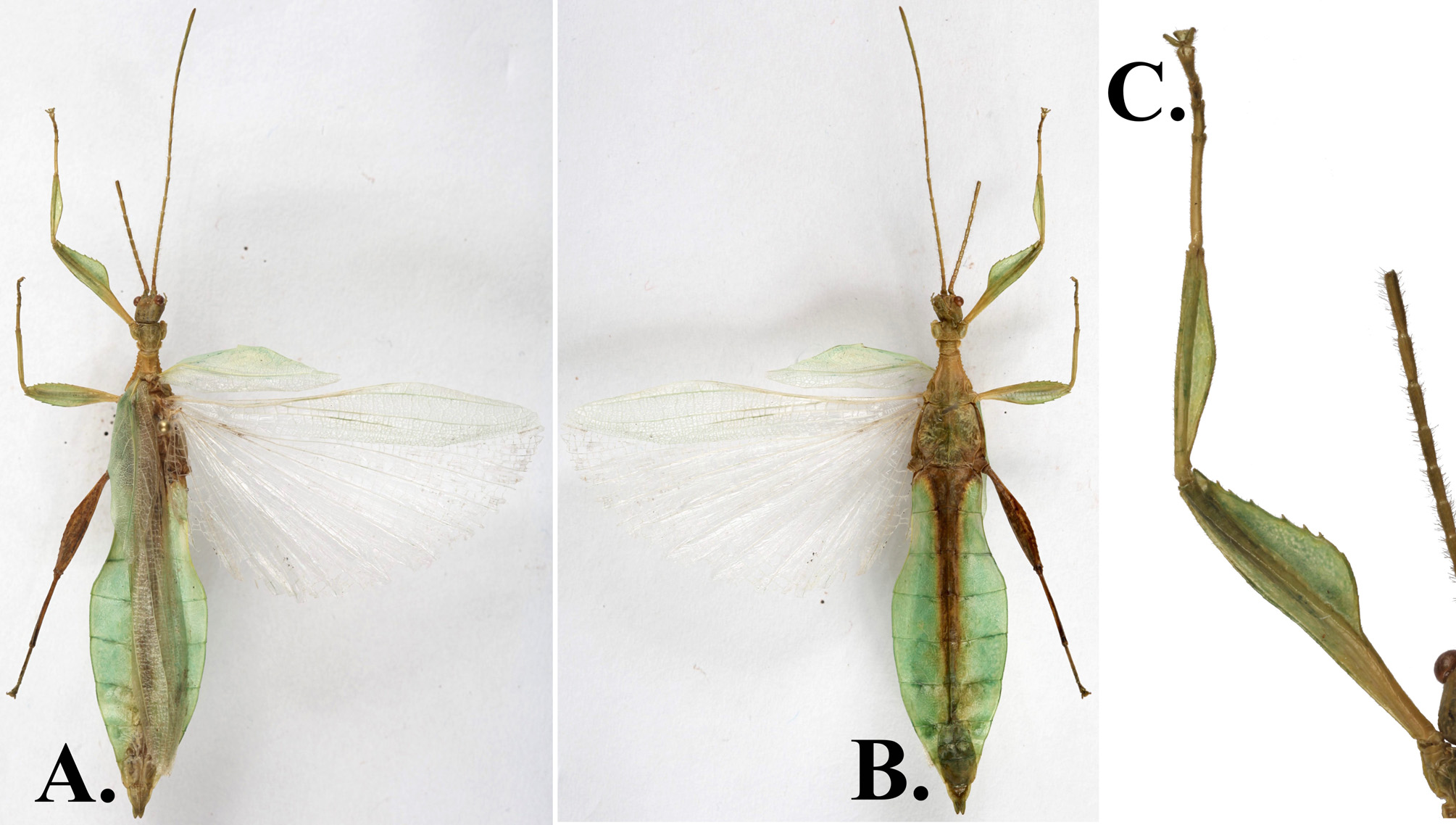
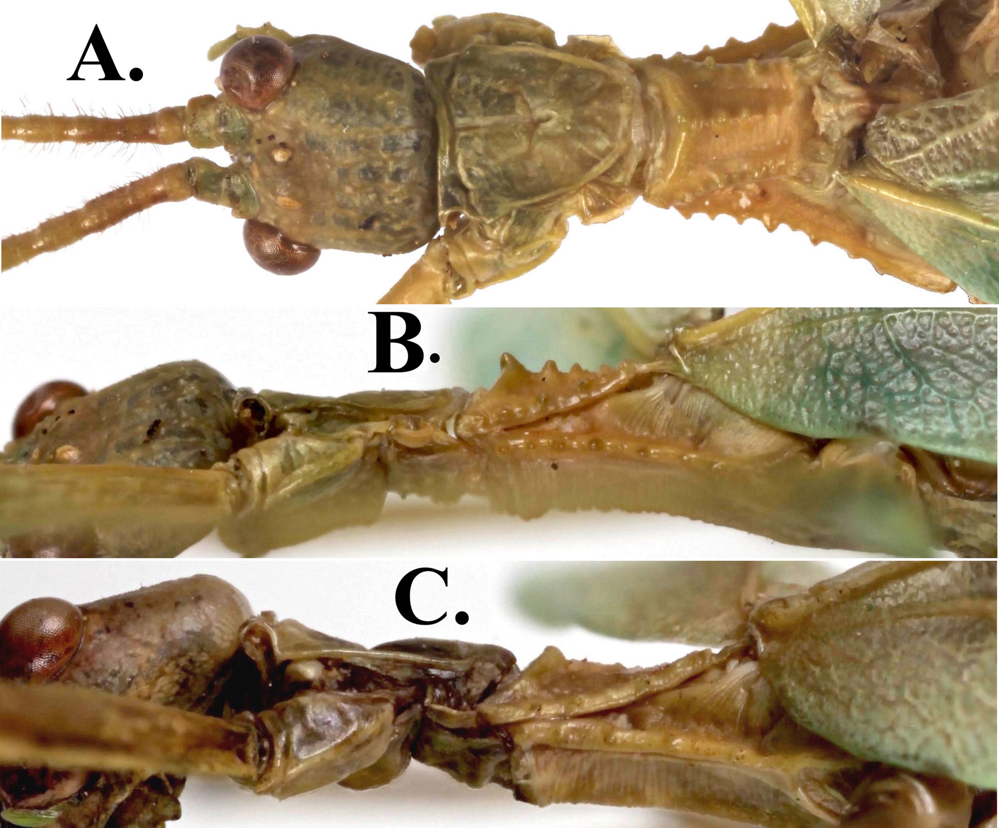
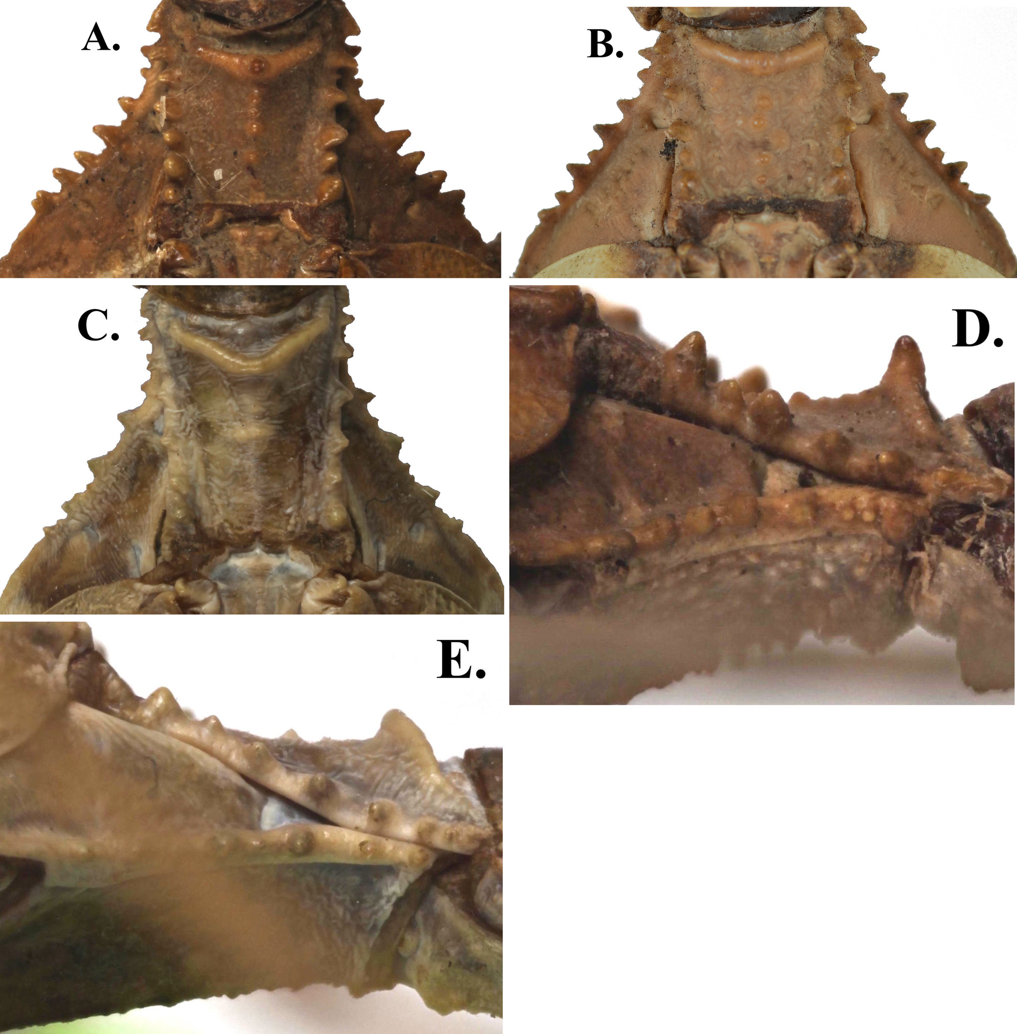
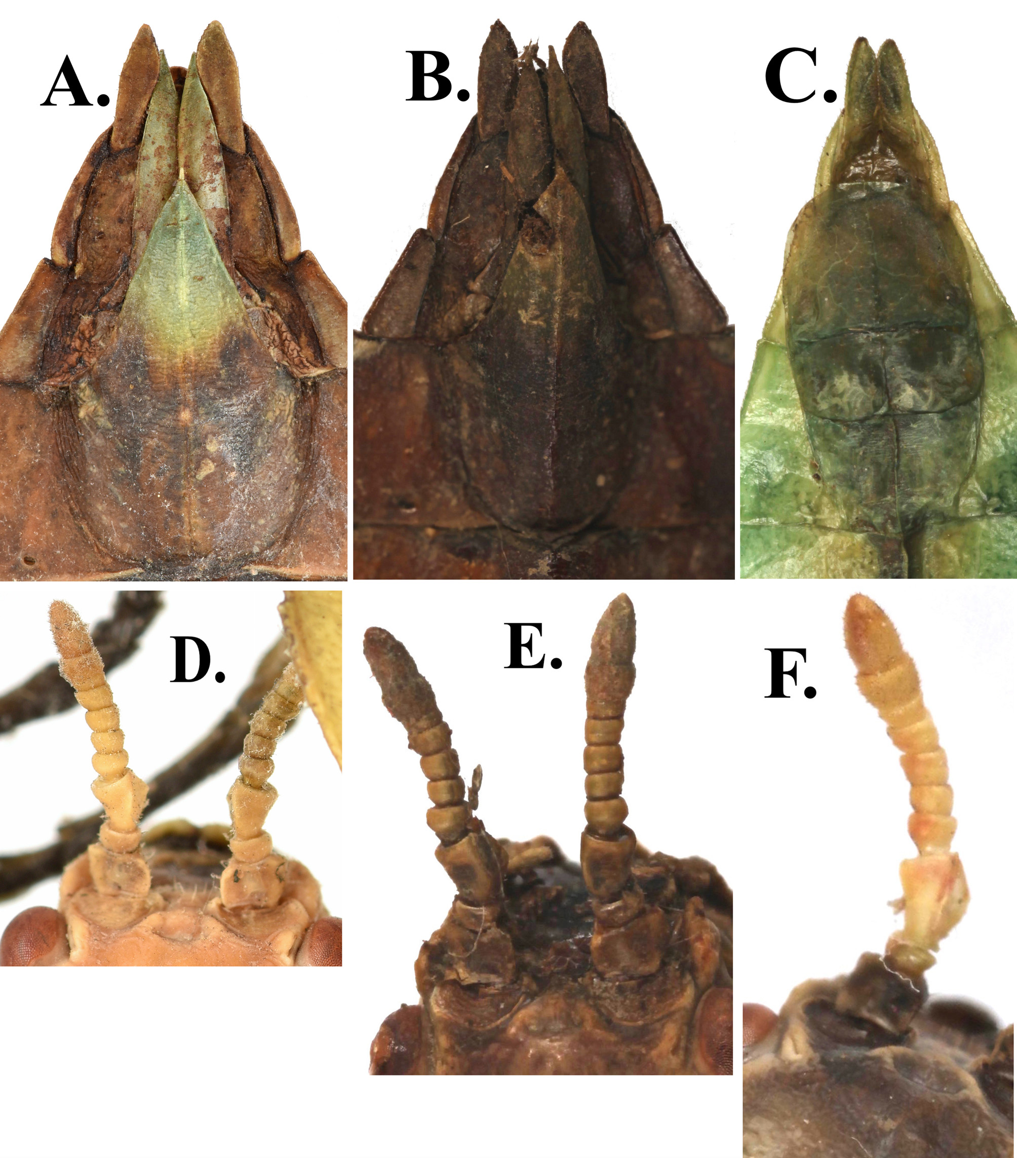
( Figs. 14A,B&C View FIGURE 14 , 15A,B,C,D&E View FIGURE15 16A,B&C View FIGURE 16 , 17A&B View FIGURE 17 , 18A,B View FIGURE 18 ,&D)
HOLOTYPE: ♀: Philippines, Luzon, Nueva Vizcaya, Kayapa , March, 2017 [Coll. RC 17-203 ]. (Deposited in the National Museum of the Philippines. PNM )
PARATYPES: 1♀: Philippines, Luzon, Nueva Vizcaya, August, 2007 [Coll. RC 17-255 ] . 1♂: Philippines, Luzon, Nueva Vizcaya, Belanue , May, 2014 [Coll. RC 16-201 ].
Coauthored with Stéphane Le Tirant, Montreal, Canada, whose collection originally contained the paratype female. Discussion. This species was originally thought to be Phyllium (Phyllium) philippinicum Hennemann et al., 2009 found outside its known range. Upon closer examination, several differences became clear. Like Ph. (Ph.) philippinicum , this new species falls under the siccifolium species-group of Hennemann, et al., 2009 because the female lacks developed alae and the male has an exterior lobe of the profemora that is more slender than the interior lobe. It is possible that future genetic analysis will find that these two populations are not conspecific, but the marked differences in the female genitalia led us to here describe them with species level status. The geographic isolation of the two populations, Ph. (Ph.) philippinicum from the Zambales Range, and Ph. (Ph.) bourquei from the Caraballo Mountains, separated by the Central Luzon Plains, also led to the decision to erect the new species.
Description. Description is based upon the HT female and the paratype male/female specimens. Coloration is based upon the dried specimens here illustrated, it is assumed the colors were lighter in life.
♂♂. Coloration. Pale green throughout (except for areas of rot), other areas (antennae and thorax in particular) paler in color, more straw colored than green. No eyespots noted on the abdomen in the male paratype.
Morphology: Head capsule length and width approximately equal ( Fig. 17A View FIGURE 17 ). Distinct antennal fields, vertex mostly smooth, only slight granulation on the posterior end and around a broad but short posteromedian tubercle. Compound eyes large and protruding, ocelli well developed. Antennal simple, and consisting of 23 segments (including scapus and pedicelus) and with long thin setae. Apical antennomere cylindrical with rounded apex, slightly more than 3x longer than wide and covered with short dense setae. Pronotum relatively smooth, with a distinct furrow and slight pit along the median plane. Anterior margin concave, lateral and posterior margins slightly convex. Shape roughly trapezoidal with the anterior length slightly less than twice that of the posterior. Anterior and lateral margins with distinct rims. Prosternum with moderate granulation. Mesopraescutum approximately as long as wide, and only slightly narrower towards the posterior. Lateral margins with 5–6 decent sized tubercles of slightly different sizes and somewhat unevenly spaced. Mesopraescutum disc raised along the median plane with a crest marked by a prominent tubercle on the anterior rim followed by three others of steadily decreasing size and even spacing with the smallest ending near the posterior rim ( Fig. 17B View FIGURE 17 ). The surface of the mesopraescutum disc on each side of the crest is free of nodes. Mesopleurae slightly diverging from mesopraescutum lateral margins for the first 1/3 and then gradually widening for the remainder. Mesopleurae margins with 5 distinct tubercles predominantly on the anterior and 3 minor tubercles intermixed on the posterior portion. Prosternum fully covered in granules of varying sizes, but none more notable than the others. Mesosternum rather smooth but with notable granules along the sagittal plane, more heavily marked on the anterior. Tegmina (length 17.9 mm, maximum width 5.2 mm), extending to the anterior margin of abdominal segment IV. Alae (length 37.7 mm), well developed, oval fan configuration with exposed section slightly sclerotized. Abdominal segment II with parallel margins, segments III – first 2/3 of segment IV gradually widening with IV marking the widest segment, posterior 1/3 of IV–X gradually tapering towards the apex, at first only slightly then more prominently creating a spade shaped abdomen. Spiracles only just visible on the ventral surface, located on the anterior margin of segments III–VIII near the median plane. Anal segment posterior half somewhat evenly rounded apex. Poculum rather stout with a straight posterior margin, slightly projecting over the posterior margin of abdominal segment IX and exposing the vomer, which is rather wide with a single stout terminal hook ( Fig. 15C View FIGURE15 ). Exterior lobe of profemora narrow, at its widest point only slightly wider than the shaft of the femur. Exterior lobe of the profemora relatively smooth, only marked by small, barely notable teeth pointing anteriorly. Interior lobe not starting until a third the way up the femur and arching in a rounded triangle marked with six to seven small anteriorly pointing teeth that are not quite evenly spaced ( Fig. 16C View FIGURE 16 ). Protibiae lacking exterior lobe, interior lobe only a smooth arch, almost triangular in shape. Exterior and interior lobes of mesofemora gently rounded and approximately equal in width and both lightly serrate. The interior lobe marked more heavily with six to seven tightly packed teeth, the exterior lobe less serrate, marked with three more widely spaced teeth. Exterior and interior lobes of metafemora gently rounded; interior lobe notably wider and with eight serrate teeth; exterior lobe lacking serration. Meso- and metatibiae simple, lacking lobes or serration.
♀♀. Coloration. Most of the holotype is discolored but a pale green is still detectable on the margins of the abdomen and forelegs. The paratype is mostly a paler color and almost completely discolored.
Morphology. Head capsule longer than wide with the posterior 1/4 sparsely granulose with a notable posteromedian tubercle at least three times the size of any other node. Antennae moderately slender and elongate, slightly longer than the postocular section of the head capsule, and consisting of ten segments ( Figs. 15D&E View FIGURE15 ). Antennae mostly lacking setae, the terminal two segments are the only ones with notable setae covering. Apical antennomere cylindrical with rounded apex, only slightly longer than the preceding segment. Pronotum relatively smooth, with a distinct furrow and slight pit along the median plane. Anterior margin concave, lateral and posterior margins slightly convex. Shape roughly trapezoidal with the anterior width approximately twice that of the posterior. Anterior margin with distinct rim, lateral margins with weak rims. Mesopraescutum only slightly narrowing towards the posterior and approximately the same length as the width. Lateral margins marked with ~6 robust major tubercles of relatively uniform size. Mesopraescutum disk with a prominent rim on the anterior margin marked by a distinct spine followed by four small but clear nodes along the sagittal plane ( Figs. 18A,B View FIGURE 18 ,&D). Mesopleurae uniformly diverging with lateral margins armed with ~7–8 robust tubercles. Mesopleurae face relatively smooth but marked with a clear pit located on the anterior third and a slight pit on the posterior third. Prosternum irregularly granulous throughout. Mesosternum mostly smooth but with notable granules along the sagittal plane, more heavily marked on the anterior. Tegmina extending slightly past the anterior margin of abdominal segment VIII. Alae rudimentary. Abdominal segments II- the first 2/3 of IV gradually widening, with segment IV marking the widest segment. The posterior third of IV–X tapering towards the apex with segments VII and VIII lobed. Anal segment at its widest, slightly wider than long, with a broad apex. Subgenital plate with a fine point reaching about half way under the anal abdominal segment ( Figs. 15A&B View FIGURE15 ). Gonapophyses rather long, slightly protruding from under the anal abdominal segment when viewed dorsally. Profemora with a widely rounded exterior lobe that has a relatively smooth outer margin. Interior lobe, slightly narrower than exterior lobe and slightly more angled, anterior portion of the margin marked with 5 small saw-like teeth of equal size and shape, but not with perfectly equal spacing. Protibiae lacking an exterior lobe and the interior lobe a rounded isosceles triangle. Exterior and interior lobe of mesofemora gently rounded; exterior lobe slightly wider than interior lobe due to the shape being more angled than the interior lobe, which is gently arching. Exterior lobe is marked with two to three widely spaced small teeth, interior lobe marked more heavily with serrate dentition (five to six teeth). Exterior and interior lobe of metafemora gently rounded with exterior lobe rather thin, interior lobe slightly wider and with seven serrate teeth, exterior lobe lacking dentition. Meso- and metatibiae simple, lacking lobes or serration.
Measurements of the type material can be found within table 6.
Etymology. This species is dedicated to Mr. Pierre Bourque. He was mayor of the City of Montreal from 1994 to 2001. Mr. Bourque was also one of the most innovative directors of the Montreal Botanical Garden from 1980 to 1994. Among other things, he was responsible for the creation of new greenhouses, the Floralies Internationales de Montréal, the Japanese Garden, the Chinese Garden, the Tree House, the Montreal Biodome and the Montreal Insectarium. Thanks to Mr. Bourque, the Montreal Botanical Garden has become the second largest in the world.
| PNM |
Philippine National Museum |
No known copyright restrictions apply. See Agosti, D., Egloff, W., 2009. Taxonomic information exchange and copyright: the Plazi approach. BMC Research Notes 2009, 2:53 for further explanation.
|
Kingdom |
|
|
Phylum |
|
|
Class |
|
|
Order |
|
|
Family |
|
|
Genus |