
Eucyclops alekseevi, Mercado-Salas & Suárez-Morales & Silva-Briano, 2015
|
publication ID |
https://doi.org/ 10.1080/00222933.2015.1061715 |
|
publication LSID |
lsid:zoobank.org:pub:2F320DE0-FF96-4E5F-8520-586303082E09 |
|
DOI |
https://doi.org/10.5281/zenodo.4332565 |
|
persistent identifier |
https://treatment.plazi.org/id/397AD47D-FFCC-FFB4-A65C-FEE3FD0577B9 |
|
treatment provided by |
Carolina |
|
scientific name |
Eucyclops alekseevi |
| status |
sp. nov. |
Eucyclops alekseevi sp. nov. Mercado-Salas and Suárez-Morales ( Figures 34 – 39 View Figure 34 View Figure 35 View Figure 36 View Figure 37 View Figure 38 View Figure 39 )
Material examined
Holotype. Adult ♂ specimen dissected, mounted in glycerin, slides sealed with Entellan (ECO-CH-Z-04640).
Allotype. Adult ♂, dissected, mounted in glycerin, slides sealed with Entellan (ECO- CH-Z-04641).
Paratypes. Five adult ♀♀, undissected, ethanol-preserved (90%) (ECO-CH-Z-04642). Samples from type locality collected 1 March 1991 by Marcelo Silva-Briano.
Type locality
Río Juchipila , Juchipila, Zacatecas, Mexico (21°24´37.59´´ N, 103°06´57.90´´W) GoogleMaps . 1250 m above sea level (asl).
Etymology
This species is warmly dedicated to Dr. Victor R. Alekseev for his valuable contributions to the knowledge of the genus Eucyclops worldwide.
Description
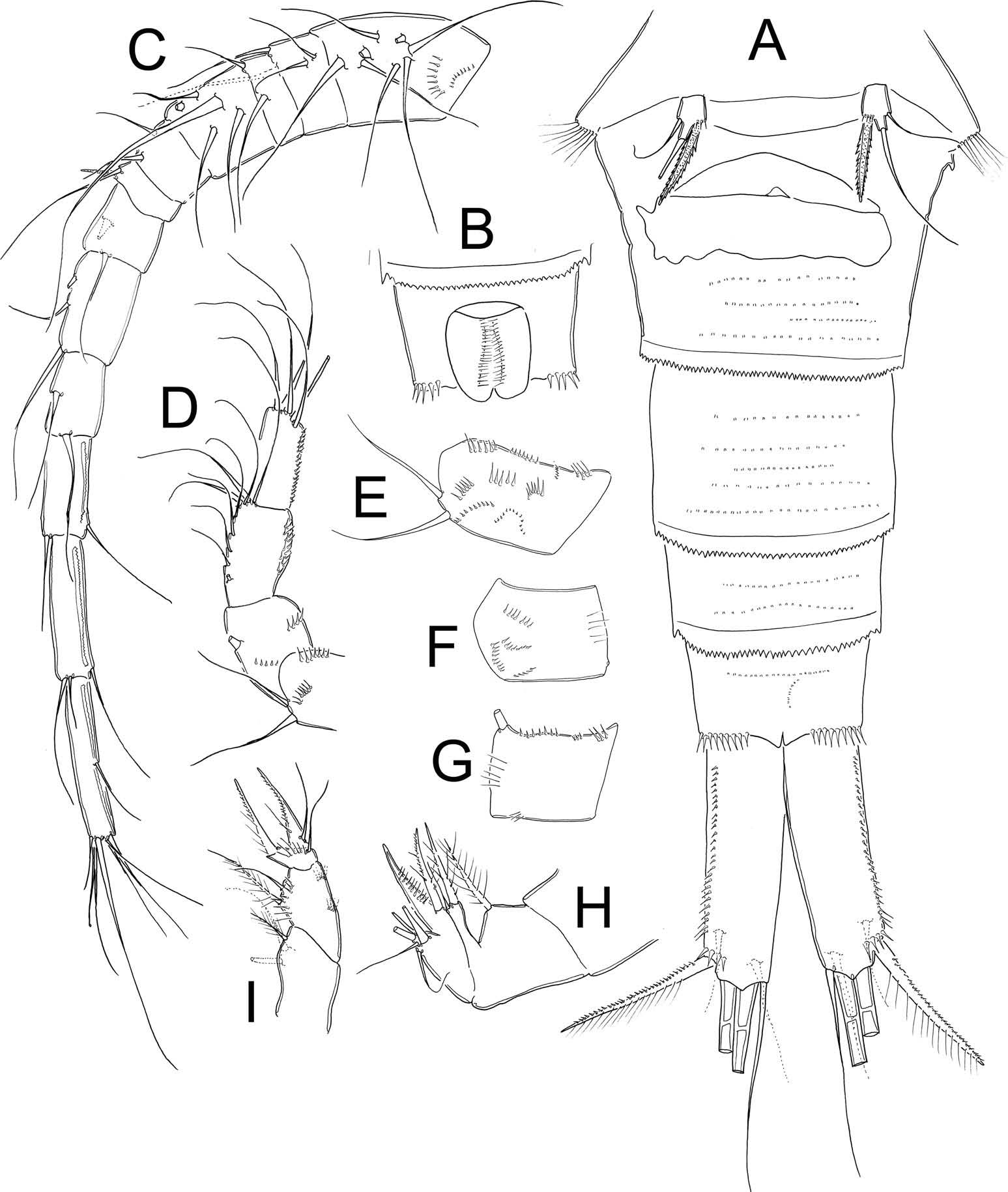
Female. Average length excluding caudal setae = 705 µm. Prosome representing 55% of total body length, symmetrical in dorsal view. Prosomal fringes finely serrate in dorsal view. Urosomal fringes strongly serrate. Genital double somite symmetrical ( Figure 36B View Figure 36 ), representing 11% of total body length; widest proximally, tapering towards distal edege. Seminal receptacle with rounded lateral arms; posterior margin with sinuous sac ( Figure 34A View Figure 34 ). Anal operculum slightly rounded, smooth ( Figure 34B View Figure 34 ). Length/width of caudal ramus = 3.5; inner margin of caudal ramus naked; strong spinules covering 60% with respect to the total length of ramus. Dorsal seta (VII) 0.5 times as long as caudal ramus, 0.8 times as long as outermost caudal seta (III). Ratio of innermost caudal seta (VI)/outermost caudal seta (III) = 1.3. Lateral caudal seta (II) inserted at 73% of caudal ramus.
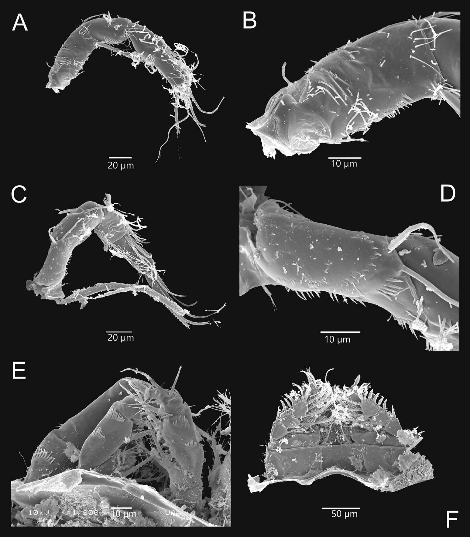
Antennule ( Figures 34C View Figure 34 , 36D–F View Figure 36 ). Tip reaching posterior margin of the fourth pediger. Armature per segment as follows: 1(8s), 2(4s), 3(2s), 4(6s), 5(4s), 6(1s+1sp), 7(2s), 8(3s), 9(2s+1ae), 10(2s), 11(3s), 12(8s). Two transverse rows of spinules on first segment, the first with strong spinules and the second below, with minute spinules. Spine on sixth segment not reaching medial margin of seventh antennular segment.
Antenna ( Figures 34E–G View Figure 34 , 37A–D View Figure 37 : Coxa (unarmed), basis (2s+Exp), plus three-segmented Enp (1s, 9s, 7s respectively). Basis with rows of spinules on frontal surface: N1(V),
N2(III), N3(6), N4(7), N5(8), N6(4), N15(4), N17(7), N18(6); on caudal surface: N7(10), N8(6), N9+10(8), N11(5), N12(7), 22(13); caudal surface of Enp1 with B2(6).
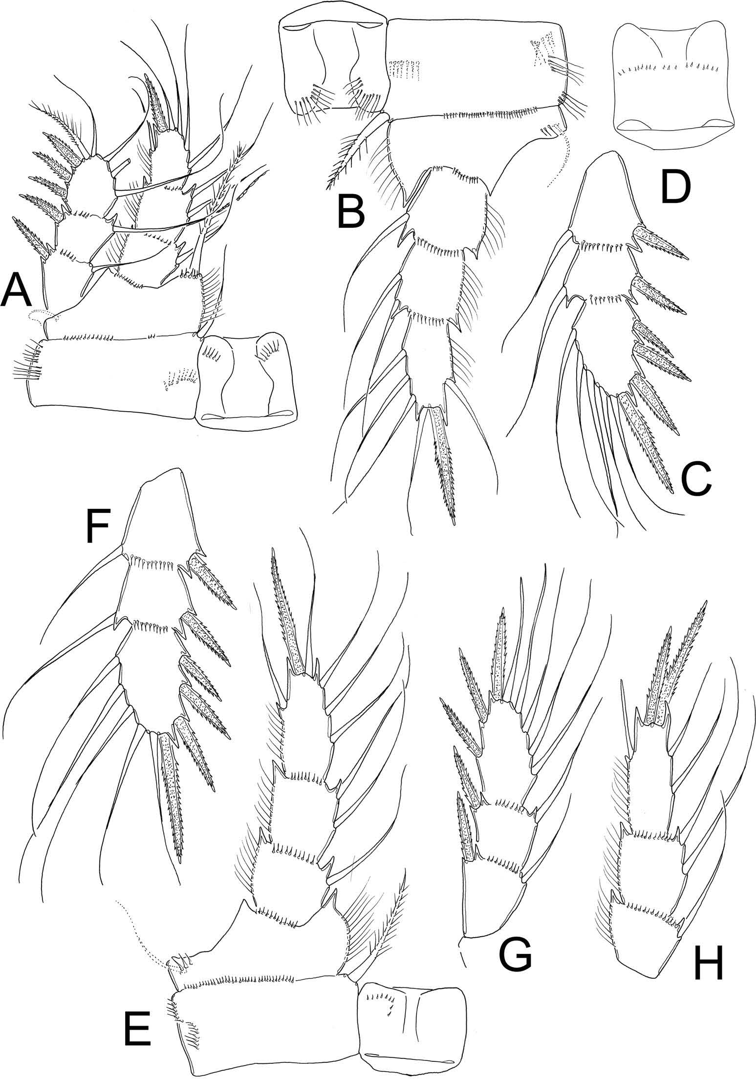
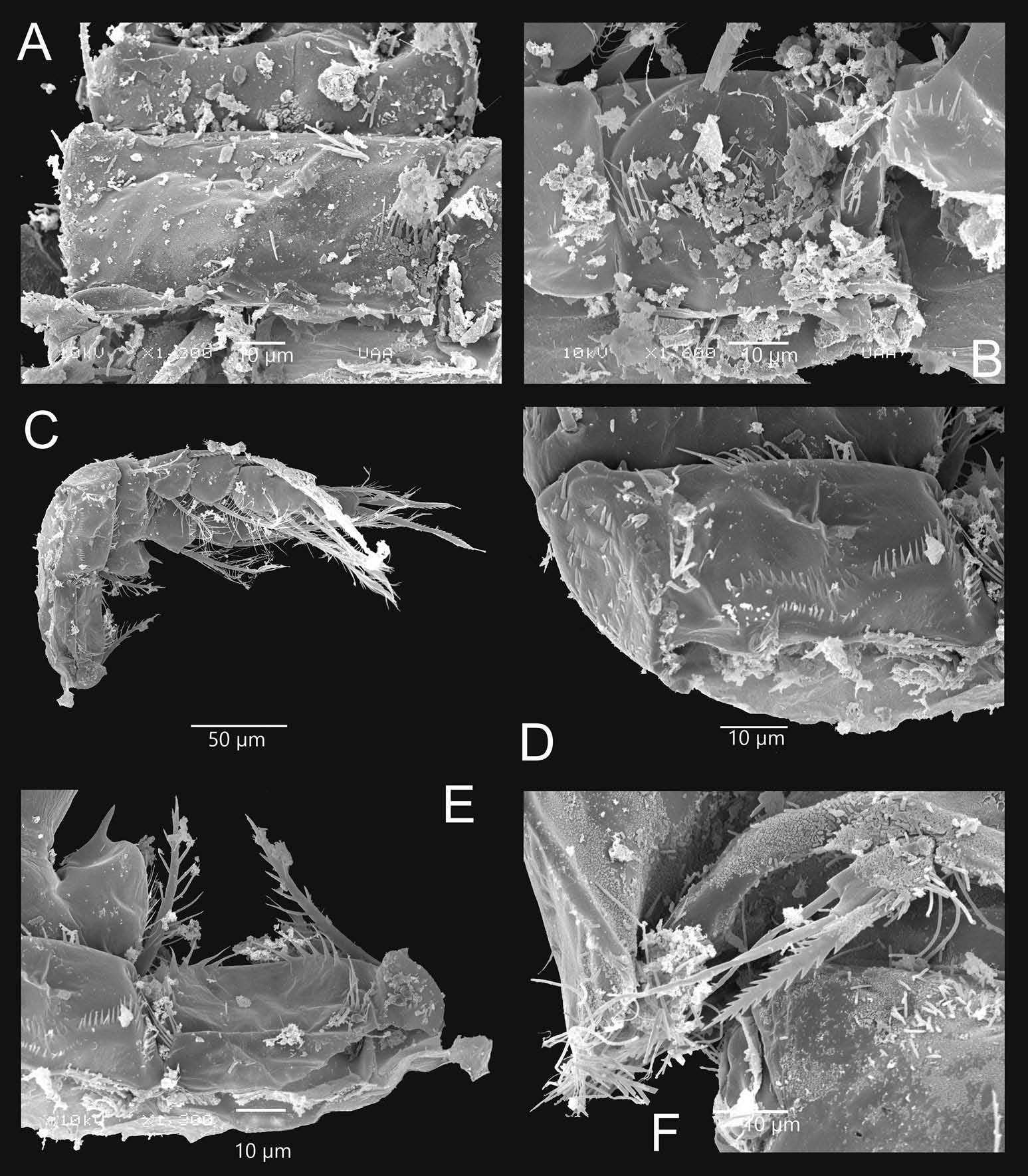
Leg 1 ( Figures 35A View Figure 35 , 37F View Figure 37 , 38A–B View Figure 38 ). Frontal surface of intercoxal sclerite with a row of strong spinules arranged in a semicircular pattern on each side, caudal surface with row II continuous, bearing 22 minute spinules, row I absent. Inner coxal seta biserially setulated, caudal coxal surface with spinule formula = A-B-C. Inner basal seta (basipodal
spine) reaching beyond midlength of Enp3, 0.9 times as long as Enp. Length/width ratio Enp3 = 1.5, apical spine of Enp3 being 1.1 times as long as Enp3.
Leg 2 ( Figure 35B–D View Figure 35 , 38C–D View Figure 38 ). Frontal surface of intercoxal sclerite with row I bearing long hair – spinules arranged in a semicircular pattern; caudal surface with row II continuous, bearing 17 small spinules, row I absent. Distal margin of intercoxal sclerite with two rounded, chitinised projections. Inner coxal seta biserially setulated, caudal coxal surface with spinule formula = A-B-C-D. Length/width ratio of Enp3 = 2.0, apical spine of Enp3 1.4 times as long as Enp3. No modified setae present.
Leg 3 ( Figures 35E–F View Figure 35 , 38E–F View Figure 38 , 39A–B View Figure 39 ). Frontal surface of intercoxal sclerite with small spinules arranged in semicircular pattern on each side. Caudal surface with row I bearing slender hair – spinules, row II continuous, with 28 strong spinules and row III continuous, with 26 long, strong spinules. Distal margin with two rounded, chitinised projections. Coxa with strong, biserially setulated inner coxal seta, basally with long hairs and distally with strong spinules along both margins. Caudal coxal surface with spinule formula = A- B-C. Length/width ratio of Enp = 2.1, apical spine of Enp3 1.2 times as long as Enp3. No modified setae present.
Leg 4 ( Figures 35G–H View Figure 35 , 39C–E View Figure 39 ). Distal margin of intercoxal sclerite with two low, rounded, chitinised projections. Frontal surface of sclerite with row I bearing small spinules arranged in a semicircular pattern, caudal surface with row I bearing long, strong spinules (gap in the middle margin), row II with strong spinules on each side of sclerite and row III with long, strong spinules on outer margins. Frontal surface of coxa with row of small spinules at insertion of Bsp. Inner coxal spine with heterogeneous ornamentation; proximal inner margin with long hairs, distal margin with strong spinules, outer margin with three spinules on distal surface and proximally smooth. Spinule formula on caudal surface = A-B-C + D-E-F-H-J. Length/width ratio Enp3 = 2.6, length ratio inner spine of Enp3/length Enp3 = 1.2; length ratio outer spine of Enp3/length Enp3 = 0.9; proportion inner/outer spines Enp3 = 1.4. Lateral seta of Enp3 inserted at 67% of segment. Modified setae in Enp and Exp.
Leg 5 ( Figure 39F View Figure 39 ). Free segment subrectangular, 1.5 times longer than wide, bearing one strong inner spine and two setae; medial seta 1.5 times longer than outer seta and 1.8 times longer than inner spine. Inner spine 2.0 times longer than segment.
Male. Prosome symmetrical in dorsal view. Urosome slightly elongated. Caudal ramus smooth along both inner and outer margins, except for strong spinules at insertion of lateral seta. Length/width ratio of caudal ramus = 2.7, dorsal seta (VII) 0.8 times as long as caudal ramus and 1.3 times as long as outermost caudal seta (III). Innermost caudal seta (VI)/outermost caudal seta (III) ratio = 2.2. Lateral caudal seta (II) inserted at 72% of ramus length.
Antennule. Armature as follows: 1(5s+2ms), 2(2s+1ms), 3(1s+1ms), 4(1s+1ms), 5(1s+2ms), 6(2s), 7(0), 8(0), 9(0), 10 (2s), 11(1sp+1s), 12(0), 13(0), 14(0), 15(3s), 16(7s).
Antenna. Basis with spinule groups on frontal surface: N1(IV), N2(2), N3(4), N4(5), N5(4), N15 (3), N17(6), N18(4) and on caudal surface: N7(10), N8(5), N9+N10 (6), N11(5), N12(6), N22(8).
Legs 1–4. Enp and Exp of all swimming legs three-segmented, armed as in females.
Leg 5. Free segment subrectangular, 1.7 times longer than wide, bearing inner spine and two setae; medial seta longer than outer seta (about 1.3 times) and inner spine (1.3 times). Inner spine as long as outer seta.
Leg 6. Represented by small, low plate adjacent to lateral margin of genital somite, armed with one strong inner spine and two unequal setae. Inner spine reaching medial margin of fourth urosomite, inner spine 1.8 times longer than medial seta and 1.5 times longer than outer seta. Small, strong spinules present at insertion of inner spine.
Remarks. Eucyclops alekseevi sp. nov. belongs to the serrulatus -group because it has the diagnostic characters established by Alekseev and Defaye (2011) to recognise members of this group: (1) longitudinal row of spinules along most of the outer margin of caudal ramus and without hair-like setae or denticles on dorsal or ventral surfaces; (2) antennules 12-segmented, with smooth membrane along three distal segments; (3) frontal side of antennal basipodite with groups N1 and N2 (both with long hairs or spinules); (4) coxopodite of P4 with strong inner spine; and (5) fifth leg with a wide, strong inner spine. Eucyclops alekseevi sp. nov. resembles other American species such as E. pectinifer , E. prionophorus and E. estherae sp. nov. (the last two species share a sinuous sac on the posterior lobe of the seminal receptacle). Eucyclops alekseevi sp. nov. can be distinguished from E. pectinifer because it has a different length/width ratio of the caudal ramus (3.5 in the new species vs 5.0 in E. pectinifer ). In addition, in E. alekseevi sp. nov. the outermost caudal seta (III) is 1.3 times longer than the innermost caudal seta (VI), while both setae are equally long in E. pectinifer . More differences are found in the caudal ornamentation of the antennal basis: in E. pectinifer rows N9 and N10 are separated, but they are fused in E. alekseevi sp. nov. Also, row N22 is present in the new species and absent in E. pectinifer , which in turn has rows N14 and N16, both absent in E. alekseevi sp. nov. The frontal surface ornamentation of P1 sclerite in E. pectinifer has row I with long hair – spinules while in the new species this row bears small, strong spinules. Both species share the absence of row I on the caudal surface of P1 but differ in the armature of row II: in E. pectinifer it has long hair-like elements and in E. alekseevi sp. nov. it has minute spinules along the medial margin. The same pattern is found in P2: in E. pectinifer row I of the frontal surface has long hair-like spinules, row I of the caudal surface is absent and row II bears long hair-like elements, while in E. alekseevi sp. nov. row I of the frontal surface bears small and strong spinules and row II bears minute spinules. The ornamentation of the P3 intercoxal sclerite differs between these species: in the new species row II of the caudal surface is armed with spinules as well as row III, while in E. pectinifer both rows have long hair – spinules. The P4 sclerite differs between these species: in E. alekseevi sp. nov. the frontal surface has row I with minute spinules and in the caudal surface rows II and III bear long spinules on the outer margins of sclerite. The pattern is different in E. pectinifer ; in the frontal surface row I bears long hair – spinules and in the caudal surface rows II and III bear spinules only along the medial margin of the sclerite. The proportion of length/width of Enp3 P 4 in E. pectinifer is about 3.4 times, while this value is 2.6 in E. alekseevi sp. nov. Also, the inner spine/length of segment of P5 ratio is 1.1 in E. pectinifer and 2.0 in E. alekseevi sp. nov. The other species resembling E. alekseevi sp. nov. is E. prionophorus , mainly in having the same length/width ratio of the caudal ramus and the presence of a sinuous sac in posterior lobe of seminal receptacle. These species can be separated by the ornamentation of the antennal basis; group N6 is absent in E. prionophorus but present in the new species, and groups N14 and N16 are absent in E. alekseevi sp. nov. but present in E. prionophorus . Other differences include the presence of row I on the caudal surface of the intercoxal sclerite of P1 and P 2 in E. prionophorus , while both rows are absent in the new species. Discussion about diffences between E. alekseevi sp. nov. and E. estherae sp. nov. are included in the remarks of the latter species.


No known copyright restrictions apply. See Agosti, D., Egloff, W., 2009. Taxonomic information exchange and copyright: the Plazi approach. BMC Research Notes 2009, 2:53 for further explanation.
|
Kingdom |
|
|
Phylum |
|
|
Class |
|
|
Order |
|
|
Family |
|
|
SubFamily |
Eucyclopinae |
|
Genus |