
Enchodelus, Thorne, 1939
|
publication ID |
https://doi.org/10.1163/156854108784513815 |
|
DOI |
https://doi.org/10.5281/zenodo.8111851 |
|
persistent identifier |
https://treatment.plazi.org/id/3A3C837A-F154-2024-60C2-F9B4FC90FC5A |
|
treatment provided by |
Carolina |
|
scientific name |
Enchodelus |
| status |
|
Enchodelus saxifragae Popovici, 1995
( Figs 3 View Fig , 5 View Fig , 6 View Fig )
MATERIAL EXAMINED
Fifty-eight females from the provinces of Almería, Granada and Jaén. In addition, four paratypes, two females and two males, from Retezat mountains , Romania, were available for study by kind courtesy of Dr Iuliana Popovici.
MEASUREMENTS
See Table 4. View Table 4
DESCRIPTION
Female
Moderately stout nematodes of medium to large size,
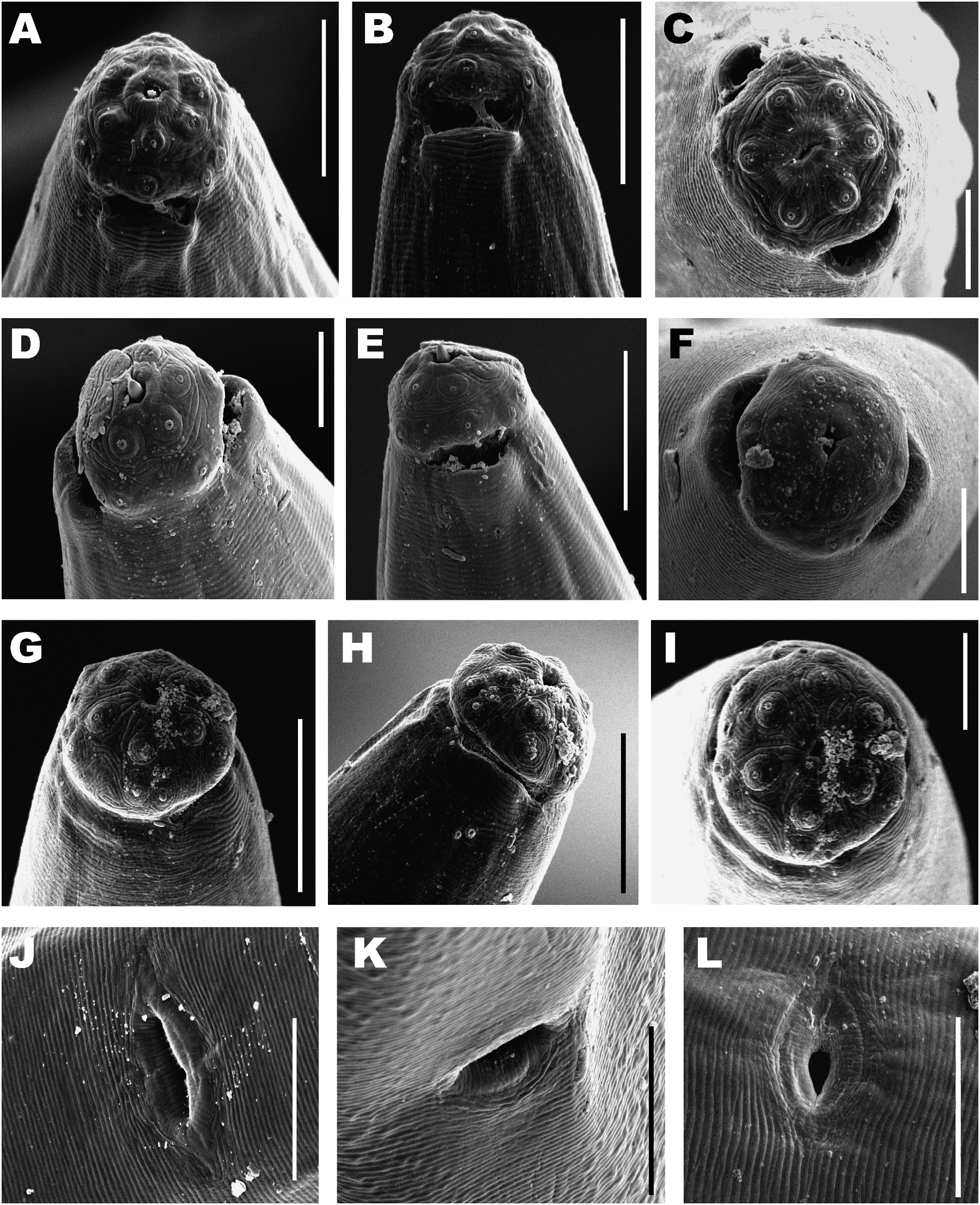
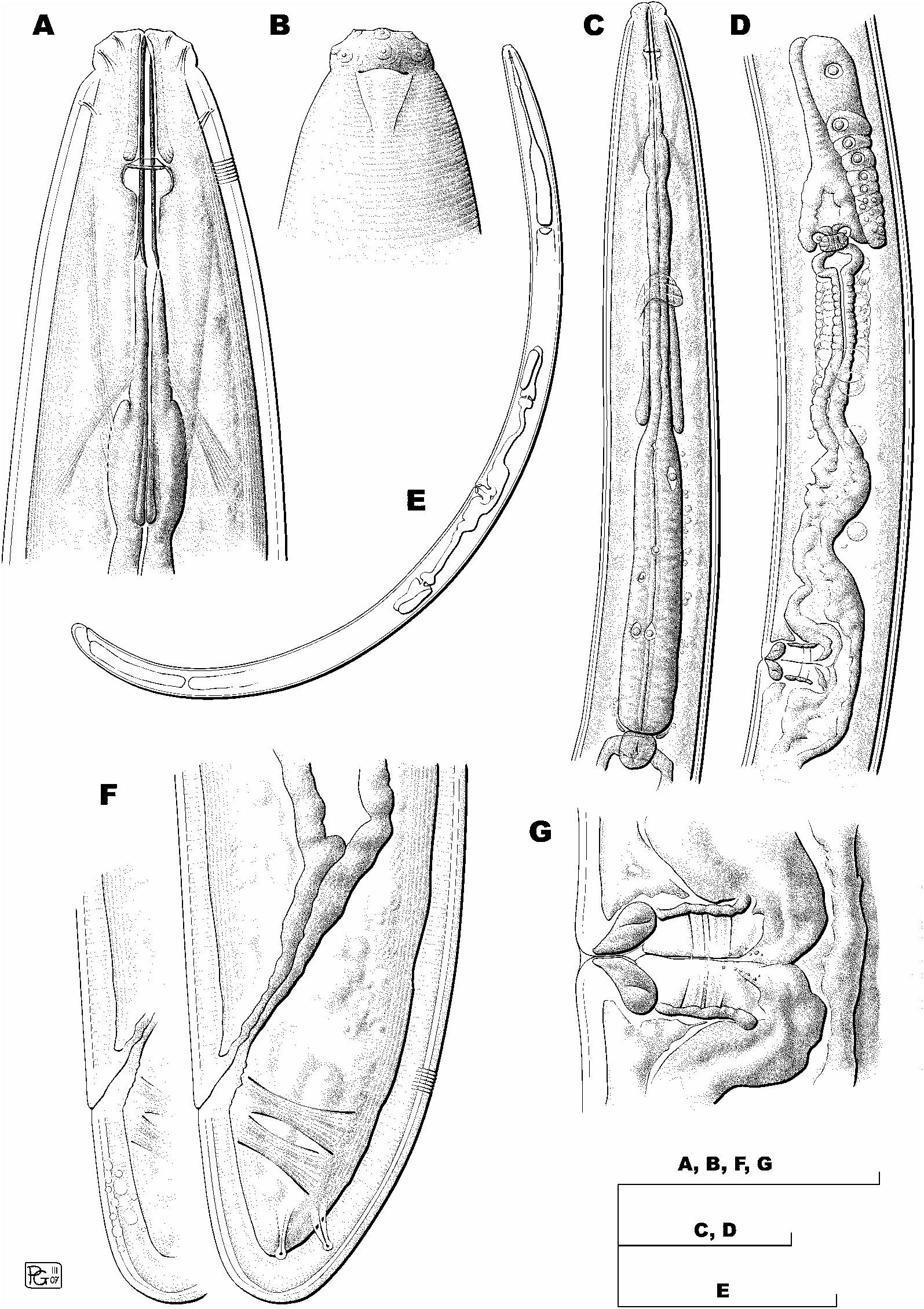
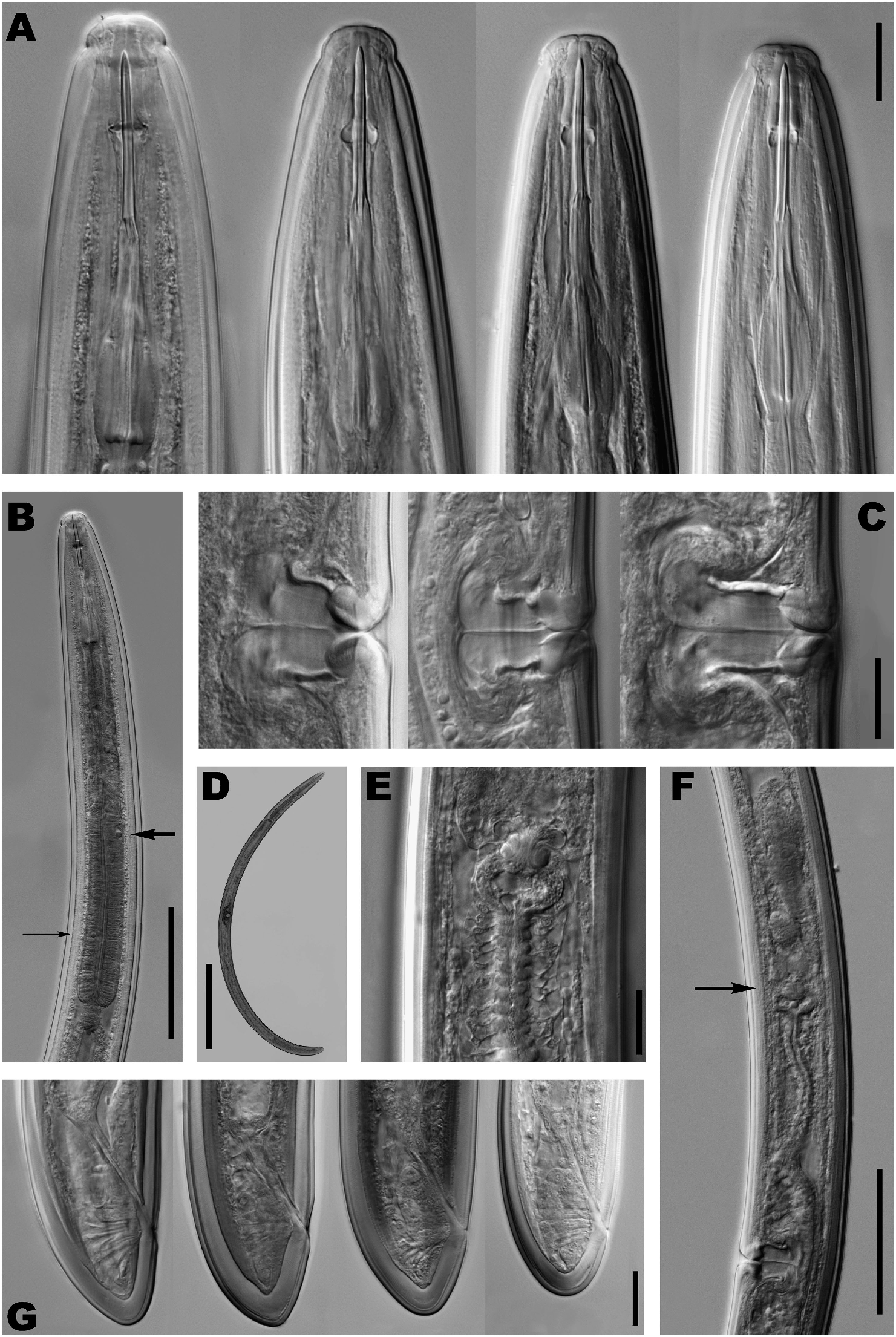
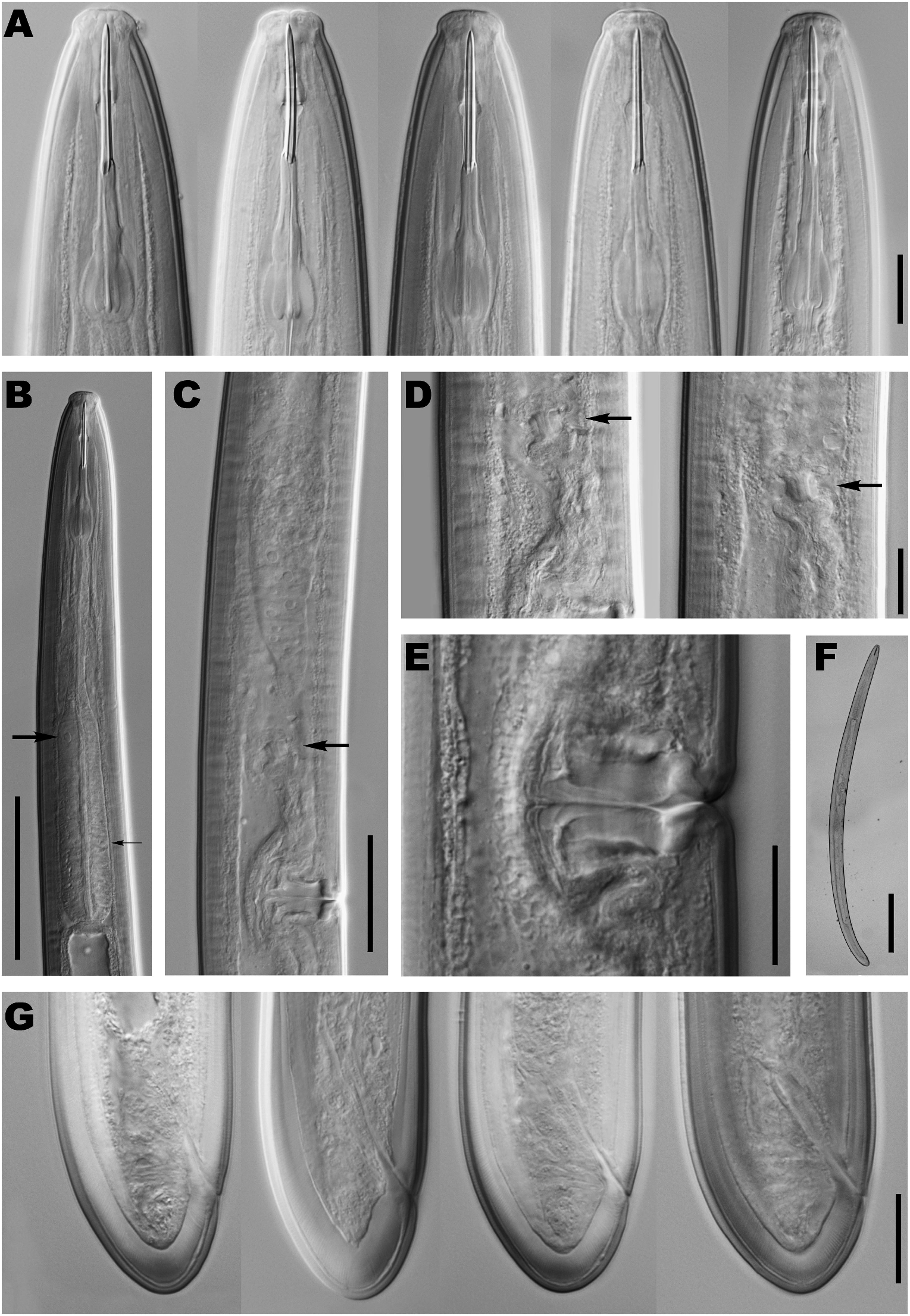
1.80-2.38 mm long. Habitus after fixation slightly curved ventrad, J- to open C-shaped. Body cylindrical, tapering towards both ends but more so towards anterior. Cuticle 3.0-6.0 µ m in anterior region, 3.5-6.0 µ m at mid-body and 6.0-10.5 µ m on tail, its outer layer bearing very fine transverse striations, and thinner than inner layer, especially at level of tail. Lateral chord relatively narrow, occupying 8-15% of corresponding body diam., lacking particular differentiation. Lateral pores obscure. Lip region rounded, offset by weak constriction, 2.3-3.4 times as broad as high and ca one-third of body diam. at neck base. Lips amalgamated; labial and cephalic papillae not interrupting cephalic contour. SEM observations showing a dorso-ventral oral aperture surrounded by a weakly differentiated elevation of perioral area, the latter containing strongly marked radial striae; both inner and outer labial, and cephalic papillae surrounded by ring-like structures, offset from lip region surface by concentric striae. Amphid fovea cup-shaped, opening at level of cephalic constriction in a slit-like aperture slightly widened at both edges, occupying 37-56% of corresponding body diam.; under SEM, two lateral pores at different levels observed on amphid fovea surface. Cheilostom cylindrical, with no particular differentiation. Odontostyle long and slender, with distinct walls and narrow lumen; 12-18 times as long as wide or 1.9-2.3 lip region diam. long; aperture small, occupying 7-14% of total length. Odontophore with thickened walls in posterior half and six distinct basal knobs, 1.0-1.4 times longer than odontostyle. Guiding ring double, located at 22-30 µ m or 1.1-1.4 lip region diam. from anterior end. Pharynx consisting of a slender, but well muscled, anterior portion expanding gradually into the basal expansion at 51-62% of total neck length, reaching full diam. at 60-69%; pharyngeal expansion occupying 35-45% of total neck length, and ca two-fifths to three-fifths of corresponding body diam. Pharyngeal gland nuclei located as follows: DN = 62-72; S 1 N 1 = 29 (n = 1); S 1 N 2 = 40 (n = 1); S 2 N 1 = 54-64; S 2 N 2 = 55-66. Base of pharyngeal expansion surrounded by weak membrane-like structure. Cardia small, rounded conoid, somewhat broader than long, 12-25 × 10-24 µ m. Greenish material found inside intestine of some specimens. Genital system didelphic-amphidelphic, with both branches equally and well developed. Ovaries relatively large, 68-286 µ m long, sometimes reaching sphincter level; oocytes first in two or more rows, then in one row. Oviduct 85-228 µ m long, or 1.0-3.1 times corresponding body diam., consisting of slender portion with prismatic cells and well developed pars dilatata with distinct lumen. Sphincter prominent, located betweenoviduct and uterus. Uterus rather long, 138- 304 µ m or 2.0-4.1 corresponding body diam.; tripartite, i.e., consisting of a wider proximal portion with distinct lumen, followed by a narrower intermediate long region with narrow lumen, refractive inner lining and surrounded by long cluster of hyaline cells, and a well developed spheroid pars dilatata distalis. Three females with one or two eggs, measuring 108-125 × 42-54 µ m. Sperm not observed in genital tract. Vagina extending inwards 40-60% of body diam.; pars proximalis longer than broad 22-36 × 15-32 µ m, with almost straight to slightly sigmoid walls, enveloped by weak circular musculature; pars refringens with (in lateral view) two drop-shaped arcuate sclerotisations with combined width of 21-29 µ m; pars distalis 1.5- 7.5 µ m long. Vulva a transverse slit frequently preceded by depression in body contour; with SEM, vulva appearing as an oval transverse aperture located in a marked depression of body surface. Prerectum 4-8 and rectum 0.7-1.5 anal body diam. long. Tail short, rounded, almost hemispheroid, to rounded conoid; terminal cuticle 7-12 µ m thick or 22-44% of total tail length. Appearance of cuticle on ventral side of tail rather variable: several saccate bodies or blister-like structures present in some individuals, but others showing no irregularities, even in same population. Two pairs subterminal caudal pores, one subdorsal, the other practically lateral.
Male
Not found.
DIAGNOSIS AND RELATIONSHIPS
Enchodelus saxifragae is distinguished by its body length of 1.80-2.38 mm, lip region distinctly offset by slight constriction and 18-22 µ m diam., odontostyle 37- 47 µ m, but almost always ca 40 µ m long or 1.9-2.3 times lip diam., odontophore flanged with distinct basal knobs and 1.0-1.4 times odontostyle length, neck 319- 443 µ m long, pharyngeal expansion 116-186 µ m long or 35-45% of total neck length, female genital system amphidelphic, uterus tripartite, pars refringens vaginae with drop-shaped sclerotisations, vulva a transverse slit (V = 44-50), tail short, rounded, hemispheroid to conoid (22-40 µ m, c = 54-92, cļ = 0.6-0.9 in females, and 30-34 µ m, c = 53-68, cļ = 0.7-0.8 in males), spicules 56-70 µ m long and presence of 13-16 regularly spaced ventromedian supplements with 1-3 posteriormost within the range of the spicules.
By having a short, rounded tail and long odontostyle (more than 35 µ m), E. saxifragae comes close to E. distinctus Ahmad & Jairajpuri, 1980, E. groenlandicus, E. macrodorus and E. microdoroides Baqri & Jairajpuri, 1974.
It can be distinguished from E. distinctus by its slightly longer odontophore (38-47 vs 36 µ m), lack of pharyngointestinal disc (vs presence of a well developed disc), uterus tripartite (vs bipartite, lacking spheroid pars distalis) and vulva pre-equatorial (vs post-equatorial); from E. groenlandicus by its lip region offset by constriction (vs shallow depression), shorter odontostyle of 37-47 µ m (but usually around 40 µ m) vs 44-53 µ m (but generally about 50 µ m), and shape of sclerotised pieces of the pars refringens vaginae (drop-shaped vs trapezoid); from E. macrodorus by its longer body (L = 1.80-2.35 vs 1.38-1.87 mm), lip region offset by constriction (vs depression), and longer and more differentiated uterus (2.0- 4.1 times longer than body diam. with long intermediate portion and well developed spheroid pars distalis vs 1.0- 2.0 times body diam. with very short intermediate region and vestigial pars distalis); finally, from E. microdoroides by its much longer body (L = 1.80-2.35 vs 1.09-1.24 mm), broader lip region (18-22 vs 13-14 µ m), relatively shorter odontostyle (1.9-2.3 times lip diam. vs 3 times lip diam.), and guiding ring located more anterior (22-30 µ m or 1.1-1.4 times lip diam. vs 37-39 µ m or 2.6-3.0 times lip diam.).
DISTRIBUTION
Of all the Enchodelus species with a rounded tail, E. saxifragae is the most frequent and widely distributed in the studied area. It has been found in 11 localities: hedgehog heath at 2450 m a.s.l., psychroxerophilous pasture at 3050 and 3350 m a.s.l. in Sierra Nevada ( Granada); meadow at 1550 m a.s.l., hedgehog heath at 1700, 1810 and 1950 m a.s.l., and pine forest with savin juniper at 1690, 1800, 1900 and 1980 m a.s.l. in Sierra Mágina (Jaén). The elevation range extends from 1550-3350 m a.s.l. The species has been found together with E. groenlandicus in one sample from Sierra Nevada.
REMARKS
This species was reported as Enchodelus cf. saxifragae from Sierra Mágina (province of Jaén) by Liébanas et al. (2002). After examination of E. saxifragae paratypes, the occurrence of this species in the Iberian Peninsula can be confirmed. However, some features need clarification. The female genital system of E. saxifragae was not accurately described in the original publication, nothing being stated in the text except for the presence of a sphincter between uterus and oviduct. Popovici’s (1995) Figure 4C View Fig depicts the posterior genital branch showing a uterus consisting of a rather simple tube. However, examination of paratypes revealed a complex structure identical to that of Iberian individuals, i.e., a tripartite uterus consisting of a wide proximal region with distinct lumen filled with sperm, a narrow intermediate tube with slightly refractive inner lining and surrounded by a cluster of hyaline cells, and a distal spheroid pars dilatata distalis, which joins the sphincter between uterus and oviduct.
Some minor morphometric differences can be highlighted. Iberian individuals can be somewhat shorter (1.80-2.34 vs 1.96-2.35 mm in females) and have slightly longer odontostyle (38-47 µ m, although only in two cases it is longer than 45 µ m, vs 37.5-40 µ m in Romanian females). The ranges of other measurements have been expanded. Finally, a remarkable difference between the Romanian and Iberian populations is the abundance of males in the former but complete absence in the latter.
No known copyright restrictions apply. See Agosti, D., Egloff, W., 2009. Taxonomic information exchange and copyright: the Plazi approach. BMC Research Notes 2009, 2:53 for further explanation.
|
Kingdom |
|
|
Phylum |
|
|
Class |
|
|
Order |
|
|
Family |