
Notogynaphallia pseudoceciliae, Lemos, Virgínia Silva & Leal-Zanchet, Ana Maria, 2008
|
publication ID |
https://doi.org/ 10.5281/zenodo.184508 |
|
DOI |
https://doi.org/10.5281/zenodo.5612006 |
|
persistent identifier |
https://treatment.plazi.org/id/3A7987BC-FFA7-BC73-FF3D-50F2FE3FCF8F |
|
treatment provided by |
Plazi |
|
scientific name |
Notogynaphallia pseudoceciliae |
| status |
sp. nov. |
Notogynaphallia pseudoceciliae View in CoL sp. nov.
Notogynaphallia sp. 6 Leal-Zanchet & Carbayo, 2000
Notogynaphallia View in CoL sp. 4 Carbayo, Leal-Zanchet & Vieira, 2001 Notogynaphallia View in CoL sp. 3 Carbayo, Leal-Zanchet & Vieira, 2002
Etymology: The specific name refers to external similarity with Notogynaphallia ceciliae Froehlich & Leal- Zanchet, 2003.
Type-material: Holotype: MZUSP PL.301: T. Fleck, leg. 25. IX. 98 – pre-pharyngeal region: transversal sections on 6 slides; pharynx: sagittal sections on 9 slides; copulatory apparatus: sagittal sections on 12 slides. Paratypes: MZU PL.00070: F. Carbayo, leg. 16. XII. 98 - pharynx: sagittal sections on 3 slides; body region between pharynx and copulatory apparatus: sagittal sections on 14 slides; copulatory apparatus: sagittal sections on 18 slides; MZU PL.00071: M. Cardoso, leg. 16. XII. 98 – anterior region in three fragments: transversal sections on 8 slides; horizontal sections on 4 slides and sagittal sections on 15 slides; pre-pharyngeal region: transversal sections on 12 slides; pharynx: sagittal sections on 10 slides; copulatory apparatus in two fragments: sagittal sections on 25 slides; MZU PL.00072: I. Fick, leg. 0 8. VII. 99 – anterior region at the level of the ovaries: sagittal sections on 15 slides; pre-pharyngeal region: transversal sections on 5 slides; pharynx: sagittal sections on 11 slides; copulatory apparatus: sagittal sections on 12 slides.
Type-locality: São Francisco de Paula, state of Rio Grande do Sul (RS), Brazil.
Distribution: São Francisco de Paula, Rio Grande do Sul, Brazil.
Diagnosis: Dorsum light-yellow with five dark longitudinal stripes, thin but distinct; wide marginal zone, free of stripes; eyes dorsal, without clear halos; typical glandular margin absent; mc:h, 18-21%; pharynx cylindrical with dorsal insertion posteriorly displaced, folded margins; esophagus absent; foremost testes posterior to ovaries, most posterior ones near root of pharynx; efferent ducts open laterally into distal portion of tubular branches of prostatic vesicle; extrabulbar prostatic vesicle, with two long tubular branches exceeding the posterior end of pharyngeal pouch; male atrium long and highly folded, with histologically differentiated proximal and distal portions; ejaculatory duct opening through a small projection into the proximal portion of male atrium; oviducts emerging dorsally from median third of ovaries, and ascending posteriorly to gonopore; common glandular oviduct dorsal to female atrium; vagina directed dorsally and forwards; female atrium oval-elongate, almost as long as the male atrium, presenting epithelium with lacunose multilayered aspect.
Description
External morphology: Body elongate with parallel margins ( Fig. 1 View FIGURES 1 – 2 ), anterior end obtuse and posterior pointed. When creeping, maximal length may reach 67 mm ( Table 1 View TABLE 1 ). Mouth distance from anterior tip varies from 64% to 77% relatively to body length, gonopore from 79% to 88% ( Table 1 View TABLE 1 ). Dorsal and ventral ground colour yellowish. Dorsum with five dark brown longitudinal stripes, one median, two paramedian, and two lateral ( Figs. 1 View FIGURES 1 – 2 , 3 View FIGURES 3 – 4 A-B). In paratype MZU PL.00071, median stripe begins, discontinuously, at 2 mm from anterior tip (ca. 3% of body length), slightly behind the beginning of paramedian stripes (1 mm from anterior tip or 2% of body length) and ahead of lateral stripes (ca. 3 mm from anterior tip or 5% of body length) ( Fig. 3 View FIGURES 3 – 4 A). Near posterior end, on each side of body, paramedian and lateral stripes converge; paramedian stripes become discontinuous at approximately 6 mm from posterior tip, whereas lateral ones almost reach this tip ( Fig. 3 View FIGURES 3 – 4 B). Median stripe ends at approximately 2 mm from posterior tip (96% of body length). At median third of body of paratype MZU PL.00071, lateral stripes located at 0.7 mm from the body margins (one-fifth of body width), so that a wide marginal zone remains free of stripes. Median stripe is the thinnest (approx. 0.07 mm or 2% of body width), followed by paramedian (approx. 0.11 mm or 3% of body width) and lateral (approx. 0.14 mm or 4% of body width) stripes.
Eyes, initially marginal and uniserial, surround the anterior tip. In paratype MZU PL.00071, eyes become pluriserial after the second millimeter (approx. 4% of body length). Between approximately 4 mm and 9 mm (ca. 7% and 14% of body length) from the tip they become dorsal, forming two to three lateral irregular series ( Fig. 3 View FIGURES 3 – 4 A). Sparser backwards, become limited to body margins, occurring up to near the posterior tip ( Fig. 3 View FIGURES 3 – 4 B). No clear halos.
Internal morphology
Epidermis and musculature at pre-pharyngeal region: Creeping sole, 64% and 58% of body width, respectively, in holotype and paratype MZU PL.00072 ( Table 1 View TABLE 1 ).
Three types of secretory cells discharge through dorsal epidermis and body margins: (1) abundant rhabditogen cells with xanthophil secretion (2) cells with coarse erythrophil secretion; (3) few cells with cyanophil amorphous secretion. The erythrophil cells and a fourth type of cell, with fine granulous xanthophil secretion, are more abundant towards body margins, but do not form a typical glandular border ( Fig. 5 View FIGURES 5 – 6 ). Creeping sole receives necks of few rhabditogen cells, cells with fine granulous erythrophil secretion, and numerous cells with amorphous cyanophil secretion.
Cutaneous musculature with the usual three layers, longitudinal layer with thick bundles, being approximately four times higher than the other two. At the sagittal plane, ventral musculature higher than dorsal ( Table 2 View TABLE 2 ), the latter becoming higher paramedianly. Towards body margins cutaneous musculature progressively lower ( Fig. 5 View FIGURES 5 – 6 ). Mc:h 18% to 21% ( Table 2 View TABLE 2 ).
Mesenchymatic musculature composed of four layers: dorsal subcutaneous with oblique fibers variously oriented (ca. 3 fibers thick); supra-intestinal transversal (approx. 4-6 fibers thick); sub-intestinal transversal (4-5 fibers thick); and subneural transversal (ca. 4-5 fibers thick) ( Fig. 5 View FIGURES 5 – 6 ). In addition, scattered ventral subcutaneous oblique fibers as well as dorsoventral ones are present. If existent, longitudinal fibers are indiscernible, few and very scattered.
Holotype Paratype Paratype Paratype MZUSP PL.301** MZU PL.00070** MZU PL.00071 MZU PL.00072 Pharynx ( Fig. 7 View FIGURES 7 – 8 ): Pharynx of cylindrical type with dorsal insertion posteriorly displaced, but still in anterior third of pharyngeal pouch, and with folded margins. Mouth in median third of pharyngeal pouch. No esophagus. Pharyngeal glands with cell bodies located in mesenchyme, mainly anterior and posteriorly as well as lateral to pharyngeal pouch. Three secretory cell types: cells with coarse granulous xanthophil secretion, cells with fine granulous erythrophil secretion, and cells with cyanophil amorphous secretion. Outer musculature of pharynx (ca. 18µm thick) constituted of thin longitudinal subepithelial layer, followed by a thicker circular one, mixed internally with few longitudinal fibers. Towards pharyngeal tip, circular layer becomes as thin as longitudinal one. Inner pharyngeal musculature (ca. 15µm thick) composed of thick circular subepithelial layer, mixed mainly externally with some longitudinal fibers. Inner musculature gradually thins down outwards, and, though mainly dorsally, also inwards.
Specimens Holotype Paratype Holotype Paratype Paratype Paratype
MZUSP MZU MZUSP MZU MZU MZU PL.00079 PL.301 PL.00072 PL.302 PL.00073 PL.00075
Reproductive apparatus: Foremost testes posterior to ovaries; most posterior ones, near root of pharynx, slightly posteriorly to or at same transversal level as ventral insertion of pharynx ( Table 1 View TABLE 1 ). Testes in two irregular rows beneath the dorsal transversal mesenchymatic muscles dorsally and intersticially to the intestinal branches on each side of the body ( Fig. 5 View FIGURES 5 – 6 ). Efferent ducts run dorsally to oviducts, sometimes laterally displaced. Behind pharynx, the right and left efferent ducts form false seminal vesicles, and enter prostatic vesicle laterally in distal region of forked portions, respectively, an approximate distance of 1.3 mm and 1.5 mm from penial bulb ( Fig. 9 View FIGURE 9 A). Prostatic vesicle ( Table 1 View TABLE 1 , Figs. 10, 13, 14 View FIGURES 10 – 15 ), tubular and forked. The two long tubular extrabulbar branches extend anteriorly near to or a little beyond posterior end of pharyngeal pouch. Ca. 1 mm from bulbar muscular coat, paired branches of prostatic vesicle unite, forming a single canal which enters the bulbar muscular coat and forms a sinuous and ample ejaculatory duct. The latter opens, through a small projection, into proximal portion of male atrium ( Figs. 9 View FIGURE 9 B, 11). Male atrium oval-elongate ( Table 1 View TABLE 1 , Figs. 9 View FIGURE 9 B, 11, 14) with numerous folds, irregular in shape, which are higher ectally.
Lining epithelium of efferent ducts cuboidal ciliated; thin muscularis (ca. 3Μm thick) mainly constituted of circular fibers. Prostatic vesicle lined with columnar to pseudostratified ciliated epithelium, receiving abundant erythrophil granulous secretion from secretory cells with bodies lying in mesenchyme, mainly around vesicle. Muscularis of vesicle (10-15µm thick) constituted of interwoven longitudinal and circular fibers. Ejaculatory duct lined with columnar ciliated epithelium, without openings from secretory cells, being coated with weakly developed muscularis (ca. 3-5µm thick) constituted of mixed longitudinal and circular fibers.
Epithelial lining of male atrium, columnar non-ciliated. Four types of secretory cells, with cell bodies external to common muscle coat, empty through the epithelium: cells with fine granulous slightly erythrophil secretion; cells with granulous heavily stained erythrophil secretion; cells with coarse granulous xanthophil secretion; and cells with cyanophil amorphous secretion. Openings from xanthophil and heavily stained erythrophil cells are more numerous into the ectal portion of male atrium; those from slightly erythrophil cells into the ental portion of male atrium. Muscularis weakly developed (3-7µm) throughout male atrium, but thicker (approx. 40µm) in the most ectal folds; mainly composed of circular layer with some mixed longitudinal fibers.
Ovaries oval-elongate in shape, measuring 0.3 mm anterior-posteriorly and 0.05 mm dorso-ventrally in paratype MZU PL.00072. Oviducts emerging dorsally from median third of ovaries, lead backwards immediately dorsal to nerve plate. Behind gonopore, oviducts ascend posterior and medially inclined, to unite dorsally to the female atrium, thus forming the common glandular oviduct ( Figs. 9 View FIGURE 9 B, 15). The latter, a long canal, dorsal to female atrium, that leads backward to communicate with vagina. Vagina as a terminal dorsally and forwardly directed diverticulum of female atrium. Female atrium, oval-elongate in shape and with a narrowed lumen, almost as long as male one ( Table 1 View TABLE 1 , Figs. 9 View FIGURE 9 B, 12, 15).
Paired oviducts lined with cuboidal ciliated epithelium which becomes, near copulatory apparatus, columnar ciliated, as is that of common glandular oviduct. Paired oviducts as well as common glandular oviduct coated with thin circular muscle layer with some interposed longitudinal fibers. Abundant shell glands with xanthophil secretion empty into distal third of ascending portion of paired oviducts, besides into common glandular oviduct ( Figs. 9 View FIGURE 9 B, 15).
Lining of female atrium very thick with multilayered aspect ( Figs. 9 View FIGURE 9 B, 12, 15). It presents many lacunae containing cyanophil secretion and others with either xanthophil or erythrophil secretion. Near to gonopore, the thick epithelial lining is substituted by a columnar epithelium. Vagina lined ectally with a thinner epithelium than that of female atrium, showing multilayered aspect, and entally with a columnar epithelium ( Fig. 12 View FIGURES 10 – 15 ). Erythrophil cells with granulous secretion and cells with cyanophil amorphous secretion discharge into vagina and female atrium. Cell bodies of cyanophil glands are external to the muscle common coat and those of erythrophil cells are internal to the muscle common coat. Muscularis (15-17µm) mainly constituted of circular fibers mixed with a number of longitudinal fibers.
Straight gonopore canal. Dorsal fold, approximately at same transversal level as gonopore canal, leads to venter, inclining posteriorly, and fuses with ventral wall of female atrium ( Figs. 9 View FIGURE 9 B, 15).
Common muscle coat with circular, longitudinal and oblique fibers, thicker around ental portion as well as along dorsal wall of male than around female atrium. Between atrial muscularis and common muscle coat, a stroma with muscle fibers variously oriented.
Remarks: Vitellaria are inconspicuous in the holotype and paratype MZU PL.00072, although well developed in paratypes MZU PL.00070 and MZU PL.00071. Paratype MZU PL.00072, probably the younger worm, and paratype MZU PL.00071, highly contracted, reveal a circular fold in the male atrium, similar to a temporary penis papilla, this being traversed by the ejaculatory duct ( Fig. 13 View FIGURES 10 – 15 ). In both the holotype and paratype MZU PL.00071, there is abundant holocrine secretion, produced by cells of the multilayered-like lining, in the lumen of female atrium. Secretory cells of the copulatory apparatus have been described based on paratype MZU PL.00070.
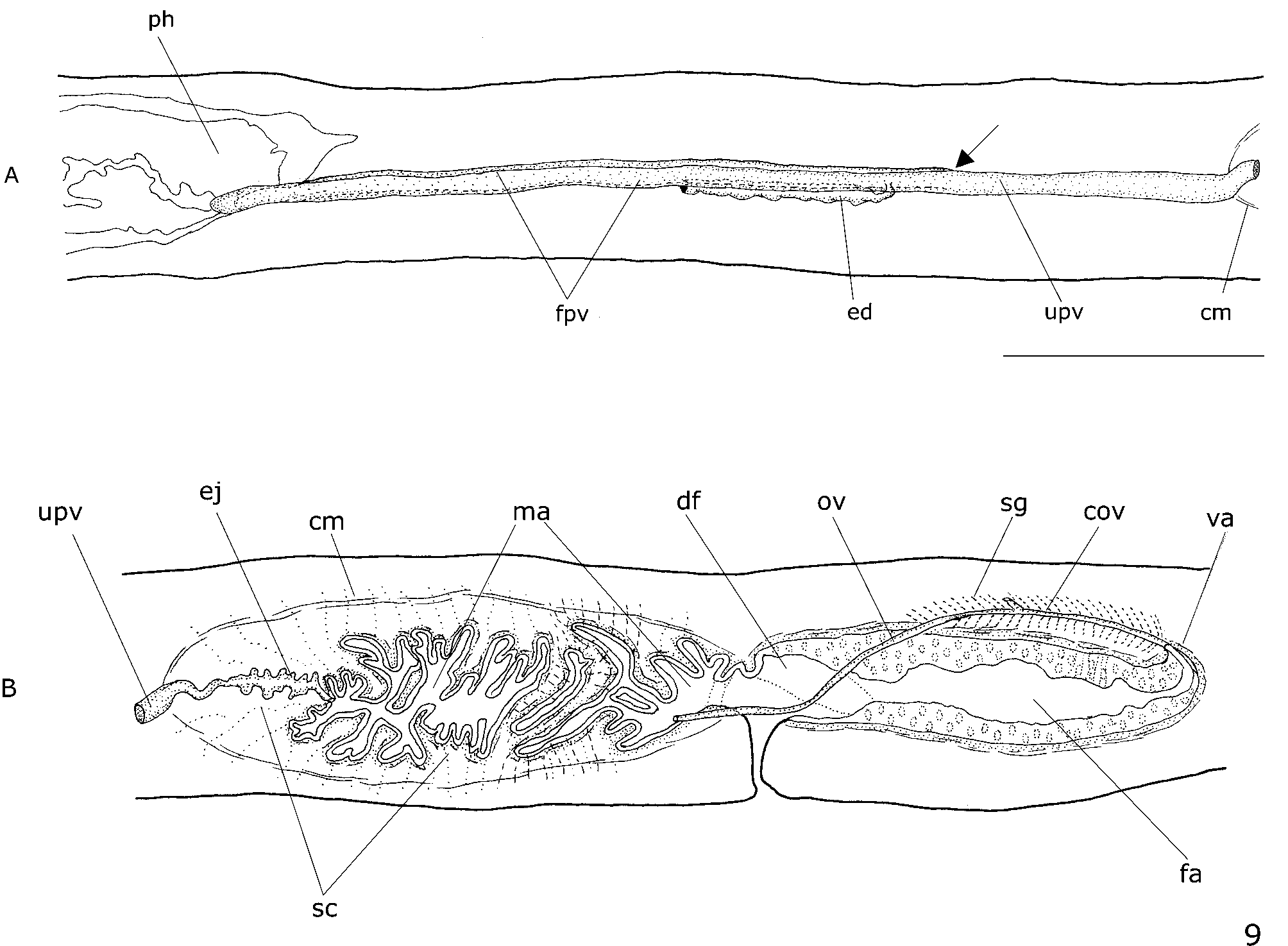

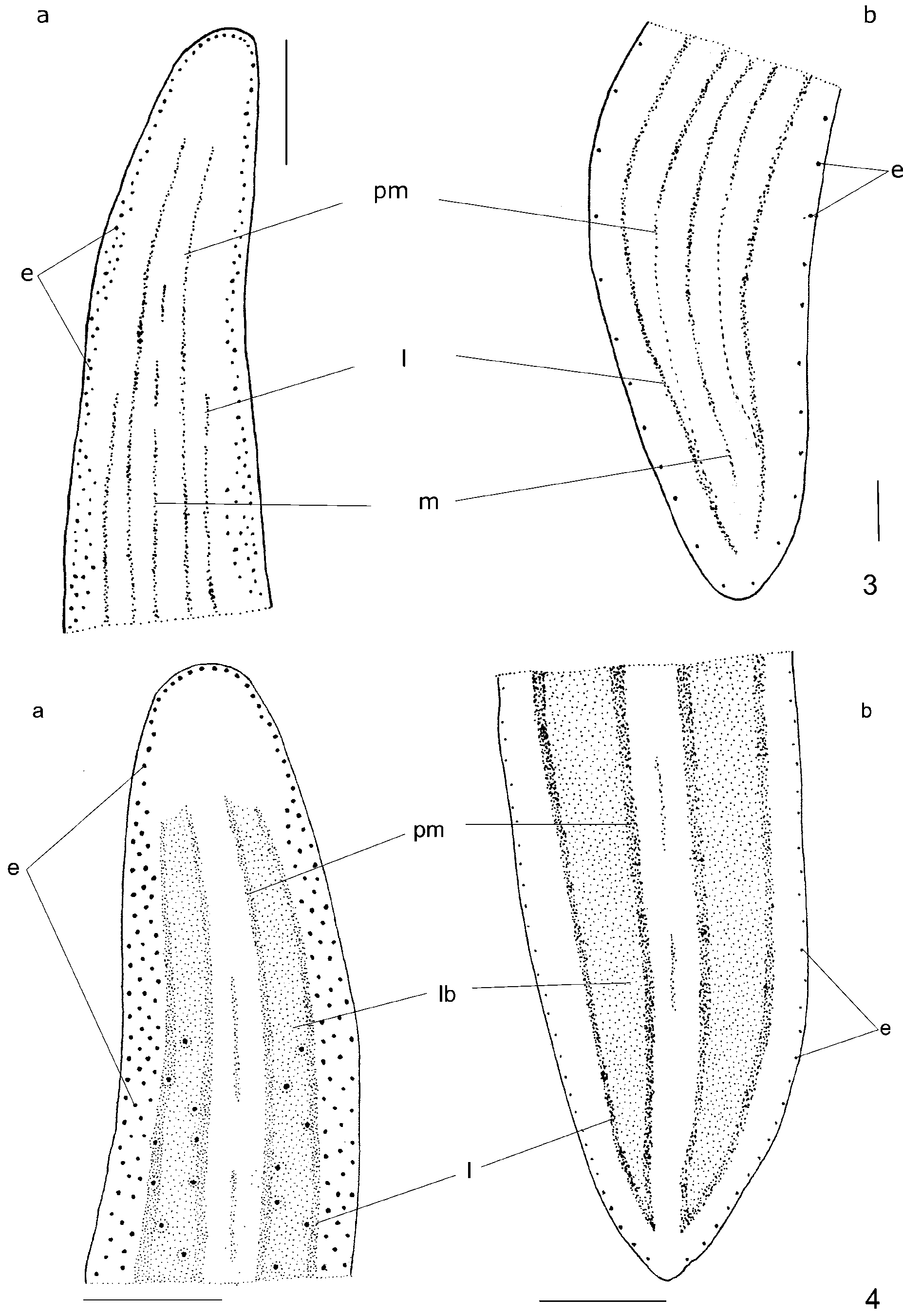
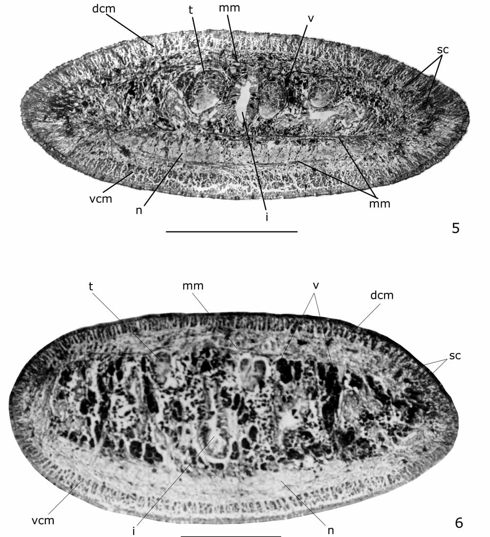
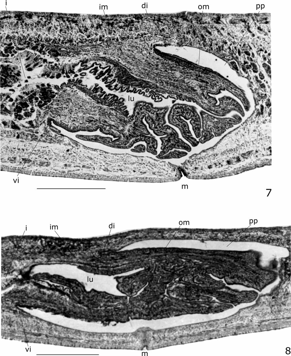
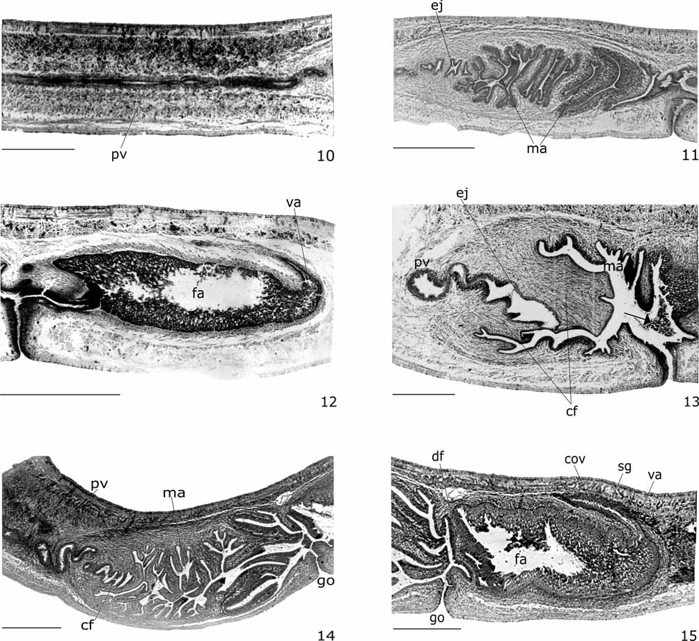
TABLE 1. Measurements, in mm, of type-specimens of N. pseudoceciliae sp. nov. –: not measured; *: After fixation; ** Specimens with damaged anterior tip (lost or regenerating); DG: distance of gonopore from anterior end; DM: distance of mouth from anterior end; DMG: distance between mouth and gonopore; DPVP: distance between prostatic vesicle and pharyngeal pouch. The numbers given in parentheses represent the position relative to body length.
| Maximum length in extension 40 Maximum width in extension 2 | 35 2.5 | 67 3.5 | 45 2.5 |
|---|---|---|---|
| Length at rest 22 | 22 | 45 | 27 |
| Width at rest 3.5 Length* 38 | 3 21 | 4.5 57 | 3.5 47 |
| Width* 2.5 | 2.5 | 3.5 | 3 |
| DM* – DG* – | – – | 44 (77) 50 (88) | 30 (64) 37 (79) |
| DMG* 8.5 | – | 6 | 7 |
| DPVP* 0 Creeping sole % 64 | 0 – | 0 – | – 58 |
| Ovaries 11 (23) | – | – | – |
| Anteriormost testes 13 (28) Posteriormost testes 25 (66) | – – | – – | – 30 (64) |
| Prostatic vesicle 3.9 | 5.4 | 2.6 | – |
| Male atrium 1.6 Female atrium 1.5 | 1.9 1.5 | 1.1 0.8 | – – |
TABLE 2. Cutaneous musculature, in µm, in the median region of a transversal section of the pre-pharyngeal region, and ratio of the height of cutaneous musculature to the height of the body (mc: h index) of specimens of N. pseudoceciliae and N. arturi.
| Dorsal musculature 70 | 66 | 71 | 112 | 62 | 62 |
|---|---|---|---|---|---|
| Ventral musculature 86 | 75 | 63 | 108 | 74 | 56 |
| Mc:h 21% | 18% | 12% | 20% | 17% | 14% |
No known copyright restrictions apply. See Agosti, D., Egloff, W., 2009. Taxonomic information exchange and copyright: the Plazi approach. BMC Research Notes 2009, 2:53 for further explanation.