
Acrogonia filiformis, Silva, Roberta Dos Santos Da, Cavichioli, Rodney R., Takiya, Daniela M. & Mejdalani, Gabriel, 2017
|
publication ID |
https://doi.org/10.11646/zootaxa.4244.4.4 |
|
publication LSID |
lsid:zoobank.org:pub:D3D6480B-9EC4-4BA2-804B-735E1ED163D5 |
|
DOI |
https://doi.org/10.5281/zenodo.6016098 |
|
persistent identifier |
https://treatment.plazi.org/id/B24E7412-3142-4044-808C-653434AE544E |
|
taxon LSID |
lsid:zoobank.org:act:B24E7412-3142-4044-808C-653434AE544E |
|
treatment provided by |
Plazi |
|
scientific name |
Acrogonia filiformis |
| status |
sp. nov. |
Acrogonia filiformis View in CoL sp. nov.
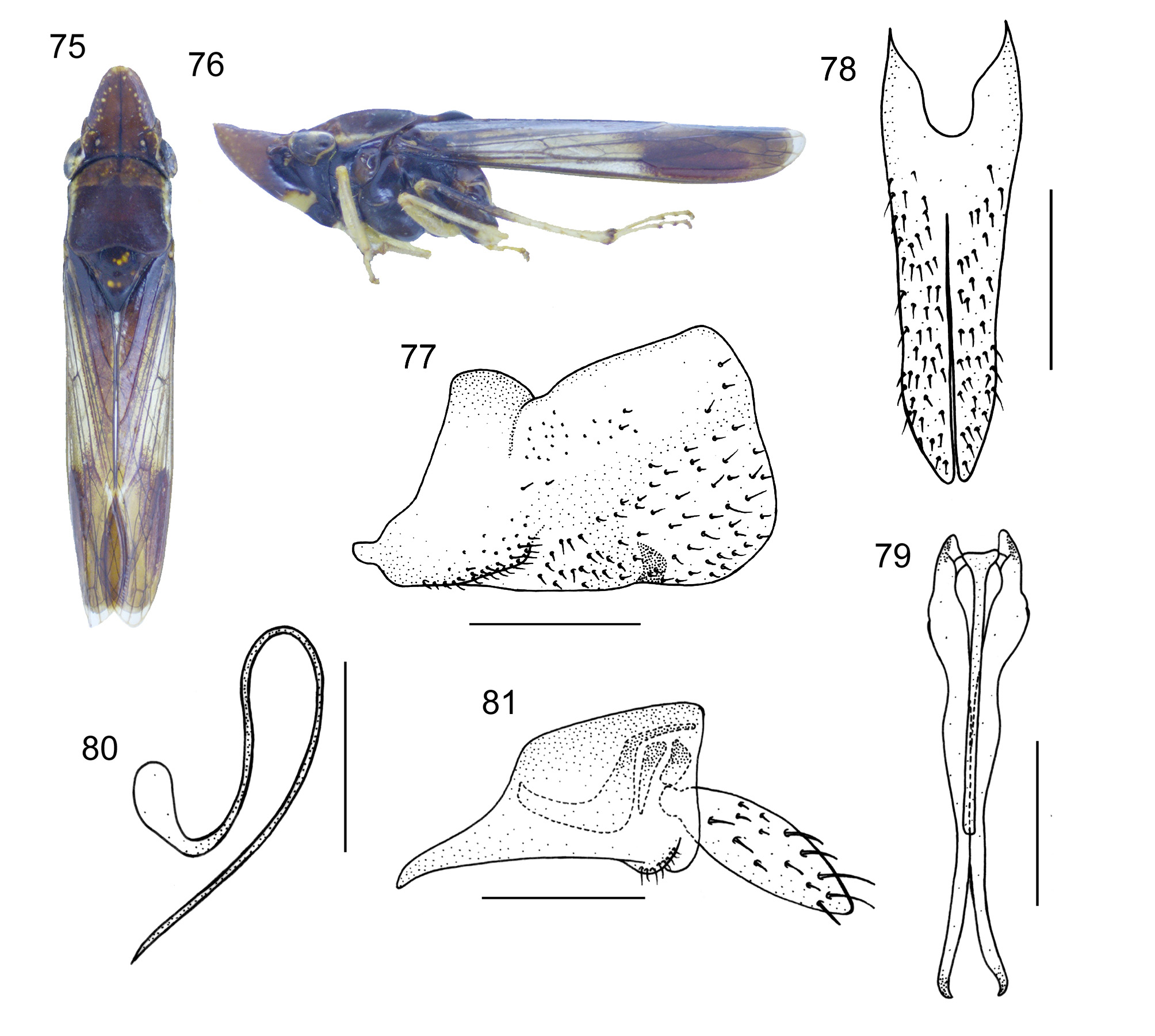
( Figs. 75–92 View FIGURES 75 – 81 View FIGURES 82 – 92 )
Total length. Male holotype 10.5 mm; male paratype 10.0 mm; female paratype 11.4 mm.
Holotype description. Head and thorax. Structural features of head and thorax ( Figs. 75–76 View FIGURES 75 – 81 ) much as described above for A. amazonensis sp. nov., except for median length of crown approximately equal to interocular width and 7/10 transocular width; forewings ( Figs. 75–76 View FIGURES 75 – 81 ) mostly translucent.
Color. Crown ( Fig. 75 View FIGURES 75 – 81 ) brown with yellow dots and marks bordering anterior margin from apical portion to anterior eye angles; other small yellow markings also present; frontogenal, temporal and coronal sutures dark brown to black. Posterior 2/3 of pronotum and mesonotum ( Fig. 75 View FIGURES 75 – 81 ) dark brown; anterior 1/3 of pronotum brown with irregular yellow marks; lateral portions of pronotum yellowish; mesonotum with pair of lateral brownishyellow marks, apex pale yellow. Forewings ( Figs. 75–76 View FIGURES 75 – 81 ) mostly translucent with brown areas and dots on clavus and apical third of corium. Face ( Fig. 76 View FIGURES 75 – 81 ) dark brown; frons superiorly brown with yellow marks, yellow inferiorly. Lateral and ventral portions of thorax ( Fig. 76 View FIGURES 75 – 81 ) mostly dark brown. Legs ( Fig. 76 View FIGURES 75 – 81 ) brownish-yellow to brown.
Male genitalia. Pygofer ( Fig. 77 View FIGURES 75 – 81 ), in lateral view, well produced posteriorly; without basiventral projection; with sclerotized projection directed inward on distal third of ventral margin; disc with dispersed setae. Subgenital plates ( Fig. 78 View FIGURES 75 – 81 ), in ventral view, subtriangular, basal portion slightly expanded laterally, then narrowing slightly toward distal fourth, latter more strongly narrowed; median portion of lateral margin slightly concave; in lateral view, plates elongate, extending almost as far posteriorly as pygofer apex; plate surface with scattered setae. Connective ( Fig. 79 View FIGURES 75 – 81 ), in dorsal view, T-shaped; arms short; stalk narrow, very elongate, with median keel. Style ( Fig. 79 View FIGURES 75 – 81 ), in dorsal view, extending much farther posteriorly than apex of connective; outer margin sinuous; without preapical lobe; apex acute. Aedeagus ( Fig. 80 View FIGURES 75 – 81 ) symmetrical, filiform, elongate; shaft curved dorsally at base and curved ventrally at basal third; without processes. Anal tube ( Fig. 81 View FIGURES 75 – 81 ) without processes.
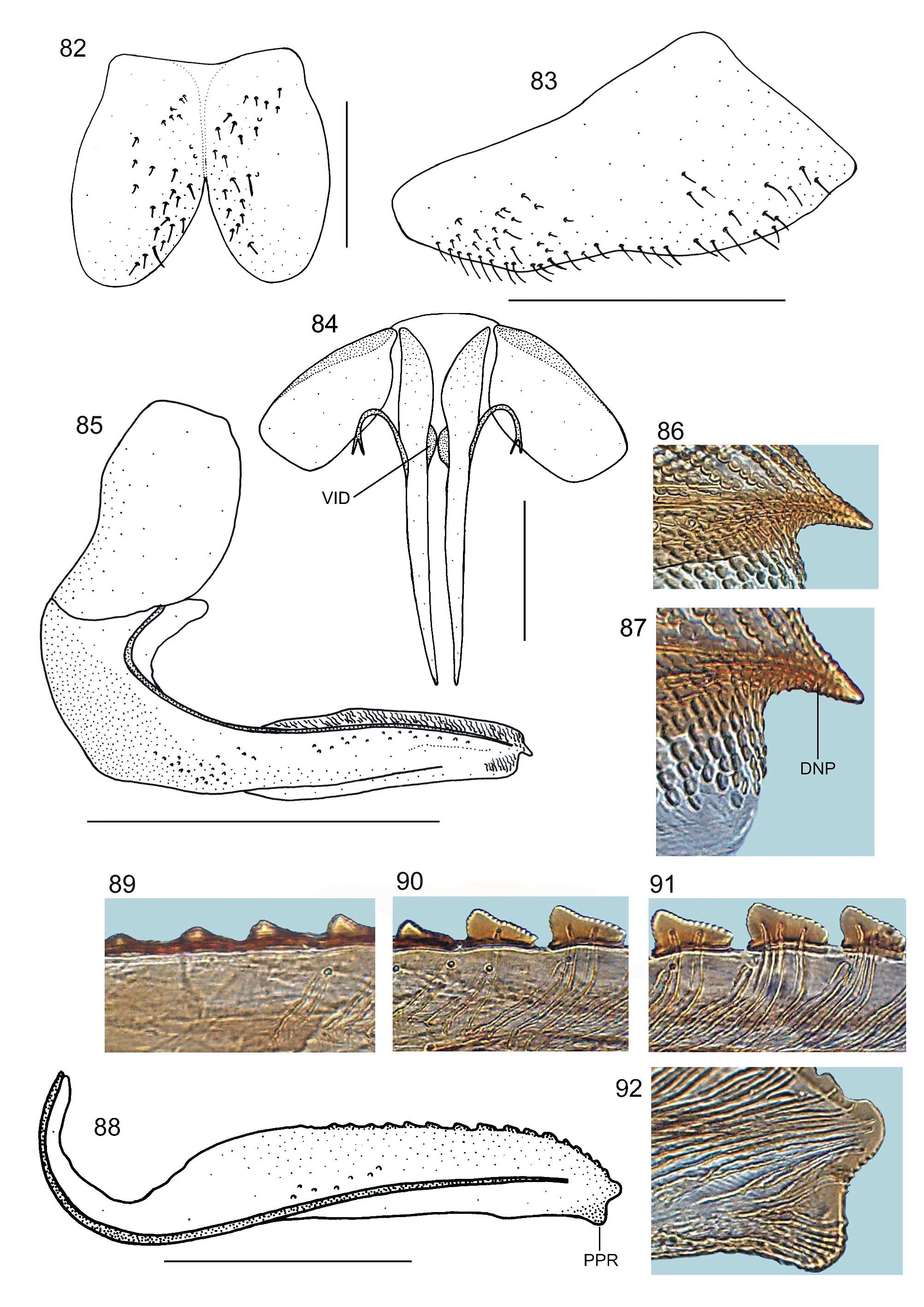
Female. Genitalia. Sternite VII ( Fig. 82 View FIGURES 82 – 92 ), in ventral view, bilobed; with deep V-shaped posterior emargination. Pygofer ( Fig. 83 View FIGURES 82 – 92 ), in lateral view, well produced posteriorly; apex obtuse; setae distributed mostly on posterior portion and extending anteriorly along ventral margin. First valvifers ( Figs. 84–85 View FIGURES 82 – 92 ), in lateral view, subrectangular. First valvulae ( Fig. 84 View FIGURES 82 – 92 ), in ventral view, slightly expanded basally; in lateral view ( Fig. 85 View FIGURES 82 – 92 ), with slight projection on basiventral half; apex acute, dentiform ( Figs. 85–87 View FIGURES 82 – 92 —DNP); ventral interlocking device ( Fig. 84 View FIGURES 82 – 92 —VID) located on basal half of blade, small, lobate; dorsal sculptured area extending from basal portion of blade to apex, formed mostly by scale-like processes arranged in oblique lines; ventral sculptured area ( Fig. 87 View FIGURES 82 – 92 ) restricted to apical portion of blade, formed mostly by scale-like processes. Second valvulae ( Fig. 88 View FIGURES 82 – 92 ), in lateral view, distinctly expanded beyond basal curvature; dorsal margin convex; ventral margin approximately straight; preapical prominence strong ( Figs. 88 View FIGURES 82 – 92 —PPR, 92); apex obtuse ( Figs. 88, 92 View FIGURES 82 – 92 ); about 16 teeth ( Figs. 88–91 View FIGURES 82 – 92 ) distributed on dorsal expanded portion of blade; most teeth ( Figs. 90–91 View FIGURES 82 – 92 ) triangular, armed with denticles, with ascending portion (= anterior edge) short, descending portion (= posterior edge) long; basalmost two or three teeth small and inconspicuous; blade with ducts ( Figs. 90–92 View FIGURES 82 – 92 ) extending to apical portion and to teeth or terminating below latter; few basal teeth ( Fig. 89 View FIGURES 82 – 92 ) without ducts. Gonoplacs, in lateral view, with basal half narrow and apical half distinctly expanded; apex obtuse.
Intraspecific variation. The color of the anterior pronotal margin varies from brown to brownish-yellow. The mesonotum may be entirely brown or brown with yellow dots. Some variation may be due to fading of coloration in preserved specimens.
Etymology. The name of the new species, filiformis , refers to the thread-like shape of the aedeagus ( Fig. 80 View FIGURES 75 – 81 ).
Type material. Holotype: male, “Sinop-MT [state of Mato Grosso]\ BR X-1976 \ M. [ Moacir] Alvarenga ” ( DZUP) . Paratypes: one male, “ Brasil Pará \ Serra Norte \ EST. NIANGANÊS\ Col. NOTURNA \ 26-X.1984 ” ( MPEG) ; one female, “ Sinop - MT\ X.1976 \ M. Alvarenga col” ( DZUP) .
No known copyright restrictions apply. See Agosti, D., Egloff, W., 2009. Taxonomic information exchange and copyright: the Plazi approach. BMC Research Notes 2009, 2:53 for further explanation.
|
Kingdom |
|
|
Phylum |
|
|
Class |
|
|
Order |
|
|
Family |
|
|
SubFamily |
Cicadellinae |
|
Tribe |
Proconiini |
|
Genus |