
Austrocarabodes (Austrocarabodes) planisetus Mahunka and Mahunka-Papp, 2011
|
publication ID |
https://doi.org/ 10.24349/acarologia/20204373 |
|
publication LSID |
lsid:zoobank.org:pub:26B249D6-B764-4936-BD6B-2BD704DA5711 |
|
DOI |
https://doi.org/10.5281/zenodo.4536395 |
|
persistent identifier |
https://treatment.plazi.org/id/3B6C87B0-FFFE-FFE9-66ED-C0AE3D3CFDD3 |
|
treatment provided by |
Felipe |
|
scientific name |
Austrocarabodes (Austrocarabodes) planisetus Mahunka and Mahunka-Papp, 2011 |
| status |
|
Austrocarabodes (Austrocarabodes) planisetus Mahunka and Mahunka-Papp, 2011 View in CoL
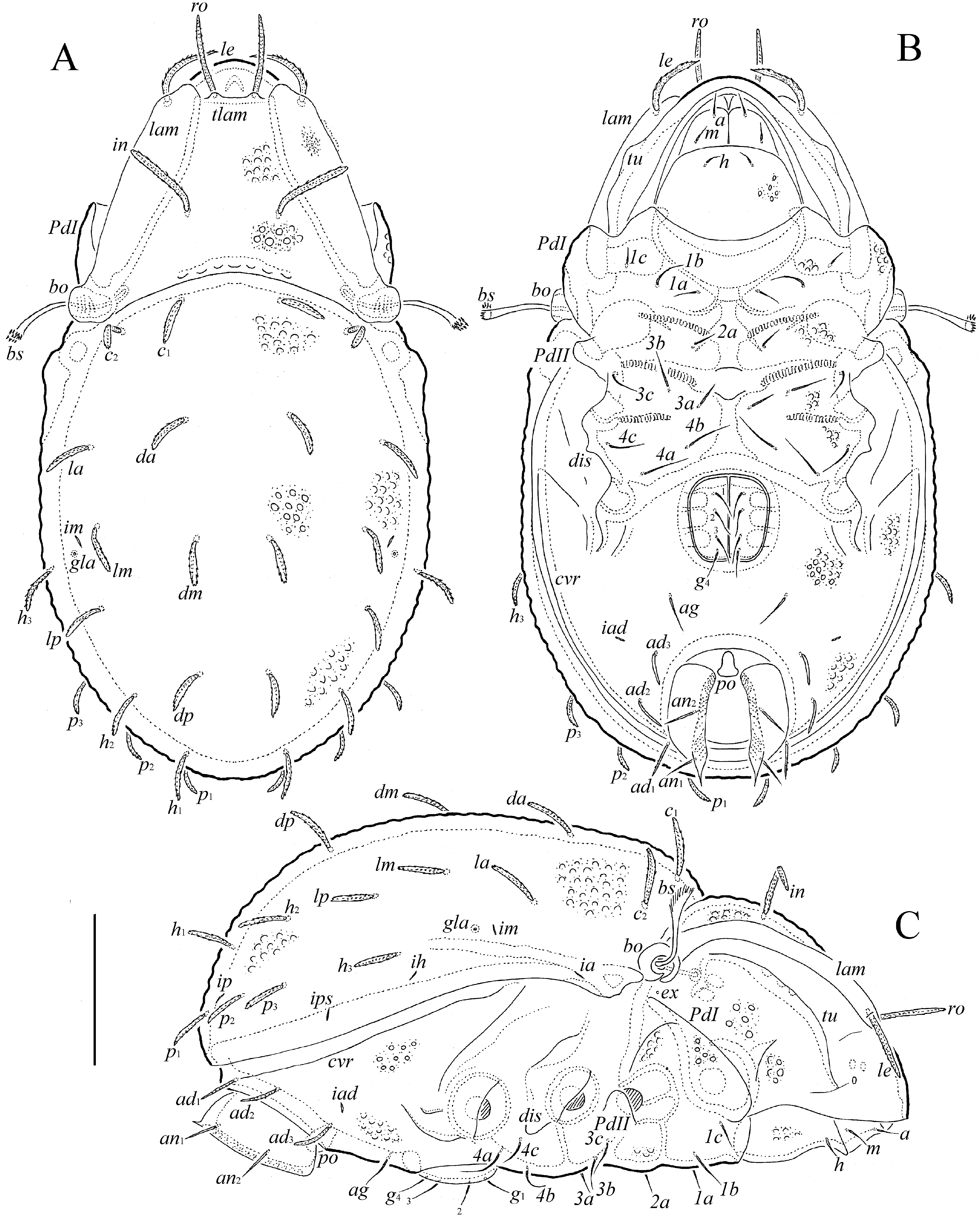
( Figures 8–10 View Figure 8 View Figure 9 View Figure 10 )
Supplementary description — Measurements – Species of medium size. Body length: 464–514 (seven specimens, two males and five females); notogaster width: 249–298 (seven specimens). Body ratio (length/width): 1.7–1.8.
Integument ( Figs 8 View Figure 8 a-c, 9a-d, 10a, 10b, 10d, 10f) – Body color light brown to brown. Body covered by thick layer of gel-like cerotegument and cerotegumental microridges and some microgranules. Prodorsum (between lamellae), notogaster, epimeres (poorly visible under light microscope) and anogenital region densely tuberculate (diameter of tubercles up to 8); subcapitular mentum, lateral sides of prodorsum (between lamellae and tutoria and on tutoria), anterolateral parts of ventral plate (pedotecta I and regions close to subcapitular mentum), and antiaxial sides of all leg femora and of trochanters III, IV foveolate (diameter of foveolae up to
8). Epimeral region with some muscle sigillae.
Prodorsum ( Figs 8 View Figure 8 a-c, 9a-d, 10a-d) – Rostrum broadly rounded. Lamellae long (slightly shorter than prodorsum), rounded distally. Translamella slightly developed. Tutoria long, ridgelike. With elongate depression (separated by transverse ridge) between lamellae and tutoria, and one depression ventrally to tutoria. Rostral setae (49–53) weakly phylliform, erect, barbed, inserted on tubercles located on translamella. Lamellar setae (49–53) narrowly phylliform, with strong spines and small barbs, directed anteromedial. Interlamellar setae (49–53) narrowly phylliform, barbed, in directed lateral. Bothridial setae (49–53) with elongate, unilaterally dilated and heavily spinose heads (sometimes setae appear clavate in dorsal view). Bothridia slightly interrupted ventrally, with inner tooth. Exobothridial setae represented by alveoli.
Notogaster ( Figs 8a, 8c View Figure 8 , 9a, 9c View Figure 9 , 10 View Figure 10 a-c) – Anterior notogastral margin slightly convex medially. Humeral processes poorly developed. Fourteen pairs of notogastral setae (p 1 – p 3, h 1 – h 3, 24–32; others 36–45) narrowly phylliform, barbed. Lyrifissures and opisthonotal gland openings distinct.
Gnathosoma ( Figs 8b, 8c View Figure 8 , 9b, 9c View Figure 9 ) – Generally, similar to Austrocarabodes madagascarensis n. sp. Subcapitulum longer than wide (106–114 × 82–86). Subcapitular setae (12–16) setiform, roughened. Postpalpal setae (10) bacilliform, slightly barbed mediodistally. Palps (57–61) with setation 0–2–1–3–9(+ω). Solenidion of palptarsi long, bacilliform. Chelicerae (131–143) with two setiform, barbed setae (cha, 45; chb, 20). Trägårdh’s organ of chelicerae elongate triangular.
Lateral podosomal and epimeral regions ( Figs 8b, 8 View Figure 8 с, 9b, 9c, 10d) – Pedotecta II trapezoid in ventral view. Discidia triangular, rounded distally. With typical epimeral setation 3–1–3–3. Epimeral setae 1a, 1c, 2a and 3a (16–20) shorter than 3b, 3c and 4a (24–28) and 1b, 4b and 4b (32–36), all setiform, slightly barbed.
Anogenital region ( Figs 8b, 8c View Figure 8 , 9b, 9c View Figure 9 , 10f View Figure 10 ) – With one pair of short, longitudinal ridges lateral to genital aperture and posterior to epimere IV. Four pairs of genital (24–28) and one pair of aggenital (28) setae setiform, slightly barbed. Three pairs of adanal setae (24–36) weakly phylliform, barbed. Two pairs of anal setae (20–24) setiform, erect, slightly barbed. Adanal lyrifissures visible, removed from anal aperture and located anterolateral to adanal setae ad 3. Circumventral ridge developed.
Legs ( Figs 9b, 9c View Figure 9 , 10a View Figure 10 ) – Generally, similar to Austrocarabodes madagascarensis n. sp. ( Table 1).
Material examined — Seven specimens (two males and five females): North Madagascar, Montagne d’Ambre National Park, circuit Ampijoroana, evergreen rain forest, 12°31’28’‘S, 49°09’52”E, 950 m a.s.l., sifting of leaf litter sample under big unidentified tree, Winkler apparatus extraction, 13.I.2014 ( R. Ravebolun and L. Rabotenoson).
Material deposition — All specimens (preserved in ethanol with a drop of glycerol) are deposited in the collection of the Tyumen State University Museum of Zoology, Tyumen, Russia.
Remarks – Our specimens of A. planisetus from Madagascar are similar in general appearance to those according to the original description ( Mahunka and Mahunka-Papp, 2011). However, some differences were observed:
1. Mahunka and Mahunka-Papp (2011) noted, that “…epimeral surface smooth…” (p. 130). We studied our specimens under a light microscope and also found no tubercles (only some unclear tubercles were poorly visible in lateral view). However, the SEM of mites showed that epimeres are in fact densely tuberculate ( Fig 9b View Figure 9 ).
2. Mahunka and Mahunka-Papp (2011) did not discuss surface of the anogenital region. In our specimens this region has tubercles.
Thus, based on the supplementary description and original description ( Mahunka and Mahunka-Papp, 2011), the diagnosis for A. planisetus is as follows:
Body size: 421-540 × 249–346. Prodorsum, notogaster, epimeres and anogenital region densely tuberculate. Translamella present. Rostral setae weakly phylliform, erect, lamellar setae narrowly phylliform, with spines and barbs, interlamellar setae narrowly phylliform, barbed; all setae comparatively long, similar in length, barbed, ro inserted on translamella. Bothridial setae unilaterally dilated and spinose. Notogastral setae comparatively short (posterior setae shortest), narrowly phylliform, barbed. Epimeral setae 1a, 1c, 2a and 3a shortest, 4a and 4b longest, all setiform, slightly barbed. Genital and aggenital setae setiform, slightly barbed. Adanal setae weakly phylliform, barbed. Anal setae setiform, erect, slightly barbed. Lateral phylliform seta l” present only on genua I, II.


| R |
Departamento de Geologia, Universidad de Chile |
No known copyright restrictions apply. See Agosti, D., Egloff, W., 2009. Taxonomic information exchange and copyright: the Plazi approach. BMC Research Notes 2009, 2:53 for further explanation.