
Watersipora subovoidea sensu Harmer, 1957
|
publication ID |
https://doi.org/10.1080/00222930601062771 |
|
persistent identifier |
https://treatment.plazi.org/id/3C0487C6-FFAD-9418-BA0A-C406FC103CDD |
|
treatment provided by |
Felipe |
|
scientific name |
Watersipora subovoidea sensu Harmer, 1957 |
| status |
|
Watersipora subovoidea sensu Harmer, 1957 View in CoL
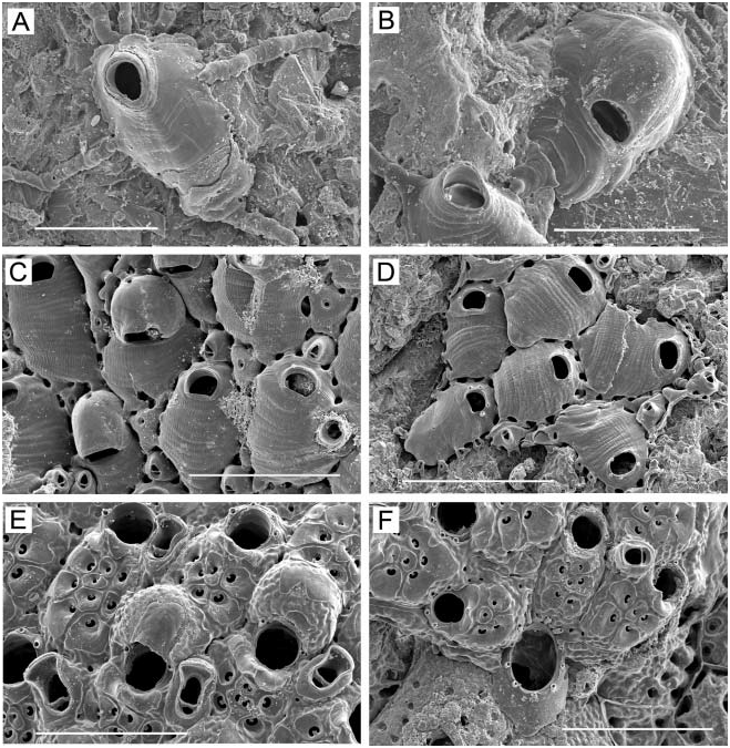
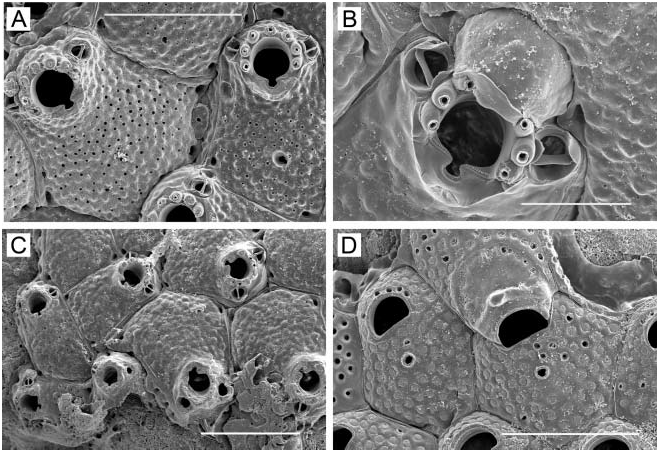
( Figure 9E, F View Figure 9 )
Dakaria subovoidea: Harmer 1957, p 1022 , Plate 69, Figure 12 View Figure 12 (in part).
? Watersipora edmondsoni Soule and Soule 1968, p 215 View in CoL , Plate 2, Figure 6 View Figure 6 ; Soule and Soule 1975, p 307, Plate 1, Figure 7 View Figure 7 , Plate 4, Figure 6 View Figure 6 .
Watersipora edmondsoni: Winston and Heimberg 1986, p 17 View in CoL , Figures 38–40.
Watersipora subovoidea sensu Harmer View in CoL : Tilbrook et al. 2001, p 75, Figure 18F.
Measurements ZL, 1.03–1.60 (1.179¡0.175). ZW, 0.55–0.75 (0.620¡0.060). OrL, 0.15–0.18
(0.163¡0.009). OrW, 0.20–0.25 (0.223¡0.012).
Description
Unilaminar, encrusting; colour iridescent black when alive, iridescent black or dark grey tinged with reddish brown when dried; operculum shiny, opaque black. Zooids ( Figure 9E View Figure 9 ) large, delineated by a raised line of calcification; variable in shape, irregularly hexagonal to long-oval or spindle-shaped, sometimes quite elongate. Frontal wall ( Figure 9E View Figure 9 ) slightly convex, covered uniformly with small pores, the distance between pores roughly two to three pore diameters; imperforate in a rugose lunate zone ( Figure 9F View Figure 9 ) of varying width proximal to orifice; each frontal pore surrounded by a slightly raised margin that is broader on distal side, giving frontal wall a scaled appearance. There may be one or two larger areolar pores on each side lateral to margin, though some zooids lack them; proximolateral angles of zooids often sunken, appearing as enlarged areolae. Orifice ( Figure 9F View Figure 9 ) terminal, slightly raised from level of frontal wall, the orificial margin a raised, rounded rim; orifice clearly broader than long, the large anter semicircular, with a deep, distinctly U-shaped proximal sinus occupying about the middle third of the proximal margin; condyles flattened on broad condylar shelves flanking sinus. Spines and ovicells lacking.
Remarks
Characters common to colonies from Hawaii (this study), Bali ( Winston and Heimberg 1986), and Vanuatu ( Tilbrook et al. 2001) include an orifice clearly broader than long, with a U-shaped sinus sharply defined by prominent, shelf-like condyles; a raised orificial collar; an imperforate area proximal to the orifice that is often ornamented with smooth tuberculation, especially on the collar; asymmetrically marginated pores that impart a slightly scaled appearance to the frontal wall; and an iridescent, black colour in living and dried colonies. Our specimens thus appear to be conspecific with material from these Indo- West Pacific sites. However, the identity of this species is unclear. It differs from the reddish to light tan colonies and the pattern of the operculum originally described for W. edmondsoni from the Hawaiian Islands ( Soule and Soule 1968), and with which Winston and Heimberg (1986) identified specimens from Bali. Harmer (1957) noted a black or grey cuticle and operculum, numerous pores, and an orifice similar in shape to that in our material for what he considered to be Watersiopora subovoidea ; however, the extensive synonymies he listed for this nominal species indicate a virtually circumtropical distribution with much variation. The genus Watersipora badly needs worldwide taxonomic revision (see Ryland 1974c for a discussion of some of the problems), ideally based on both morphological and molecular data.
Distribution
Bali, Hawaiian Islands, Vanuatu.
Superfamily SCHIZOPORELLOIDEA Jullien, 1883 Family STOMACHETOSELLIDAE Canu and Bassler, 1917 Genus Junerossia new genus
Type species
Junerossia copiosa n. sp., this study.
Etymology
The genus is named in honour of Professor June R. P. Ross for her significant contributions to the knowledge of fossil and Recent Bryozoa.
Diagnosis
Frontal wall cryptocystidean, sparsely perforated over the entirety except in a zone proximal to orifice that represents an umbonuloid component of the shield, the margin of this zone delineated externally by a semicircle of pores and internally by a ring scar. The frontal pores are tiny pseudopores identical in form to the uniporous septula in the vertical walls; frontal pores marginated by thickening of frontal wall. Orifice subterminal, the proximal and distal margins broadly arcuate; condyles not evident from external view, but present internally near junction of ring scar and distal semicircle of orifice. Peristome a thick, moderately tall rim around orifice, often with several nodules on top. Ovicell opens into peristome, not closed by zooidal operculum; imperforate; covered with interior-walled calcification like frontal wall. Frontal budding occurs. Basal wall heavily calcified. Zooids interconnect by uniporous septula. Ancestrula similar in form to later zooids but imperforate except for two or three frontal pores on each side proximolateral to orifice; gives rise to five daughter zooids.
Remarks
The familial placement of this genus is difficult. Gordon (2000) discussed a number of species across several ‘‘lepraliomorph’’ ( Gordon 1989a) families, including families of Schizoporelloidea , that have a frontal shield with extensively or moderately developed umbonuloid components; thus the composition of the frontal shield in Junerossia does not in itself seem diagnostic. Three families include combinations of some of the characters seen in Junerossia , but placement is not entirely satisfactory in any of them, and depends upon which characters are given weight at the familial level. One family is the Cyclicoporidae , represented by the genus Cyclicopora Hincks, 1884 , which is quite similar to Junerossia in the following characters: a perforate frontal shield with a crescentic imperforate area proximal to the orifice suggestive of an umbonuloid component; a subcircular orifice lacking condyles; spines and avicularia lacking; and a globose ovicell covered with finely tubercular interior-walled calcification. However, the ovicell is perforate in Cyclicopora , and Canu and Bassler (1920, Figure 125) indicated a spinocystally or kenozooidally derived frontal wall quite different internally from that of Junerossia , and an orifice lacking even internal condyles.
Members of the monogeneric family Pacificincolidae , recently erected by Liu and Liu (1999), are similar to Junerossia in the following characters: an often unilaminar, encrusting growth form; evenly perforated cryptocystidean frontal wall; hyperstomial, imperforate ovicells covered with tuberculate calcification; avicularia usually absent; and no oral spines. However, in contrast to Junerossia , pacificincolids have an orifice usually referred to as ‘‘bell-shaped’’, with distinct condyles evident at most stages of zooidal ontogeny; a small suboral heterozooid of unknown function ( Nielsen 1981); large, multiporous interconnecting septula; ovicells closed by the zooidal operculum; and an ancestrula that characteristically buds a triplet of daughter zooids.
Another family is the Stomachetosellidae . Species of Stomachetosella lack oral spines and avicularia, and nodular or tubercular sculpturing on the rim of the peristome is evident in some species (e.g. S. tuberculata Androsova, 1958 ). Also within Stomachetosella are species with a transversely elliptical orifice lacking at least overt condyles (e.g. S. abyssicola Osburn, 1952 ). Furthermore, there are species with an imperforate ovicell covered with coarsely granulate interior-walled calcification (e.g. S. normani Hayward, 1994 ); the single pore (e.g. S. sinuosa Busk, 1860 ) or elongate foramen (e.g. S. tuberculata Androsova, 1958 ) in the ovicell of some species seems to reflect incomplete closure of a fundamentally imperforate ovicell. In addition, species of Stomachetosella can have either pore chambers or septula ( Kluge 1975). However, unlike Junerossia , species of Stomachetosella also typically have a cormidial secondary orifice, and ovicells are partly or entirely immersed.
At present, the sum of characters of Junerossia seems to favour placement in the Stomachetosellidae . However, with its globose hyperstomial ovicell, raised peristome, frontal shield with a significant umbonuloid component marginated by frontal pores, and uniporous septula, it is distinct from other stomachetosellid genera and, indeed, from other cheilostome genera. It will be interesting to determine whether the frontal walls of other stomachetosellids have mixed lepralioid–umbonuloid frontal shields.


No known copyright restrictions apply. See Agosti, D., Egloff, W., 2009. Taxonomic information exchange and copyright: the Plazi approach. BMC Research Notes 2009, 2:53 for further explanation.
|
Kingdom |
|
|
Phylum |
|
|
Class |
|
|
Order |
|
|
Family |
|
|
Genus |
Watersipora subovoidea sensu Harmer, 1957
| Dick, Matthew H., Tilbrook, Kevin J. & Mawatari, Shunsuke F. 2006 |
Watersipora subovoidea sensu
| Tilbrook KJ & Hayward PJ & Gordon DP 2001: 75 |
Watersipora edmondsoni : Winston and Heimberg 1986 , p 17
| Winston JE & Heimberg BF 1986: 17 |
Watersipora edmondsoni
| Soule DF & Soule JD 1975: 307 |
| Soule DF & Soule JD 1968: 215 |
Dakaria subovoidea :
| Harmer SF 1957: 1022 |