
Paranura reticulata, Smolis, Adrian & Deharveng, Louis, 2015
|
publication ID |
https://doi.org/ 10.11646/zootaxa.4033.2.2 |
|
publication LSID |
lsid:zoobank.org:pub:44006C5D-66C6-4C47-BB80-E29C2778F417 |
|
DOI |
https://doi.org/10.5281/zenodo.6121963 |
|
persistent identifier |
https://treatment.plazi.org/id/3C0C87B7-FFB3-FFAD-DBEC-FF766CBBFEC6 |
|
treatment provided by |
Plazi |
|
scientific name |
Paranura reticulata |
| status |
sp. nov. |
Paranura reticulata sp. nov.
Figs 1–14 View FIGURES 1 – 5 View FIGURES 6 – 14 , Tables 1–2 View TABLE 1 View TABLE 2
Etymology. The name of the species derives from the Latin word net (“reticulum”), underlining the presence of reticulations on its body.
Diagnosis. Body bluish grey. 3+3 eyes on head. Some tubercles present on dorsal side of body, underlined by reticulations. Head with chaetae O, A and E. Head with three ocular chaetae. Thorax I with 2 chaetae De. Thorax II–III with 3 chaetae Di. Thorax II–III with 3 and 4 ordinary chaetae De respectively. Abdomen V with 3+3 chaetae Di. Abdomen V distinctly longer than VI. Abdomen without clavate chaetae. Furca rudimentary with microchaetae. Male ventral organ absent. Tibiotarsi with chaetae M.
Description. Habitus as in Fig. 1 View FIGURES 1 – 5 , abdomen V very long, twice as long as the last segment of body. Buccal cone elongate. Body length (without antennae) 0.8–1.70 mm (holotype: 1.55 mm). Colour of body when alive and in alcohol bluish grey. Tubercles developed on central area of head, on abd. V-VI, and in De position on abd I-IV where they are very small, their arrangement as in Figs 1 View FIGURES 1 – 5 , 9, 10 View FIGURES 6 – 14 . Ordinary dorsal chaetae ( Figs 1 View FIGURES 1 – 5 , 9, 10, 13 View FIGURES 6 – 14 ) differentiated into short, thin, acuminate microchaetae, medium size, smooth, acuminate mesochaetae and long, smooth (without visible denticles), relatively thick, acuminate macrochaetae Ml and Mc. No plurichaetosis on body.
Head. Antennae slightly shorter than head. Antennal segment II with 12 chaetae. S-chaetae of ant. IV relatively long and thin, S1 distinctly longer than others ( Fig. 6 View FIGURES 6 – 14 ). Apical bulb distinct and trilobed ( Figs 7, 8 View FIGURES 6 – 14 ). Chaetotaxy of antennae as in Fig. 6 View FIGURES 6 – 14 and Tab. 1 View TABLE 1 . Buccal cone relatively long and rounded at apex ( Figs 3, 4 View FIGURES 1 – 5 , 11 View FIGURES 6 – 14 ). Maxilla needlelike, mandible tridentate. Chaetotaxy of labium (distally rounded) as in Fig. 4 View FIGURES 1 – 5 , labial papillae x absent. Labrum rounded apically, its chaetotaxy 4/2,2 ( Fig. 11 View FIGURES 6 – 14 ). Group Vi with 6+6 chaetae ( Fig. 4 View FIGURES 1 – 5 ). Groups Vea, Vem and Vep with 4, 3 and 4 chaetae respectively. Dorsal chaetotaxy of head as in Tab. 1 View TABLE 1 . and Figs 1 View FIGURES 1 – 5 , 9 View FIGURES 6 – 14 . Chaetotaxy of central area complete, with 3 chaetae Oc and chaetae A, B, C, D, E, F, G, O ( Fig. 9 View FIGURES 6 – 14 ). Line of chaetae Di2–De2 crosses line Di1– De1 on head (cross-type, Deharveng 1983). 3+3 large eyes, their diameter about four times as large as the diameter of chaeta Ocm socket ( Fig. 9 View FIGURES 6 – 14 ), pigmented in black. Tubercle Af divided into two large ones along midline and a small one with chaeta O ( Fig. 9 View FIGURES 6 – 14 ).
Thorax, abdomen, legs. Dorsal chaetotaxy as in Figs 1 View FIGURES 1 – 5 , 10 View FIGURES 6 – 14 and in Tab. 2 View TABLE 2 . Ventral chaetotaxy as in Tab. 2 View TABLE 2 and Figs 2, 5 View FIGURES 1 – 5 . S-chaetae very long, distinctly longer than nearby macrochaetae ( Figs 1 View FIGURES 1 – 5 , 10, 12 View FIGURES 6 – 14 ). S-chaetae formula of body: 022/11111, s-microchaeta on Dl of th. II present. Tubercles well developed on abd. V and VI, on abd. I–IV only small tubercle De present. Tubercles Di of abd. V and VI fused. Furcal remnant with 8 microchaetae and 4–5 mesochaetae ( Fig. 5 View FIGURES 1 – 5 ). Male without ventral modified chaetae (“male ventral organ”). Claw without internal tooth. Chaetotaxy of legs as in Tab. 2 View TABLE 2 . Chaeta M present on tibiotarsus. Chaetae B4 and B5 relatively long ( Fig. 14 View FIGURES 6 – 14 ).
Types. Holotype: male on slide, United States of America: Oregon, Oregon Dunes, Siuslaw National Forest, 10 km North of Florence town, Baker Beach, litter from dune-forest with sitka spruce Picea sitchensis , 7.VI. 2009, leg. A. Smolis. Holotype deposited in DIBEC. Paratypes: female (MNHN) and male (DIBEC) on slides, same data as holotype.
Other material. Male, female and 3 juveniles on slides ( DIBEC), USA: Oregon, Blue River Ranger District of Willamette National Forest, neighborhood of H. J. Andrews Experimental Forest, 6.5 km East of Blue River town, c. 520–550 m above sea level, “Cougar 1” site, old-growth forest of Tsuga heterophylla Zone , litter, 27.IX.–3.X. 2006, leg. A. Smolis.
Remarks. Distinct and characteristic shape of the end of body (trapezoidal) and well developed tuberculation place the new species very close to Paranura s-uenoi Yosii, 1955, described from the Japan island Nakanosima ( Yosii 1955). Paranura reticulata sp. nov. differs clearly from its congener in having three ocular chaetae on head (in s-uenoi two chaetae), chaeta F on head equal to chaeta A (in s-uenoi chaeta F is distinctly longer than A), four ordinary chaetae De on th. III (in s-uenoi three chaetae) and three chaetae Di in abd. V (in s-uenoi two chaetae). A more substantial comparison between these species is not currently possible, since the original description of Japan species lacks many characters used in modern taxonomy of Neanurinae . Because of the presence of well developed tubercles on the body Palacios-Vargas and Simón Benito (2007a) suggested to place P. s-uenoi within the genus Nahuanura Palacios-Vargas & Najt, 1986. In contrast to Nahuanura species, P. s - u e no i has however clearly elongated abd. V, and a quite different reticulation pattern (abd. VI reticulate versus non-reticulate, and reticulations of abd. V only lateral, versus only central). Morphological evidence therefore does not support the placement of P. s-uenoi in the genus Nahuanura.
a) Cephalic chaetotaxy––dorsal side.
b) Chaetotaxy of antennae.
Terga Legs
Di De Dl L Scx2 Cx Tr Fe TT th. I 1 2 1 – 0 3 6 13 19 th. II 3 3+s 3+s+ms 3 2 7 6 12 19 th. III 3 4+s 3+ s 3 2 8 6 11 18
Sterna
abd. I 2 3+ s 2 3 VT: 4
abd. II 2 3+ s 2 3 Ve: 5; Vel– present
abd. III 2 3+ s 2 3 –4 Ve: 5–6; Fu: 5 me, 8 mi
abd. IV 2 2+ s 3 9 –10 Vel: 4; Vec: 2; Vei: 2 Vl: 4
abd. V (3+3) 6–7+s Ag: 3; chaetae L‘ and Vl present abd. VI (7+7) Ve: 14; An: 2 mi
Biology. The species was found in litter of two completely different types of coniferous forests. One of them represents unique dune-forest with complete dominance of sitka spruce ( Fig. 15 View FIGURE 15 ), the other one is typical woods for the lower altitude of Cascade Range and was composed of three main tree taxa: Douglas fir Pseudotsuga menziessi , western red–cedar Thuja plicata and western hemlock Tsuga heterophylla . Bisexual form.
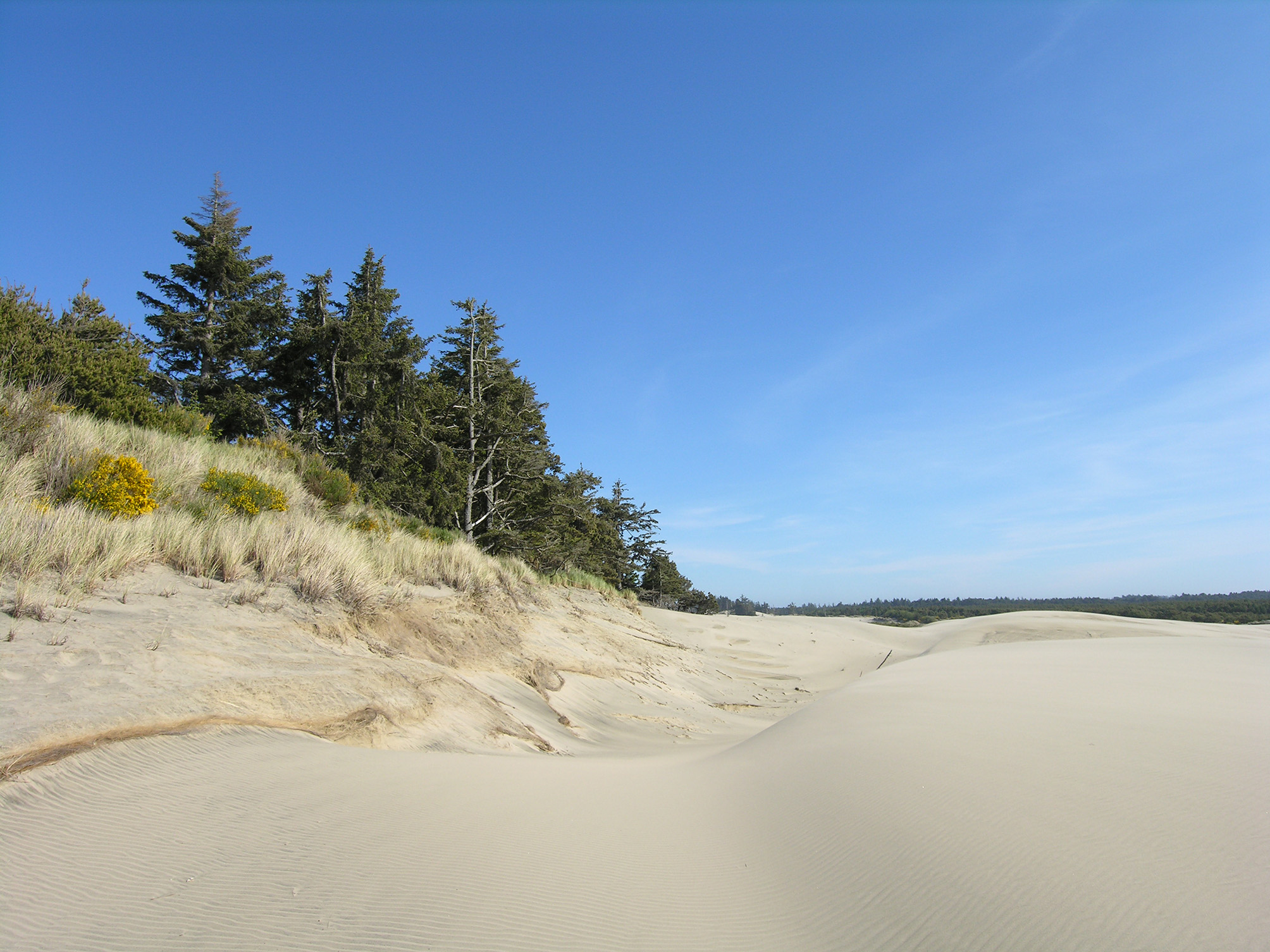
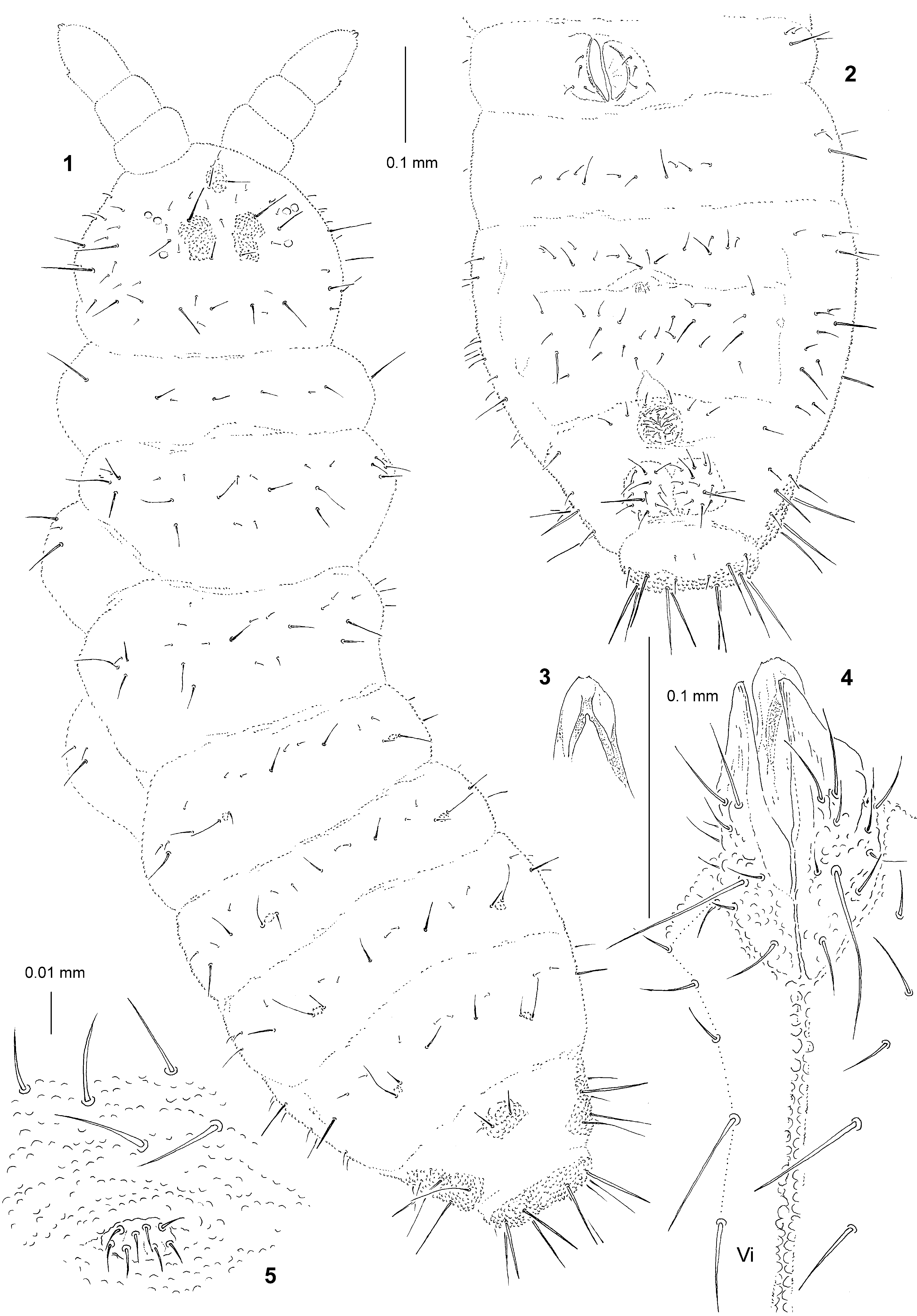
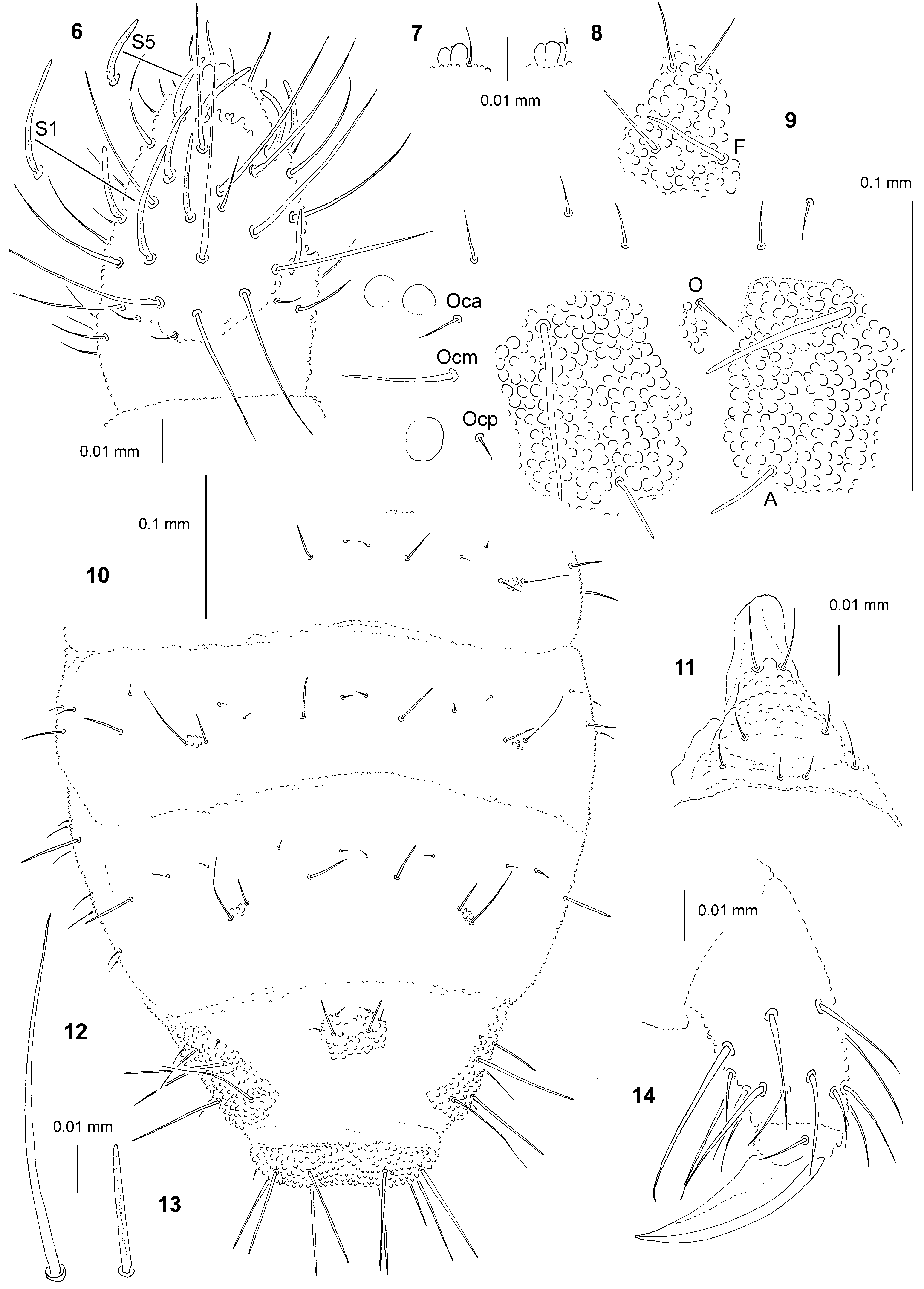
TABLE 1. Cephalic chaetotaxy of Paranura reticulata sp. nov.
| Group of chaetae | Number of chaetae | Types of chaetae | Names of chaetae |
|---|---|---|---|
| Cl | 4 | Mc me | F G |
| Af | 11 | Ml Mc me | B A D, E, C, O |
| Oc | 3 | Ml me mi | Ocm Ocp Oca |
| Di | 2 | Mc me | Di1 Di2 |
| De | 2 | Ml me | De1 De2 |
| Dl | 6 | Ml Mc me | Dl5 Dl1, Dl6 Dl2–4 |
| (L+So) | 10 | Ml me | L1, L4, So1 So2–6, L2–3 |
TABLE 2. Postcephalic chaetotaxy of Paranura reticulata sp. nov.
| Segment, Group | Number of chaetae | Segment, Group | Number of chaetae in adult |
|---|---|---|---|
| I | 7 | IV | or, 8 S, i, 12 mou, 6 brs, 2 iv |
| II | 12 | ||
| III ve | 5 S-chaetae AO III 5 | ap | 8 bs, 5 miA |
| vc | 4 | ca | 2 bs, 3 miA |
| vi | 4 | cm | 3 bs, 1 miA |
| d | 5 | cp | 8 miA, 1 brs |
No known copyright restrictions apply. See Agosti, D., Egloff, W., 2009. Taxonomic information exchange and copyright: the Plazi approach. BMC Research Notes 2009, 2:53 for further explanation.