
Spionicola mystaceus, Bjornberg & Radashevsky, 2009
|
publication ID |
https://doi.org/10.1590/S0031-10492009002000001 |
|
persistent identifier |
https://treatment.plazi.org/id/3C18D96F-2F6A-1417-FD0F-FF69FC33472B |
|
treatment provided by |
Carolina |
|
scientific name |
Spionicola mystaceus |
| status |
gen. nov. and sp. nov. |
Spionicola mystaceus gen. nov. and sp. nov.
Material: The study is based on 2 females of which one was ovigerous and 3 males collected on 31 Jan.2004 and on 13 May 2004, at 1-5 m depth off Praia do Curral in São Sebastião Island, state of São Paulo, associated with the polychaete Dipolydora armata which inhabits shells of living gastropods or shells of the same gastropods occupied by hermit crabs. The holotype female ( MZUSP 16319 View Materials ) is deposited in the Museum of Zoology of the University of São Paulo. The paratypes remained at the CEBIMar ( 1 male nº 011; 1 female nº 012). One male was lost during manipulation and the other dissected and retained in the authors collection .
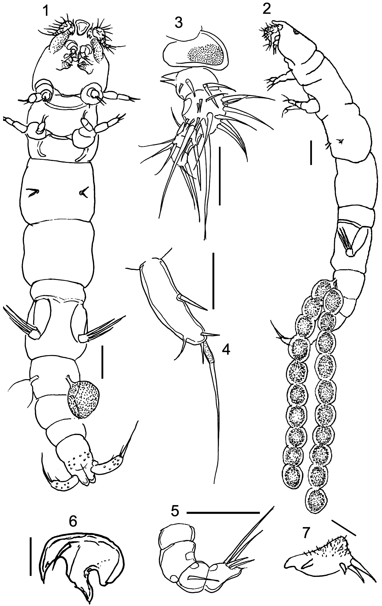
Description: Adult female 1.52 mm long, vermiform, poorly sclerotized, with two uniserial egg strings, each with 10 reddish-orange eggs ( Figs. 1-2 View FIGURAS 1‑7 ). Color: lightbrown-greenish. Rostrum pear-shaped, very protuberant, rounded anteriorly ( Fig. 3 View FIGURAS 1‑7 ). Cephalon not completely separated from first pedigerous somite. The ventrally expanded margins of the cephalothorax are densely ornamented with tiny spinules. Orange colored eye present dorsally. Pedigerous somites not distinctly separated from each other. Urosome with five somites. Caudal ramus about as long as anal somite, with 5 setae ( Fig. 4 View FIGURAS 1‑7 ). Caudal ramus 0.100 mm long and 0.025 mm wide. Paired egg strings 0.961 mm long ( Fig. 2 View FIGURAS 1‑7 ) attached to second urosomite.
Antennule five-segmented ( Fig. 3 View FIGURAS 1‑7 ); setal formula: 4, 15, 3, 3, 3. Segment 1 with two proximal ventral, pointed processes directed posteriorly. Antenna 4-segmented ( Fig. 5 View FIGURAS 1‑7 ); segment 3 with one seta; distal segment with a rounded projection and 5 setae, of which 2 longer.
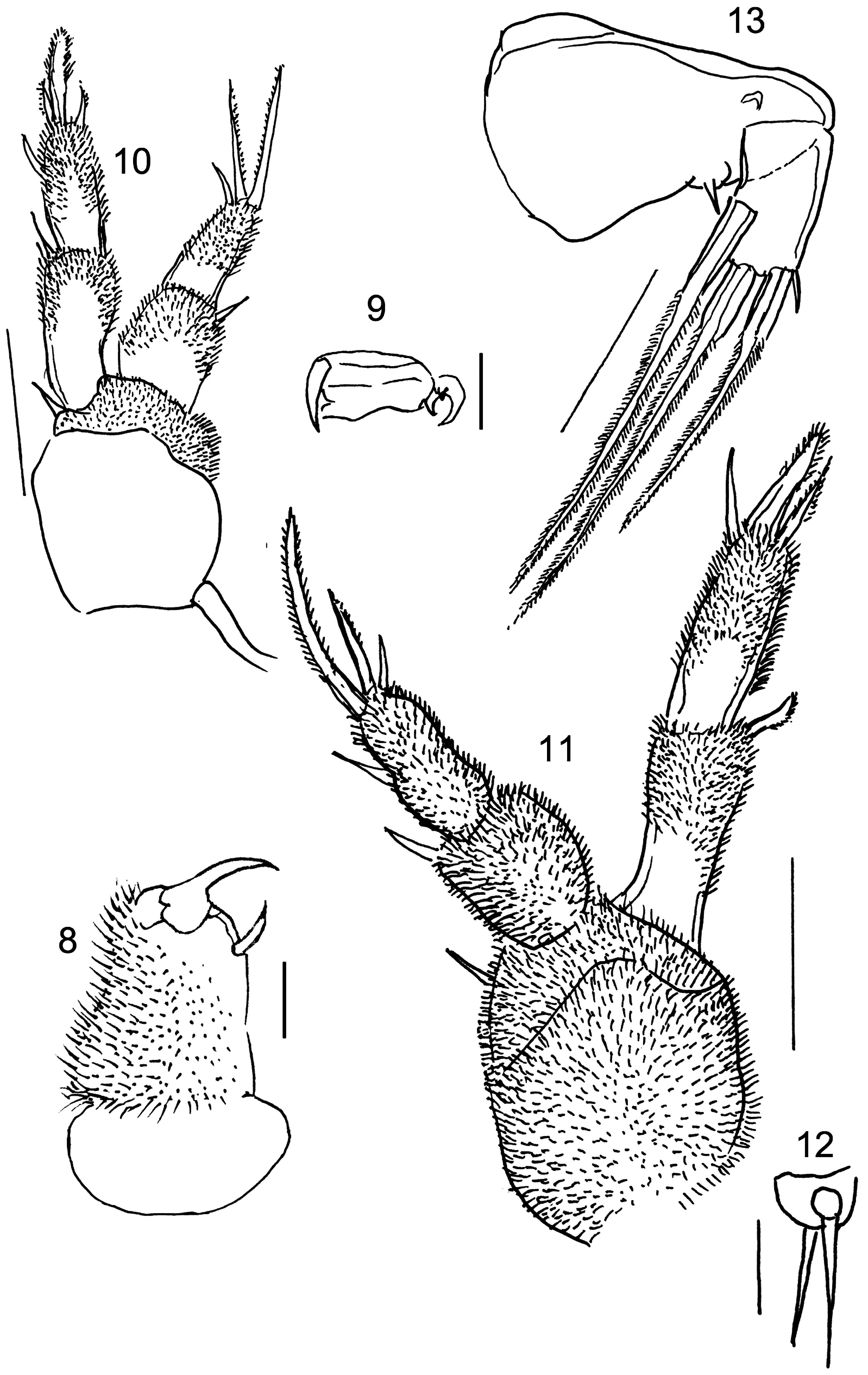
Mandible ( Fig. 6 View FIGURAS 1‑7 ) with distal pointed hook and 1 setule. Maxillule ( Fig. 7 View FIGURAS 1‑7 ): of velvety appearance, covered by microspines, with 2 digitiform internal protuberances and 3 spines. Maxilla covered with minute spines ( Fig. 8 View FIGURAS 8‑13 ); with 1 small, hook-like terminal spine, 1 cushion-like rounded projection and 2 small terminal spines. Maxilliped very small ( Fig. 9 View FIGURAS 8‑13 ), represented by small hook-like distal spine and 2 lateral minute spines on minute rectangular protuberance.
Legs ( Figs. 10-13 View FIGURAS 8‑13 ): with patches of minute setules giving them velvety appearance. Legs 1 and 2 with strong coxa, small basis with 1 outer spine, and 2-segmented rami. Exopod of leg 1: segment 1 with 1 outer spine; segment 2 with 1 outer and 3 terminal spines. Endopod of leg 1 with 3 terminal spines on segment 2 and 1 inner spine on segment 1. Exopod of leg 2: segment 1 with 1 outer spine and segment 2 with 3 terminal spines and 1 outer spine. Endopod of leg 2 with 3 terminal on segment 2 and 1 distal spine on segment 1. Leg 3 reduced to 2 spines. Fourth pair of legs absent. Fifth pair of legs 2-segmented; first segment long and wide with 1 spinule; segment 2 about half as long as first and half as wide, with 1 small spinule and 3 robust long setose spines.
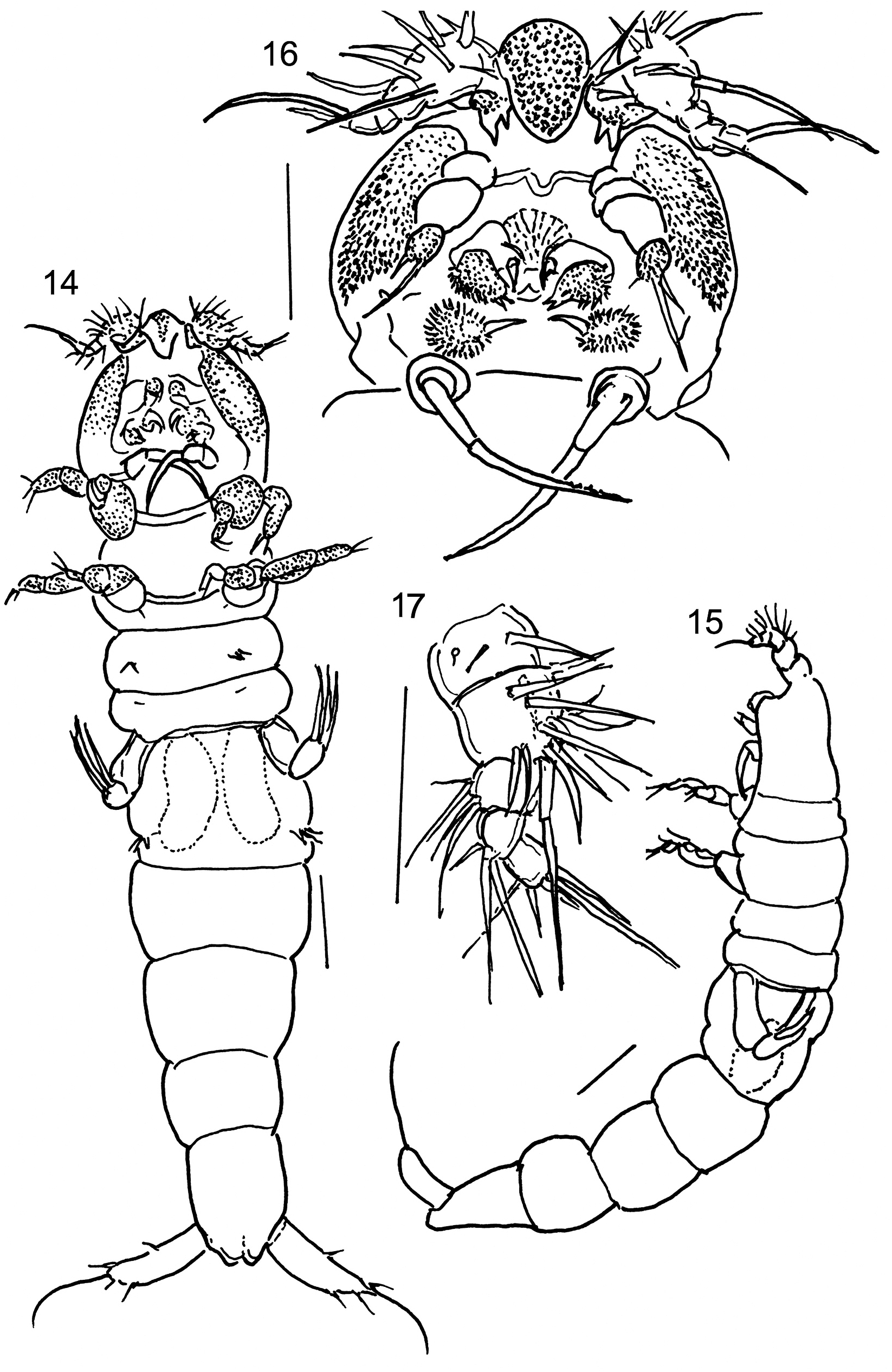
Males ( Figs. 14-15 View FIGURAS 14‑17 ): Two specimens, 0.94 mm and 1.03 mm long, vermiform, poorly sclerotized like females. Rostrum prominent, pear-shaped, tapering towards blunt posterior margin, more or less triangular in ventral view ( Fig. 16 View FIGURAS 14‑17 ). First pedigerous somite not clearly separated from cephalon. Prosome and urosome 5-segmented. First urosomite with bean-shaped spermatophores. Caudal rami about as long as anal somite, armed with 5 setae; mid-terminal seta, mounted on a pedestal, slightly longer than caudal ramus.
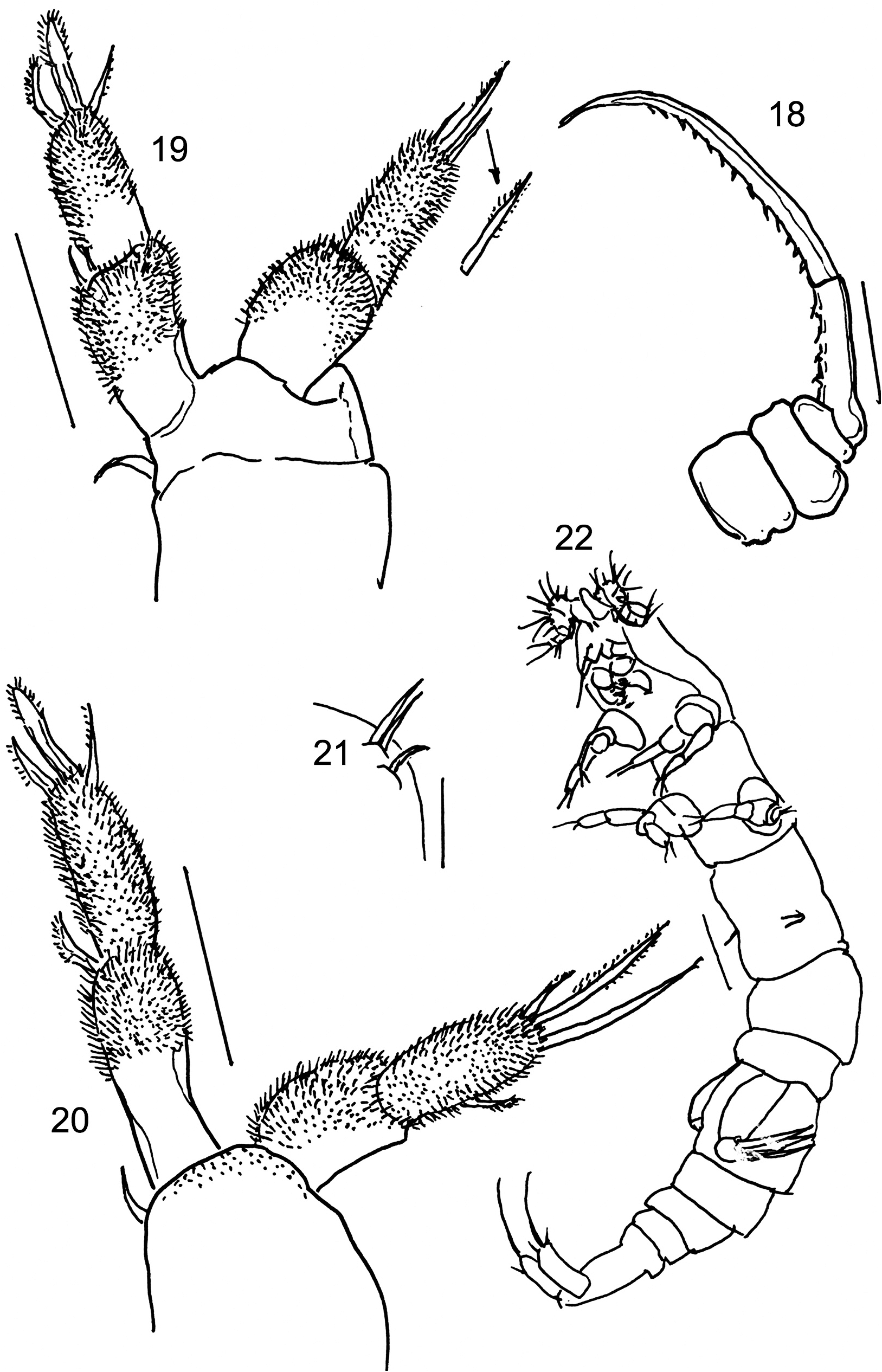
Antennule 5-segmented ( Fig. 17 View FIGURAS 14‑17 ); setal formula 3, 13, 4, 3, 5. Antenna, mandible, maxillule and maxilla as in female. Maxilliped 4-segmented ( Fig. 18 View FIGURAS 18‑22 ), with 1 strong, very long serrate claw on the distal serrate segment.
Leg 1 covered with setules with separate coxa and basis ( Fig. 19 View FIGURAS 18‑22 ); exopod 2-segmented with outer spine on segment 1 and segment two, with 3 terminal setae; endopod 2-segmented, 2 setulate spines and a thin spine on last segment. Leg 2 ( Fig. 20 View FIGURAS 18‑22 ) covered with setules, with 1 seta on coxobasis and endopod 2-segmented with 1 inner seta on segment 2, and 3 distal spines on distal segment; exopod 2-segmented, with 1 outer spine on first segment, and 3 terminal setulate spines on distal segment. Leg 3: 2 spines on pedestal. Leg 4 absent. Leg 5 as in female; Leg 6 ( Fig. 21 View FIGURAS 18‑22 ) represented by 3 small spines on each side of first urosomite.
Young female: (copepodid V?) 1.18 mm long ( Fig. 22 View FIGURAS 18‑22 ). Cephalon not separate from first pedigerous somite. Pedigerous somites 2, 3, 4 and 5 well delimited. Rostrum, antennules, antennae, mandibles, maxillules, maxillipeds and legs as in adult female. Urosome 5-segmented. Caudal rami as in adult female.
Habitat: The host polychaete, Dipolydora armata is a borer of coralline algae and shells of various mollusks (Radashevsky & Nogueira, 2003). The largest polychaetes with 35 segments, reach 5.5 mm long and 0.3 mm wide. In São Sebastião Channel, D. armata was found in the shallow subtidal in shells of a variety of live gastropods Astraea olfersii (Philippi, 1846) , Morula nodulosa (CB Adams, 1845) , Pisania auritula (Link, 1807) , Pisania pusio (Linnaeus, 1758) , Stramonita haemastoma (Linnaeus, 1757) and shells of the same gastropods occupied by hermit crabs Paguristes tortugae Schmitt, 1933 and Pagurus brevidactylus (Stimpson, 1859) . The polychaetes resided in burrows within the shells. Tens of polychaetes were found in a single shell, and up to 15/cm 2 were found on the shell surface. Of 220 shells of the gastropod Stramonita haemastoma ( 22-35 mm high), 79.1% (174 shells, including 10 live mollusks and 164 shells occupied by the hermit crab P. tortugae ) were free from D. armata ; 20.9% (46 shells, including 2 live mollusks and 44 shells occupied by hermit crabs P. tortugae ) were infested by polychaetes. Hundreds of them were examined and only 5 copepods were found. Copepods were firmly attached to the dorsal side of the middle segments of the polychaete and probably sucked from the host’s body. We can therefore conclude that Spionicola mystaceus is not a common parasite in the region where it was found.
| V |
Royal British Columbia Museum - Herbarium |
No known copyright restrictions apply. See Agosti, D., Egloff, W., 2009. Taxonomic information exchange and copyright: the Plazi approach. BMC Research Notes 2009, 2:53 for further explanation.