
Ceratophrys (Lynch, 1971)
|
publication ID |
https://doi.org/ 10.11646/zootaxa.4658.1.2 |
|
publication LSID |
lsid:zoobank.org:pub:16EDCB6E-49D1-4214-AEB3-203C19CA56A0 |
|
persistent identifier |
https://treatment.plazi.org/id/3C7387AF-FFBA-FF83-19E5-FF5F2040564C |
|
treatment provided by |
Plazi |
|
scientific name |
Ceratophrys |
| status |
|
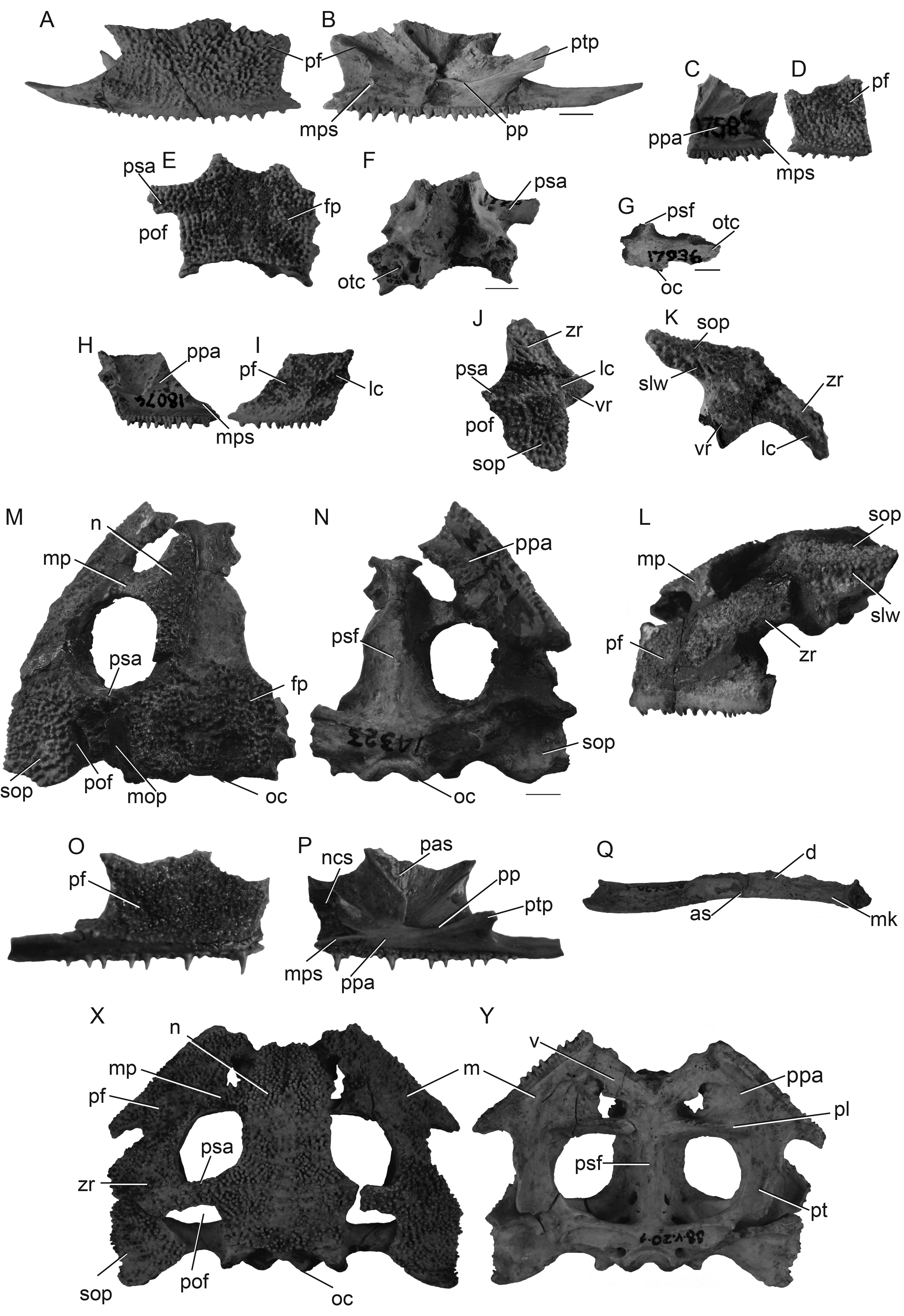
Ceratophrys View in CoL sp. (MLP 34.V.10.8; 88.V.20.1; MACN 17585; 17936; 18074) from the vicinity of the city of Mar del Plata along the Atlantic Cost of the province of Buenos Aires, Argentina ( Mercadal de Barrio & Barrio 2002)
Numerous Pleistocene fossils, especially mammals, have been collected from a series of localities along the Atlantic Cost of the province of Buenos Aires between the cities of Mar del Plata and Miramar ( Fig. 1 View FIGURE 1 , locality 8; Cione & Tonni 2005; Marshall et al. 1984). Several fragmentary remains of anurans (MLP 34.V.10.8; 88.V.20.1; MACN 17585; 17936; 18074) also were collected from localities from this area (e.g., Punta Vorohué, Arrollo Lobería, and Chapadmalal) and identified as Ceratophrys sp. by Mercadal de Barrio & Barrio (2002).
Osteological features. MLP 34.V.10.8 ( Fig. 5A, B View FIGURE 5 ) is a nearly complete right maxilla (which possesses most of the characters described below) and a posterior fragment of the left maxilla. This bone has a high, ornamented pars facilis that predominates the forward part of the maxilla anterior to the pterygoid process ( Fig. 1B View FIGURE 1 ).The lingual surface of the maxilla lacks a discrete pars palatina in its anterior half. Posteriorly, the pars palatina is oriented dorsomedially; possibly, the anterior ramus of the pterygoid, which is evident in continuity with the subtriangular pterygoid process ( Fig. 5B View FIGURE 5 ), articulated with the maxilla in this region. Others scars that would indicate bone articulations with the lingual surface of the maxilla are also evident. One is an elongated socket that reaches the level of the fourth dental position; it would have articulated with the maxillary process of the premaxilla.The second, posterior scar seems to represent the articulation of the maxilla with the planum antorbitale. The few complete teeth lack distinct crowns and pedicels.
MACN 17585 ( Fig. 5C, D View FIGURE 5 ) is the anterior portion of a left maxilla bearing a high, ornamented pars facilis; a distinguishable pars palatina is absent, but a series of articular scars are present. A long and elongated anterior socket for the maxillary process of the premaxilla is evident ( Fig. 5C View FIGURE 5 ). No teeth seem to be completely preserved.
MACN 17936 ( Fig. 5 View FIGURE 5 E–G) is two skull fragments—the posterior part of a skull roof and the posterior part of a neurocranium. The skull roof is primarily represented by a pair of ornamented, synostotically articulated frontoparietals ( Fig. 1E, F View FIGURE 1 ). A lateral process of the left frontoparietal forms a robust temporal arcade (or parieto-squamosal arch) of the postorbital fenestra. Although the posterior neurocranium is extremely fragmentary, it is clear that at least exoccipitals, prootics, parasphenoid, and probably the pterygoids are indistinguishably fused ( Fig. 1G View FIGURE 1 ).
The two bones of MACN 18074 ( Fig. 5 View FIGURE 5 H–K) are a fragmentary left maxilla and a right squamosal. The maxilla has an ornamented, high pars facilis that bears a well-developed crest posteriorly ( Fig. 5I View FIGURE 5 ). The lingual side of the maxilla lacks a distinguishable pars palatina and a long, elongated socket that probably accommodated the maxillary process of the premaxilla anteriorly ( Fig. 5H View FIGURE 5 ). A few, non-pedicellate teeth are preserved. The squamosal has a well-developed, but incompletely preserved, otic plate; thus its shape and size cannot be described ( Fig.5J, K View FIGURE 5 ). However, the plate has a concave dorsal surface and bears a lateral wall. A crest is present at the edge of the dorsolateral part of the otic plate; the crest extends anteriorly over the zygomatic ramus. A fragmentary medial projection may represent part of the squamosal portion of the temporal arcade. The posterior border of this projection seems to be complete and to have formed a postorbital fenestra.
Remarks. All these remains possess the proposed and possible synapomorphies for Ceratophryidae and Ceratophrys that can be evaluated, as follow: MLP 34-V-10-8. —exostosis, non-pedicellate teeth, anterior maxillary pars palatina absent ( Ceratophryidae ), and long maxillary processes of premaxilla ( Ceratophrys ); MACN 17585.—exostosis, anterior maxillary pars palatina absent ( Ceratophryidae ), and long maxillary processes of the premaxilla ( Ceratophrys ); MACN 17936.—exostosis, parieto-squamosal arch ( Ceratophryidae ), and postorbital fenestra ( Ceratophrys );. MACN 18074.—exostosis, anterior maxillary pars palatina absent ( Ceratophryidae ), postorbital fenestra, long maxillary processes of premaxilla ( Ceratophrys ). Thus, the foregoing should be assigned to Ceratophrys . MLP 34-V-10-8, MACN 17585, and MACN 17936 lack diagnostic characters of a less-inclusive taxon. MACN 18074 has a combination of characters present in the [ C. aurita – C. joazeirensis ] clade, C. calcarata , and C. ameghinorum (i.e., lateral crest and squamosal otic plate with lateral wall); however, this specimen is too fragmentary to justify its attribution to any of these taxa.

No known copyright restrictions apply. See Agosti, D., Egloff, W., 2009. Taxonomic information exchange and copyright: the Plazi approach. BMC Research Notes 2009, 2:53 for further explanation.