
Siconema ovicallosum, Luc, Pham Van, 2012
|
publication ID |
https://doi.org/ 10.5281/zenodo.212402 |
|
DOI |
https://doi.org/10.5281/zenodo.5671807 |
|
persistent identifier |
https://treatment.plazi.org/id/3D0087C1-FFC0-B61D-9DD6-FF03C7807538 |
|
treatment provided by |
Plazi |
|
scientific name |
Siconema ovicallosum |
| status |
sp. nov. |
Siconema ovicallosum sp. n.
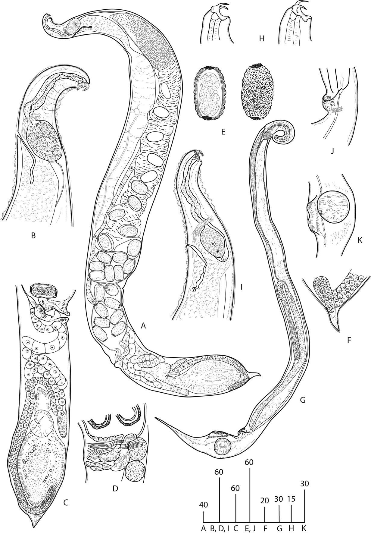
( Fig. 1 View FIGURE 1 & 2 View FIGURE 2 )
Measurements: Table 1.
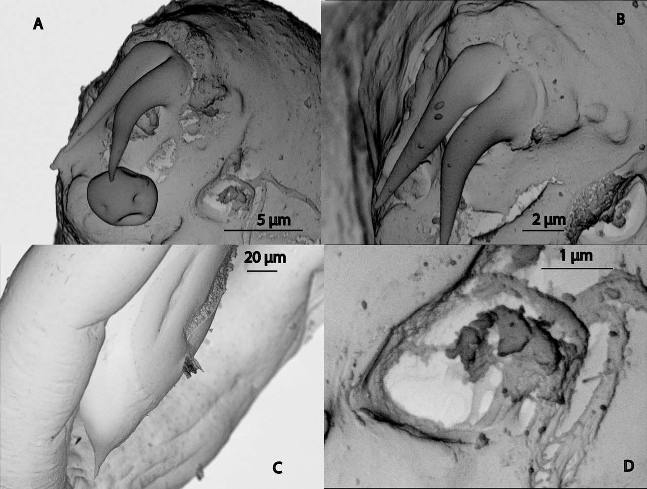
Adult: Larger ungellids. Whole body covered by loose cuticular membrane prominent on curves and bends of body. Cuticle thin, smooth. Lateral fields not discernible. Head inclined dorsally bearing pair of dorsally directed head hooks. Hook base thick; embedded in head tissue; not divided into left and right parts. Hook blades proximally close; distal tips diverging. Blades conical in shape, proximally curved then directed parallel to hook base. Mouth aperture situated beneath distal blades. Stoma tubular, incorporated in hooks base; apical portion slightly projecting from base. Amphids situated closely to hooks on lateral sides of head; pocket-like with small pouches and half-moon-like apertures. Cephalic sensilla absent. Pharynx comprising long, curved, uniformly broad, very finely muscled corpus and twice as shorter and twice as wider glandular bulb. Bulb displaced to dorsal side of body. Fine nerve ring crossing corpus anterior to bulb. Excretory pore located opposite anterior part of bulb. Excretory duct cuticularised. Excretory gland extending to mid-body; constituting from two parts, anterior of which with darker than posterior one, content. Enormous nucleus present in each part of gland. Excretory channels wide, weakly cuticularised. Cardia and intestine developed.
Female: Body tapering towards head end, wide, tail end only slightly expanded due to vast caudal organs. Short conical tail tip present. Body maximum width at level of uterus. Overall height of hooks measured from base to tip blades 11–12 µm. Hook base 6–7µm high and 15–18 µm wide; blades 11–12 µm long, proximally 4–6 µm wide. Stoma 5–7 µm long and 2–4 µm wide. Pharyngeal corpus 9–15 µm wide; bulb 50–58 µm long and 30–38 µm wide. Excretory duct 74–90 µm long and 3–5 µm wide. Monodelphic, prodelphic. Ovary tip located in caudal region. Ovary running anteriad by dorsal side of caudal organs, twisting 2–3 times posterior to vulva level then leading anteriad by ventral body side. Developing oocytes initially spherical, then elongated. Large elongated spermatheca (80–150 µm long and 40–90 µm wide) located at flexure in 243–428 µm from anterior. Globular sperm in spermatheca 3–4 µm in diameter. Oviduct thick-walled, containing a few immature eggs. Ca. 20 fully formed, not embrionated eggs in thin-walled uterus. Eggs truncate-ovoid with suberose caps ca. 12 µm in diam. covered by ca. 40 tiny papilliform tubercles ca. 1.5–2 µm high; eggshell walls 4–5 µm thick, composed of 3 layers. Surface between caps composed from low knob-like protuberances densely covered by tinier than on poles papilliform tubercles. Vagina straight, 50–70 µm long, with thick muscular walls. Vulva lips slightly inflated. No post-uterine sack present. Vulva posterior. Anal aperture not detected. Caudal organ region occupies hind portion of postvulval region. Caudal organs externally broadly elliptical, surrounded by thin rim. Surface of caudal organs lacking cuticle. Internally, body of each organ consisting of spongy tissue penetrated by sinuous channel. Anterior to elliptical portion of organ, irregularly shaped diverticulum bulging laterad present. Posterior to diverticulum, on ventral and dorsal body sides glands (or two lobes of the same gland) of unknown function visible. Intestine thin, transparent, probably ending blindly (anal aperture not detected).
Male: Smaller in size than female. Head, pharynx, excretory system and caudal organs structure as in females. Hooks’ total height 9 µm, base height 6 µm, blades 6 µm long and proximally 3 µm wide. Stoma ca. 2 x 2 µm. Amphid aperture 2 µm in diam. Pharynx 1.5 times shorter than in female with smaller bulb (20 x 10 µm) and thinner corpus (8 µm). Excretory duct 55 µm long and 1 µm wide. Monorchic. Testis 530 µm long, reflexes in 376 µm from anterior end, flexure 200 µm long. Spermatocytes spherical, in 2, then 3 rows. Va s de fe re n s as wide as testis. Immature sperm in long ejaculatory duct ca. 5 µm in diam. Prominent pericloacal elevation. No genital papillae present. Tail expanding to 50 µm shortly behind cloaca due to the presence of caudal organs. Posterior to caudal organs, tail conical, narrowing from 37 µm to 3 µm. Conical portion of tail 110 µm long. Caudal organs nearly circular externally, ca. 31 µm in diam.
Type material. Holotype female (on the slide accession No. 1125) and paratype male (on the slide accession No. 1126) deposited in the Museum of the Helminthological Collections of the Centre of Parasitology at the Severtsov Institute of Ecology and Evolution, Moscow.
Type-host and locality. Amynthas tuberculatus collected in Pu Mat Nature Reserve, prov. Nghe An, Vietnam, November 2008, by S. Spiridonov.
Etymology. The species name is derived from Latin words ‘ ovum’ —egg and ‘ callosum’ —thick-skinned and refers to the structure of eggshells.
Diagnosis and relationships. Siconema ovicallosum sp. n. is characterised by females with a wide body, an insignificantly expanded post-vulval region, caudal organs displaced to posterior extremity, a short tail tip; short and slim males with caudal organs located closely to a cloacal opening and a long, conical posterior portion of tail; cephalic hooks in both sexes with sturdy base and thin, diverging blade tips; the tubular stoma incorporated in hooks base; eggs with very thick shells and two suberose polar caps.
The species most closely resembles another Vietnamese species, S. laticaudatum Ivanova & Pham Van Luc, 1997 by having similar hooks, eggs with two polar caps and the similar position of caudal organs in both sexes. It can be reliably differentiated by males being twice shorter than females vs insignificantly shorter with much smaller, circular (ca. 31 µm in diameter) caudal organs vs elliptical, 55– 70 x 47–95 µm in size. Cloacal flaps are less prominent in the present species and no projection protruding from anal opening is present. Females of the new species differs from S. laticaudatum by the shape (truncate vs ovoid) and ornamentation of eggshells which are much thicker (4–5 vs 2 µm). In S. laticaudatum , the surface of an eggshell is evenly ovoid and heavily mammilate, caps are convex and bearing each 14–17 tubercles ca. 2–3 µm high, whether in the new species eggshells uneven, knobbly (ca. 5 x 7 µm) and covered by tubercles less than 1 µm high. Eggshell caps in the new species are flat and bear more numerous (ca. 40 vs 14–17) tubercles. Additionally, S. ovicallosum sp. n. differs from S. laticaudatum by having the longer pharynx in females (141–161 vs 85–110 µm) with the larger, elongated bulb (50 x 30 vs 30 x 30 µm) and lacking a post-uterine sack. The female tail is less expanded compared to S. laticaudatum (129–155 µm vs 185–260 µm) and caudal organs more elongated (150–193 x 114–125 µm vs 112–140 x 92 –115 µm). No transversal groove in a caudal organ of either sex is detected as well as two cuticularised channels running through the organ. Head sensilla are absent vs present. From S. micrurum Timm, 1966 females, to which S. ovicallosum sp. n. females are similar by the general appearance, they can be differentiated by the twice larger body size and wider eggs with thicker eggshells.
No known copyright restrictions apply. See Agosti, D., Egloff, W., 2009. Taxonomic information exchange and copyright: the Plazi approach. BMC Research Notes 2009, 2:53 for further explanation.
|
Kingdom |
|
|
Phylum |
|
|
Class |
|
|
Order |
|
|
Family |
|
|
Genus |