
Scydmaenus (Mascarensia) australiensis Franz
|
publication ID |
https://doi.org/ 10.11646/zootaxa.5371.1.1 |
|
publication LSID |
lsid:zoobank.org:pub:D60B50D1-280B-4403-9E5B-25C0704A43A1 |
|
DOI |
https://doi.org/10.5281/zenodo.10249301 |
|
persistent identifier |
https://treatment.plazi.org/id/3E380C57-FFD1-4A69-27AC-B435FA43E1B9 |
|
treatment provided by |
Plazi |
|
scientific name |
Scydmaenus (Mascarensia) australiensis Franz |
| status |
|
Scydmaenus (Mascarensia) australiensis Franz View in CoL
Scydmaenus (Mascarensia) australiensis Franz, 1975: 282 View in CoL .
( Figs 95–108 View FIGURES 95–96 View FIGURES 97–100 View FIGURES 101–102 View FIGURES 103–108 , 197 View FIGURES 189–205 )
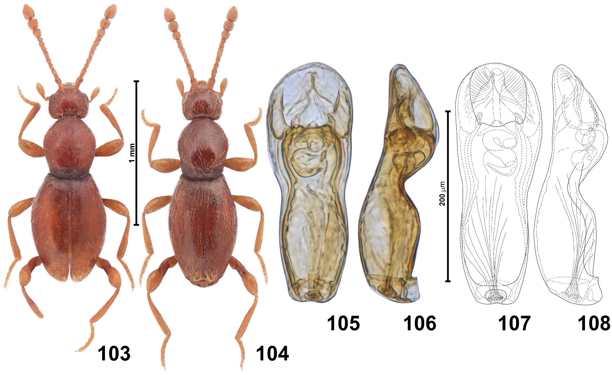
Type material studied. Holotype ( AUSTRALIA: NORTHERN TERRITORY): ♂ ( Fig. 103 View FIGURES 103–108 ), labels illustrated in Fig. 191 View FIGURES 189–205 : “ Port Darwin. / 92—2.” [brownish, with longitudinal red line at middle, printed], “4668” [brownish, printed], “ Holo- / type” [white circle with red margins, printed], “ Scydmaenus / ( Mascarensia ) / australiensis m. / det. H.Franz ” [white, handwritten and printed], “Typus” [red, handwritten] ( BNHM).
Additional material studied. QUEENSLAND: 2 ♂♂, Halensvale , 03.10.1988, Richard Bejsak, under bark ( ANIC, cPJ) .
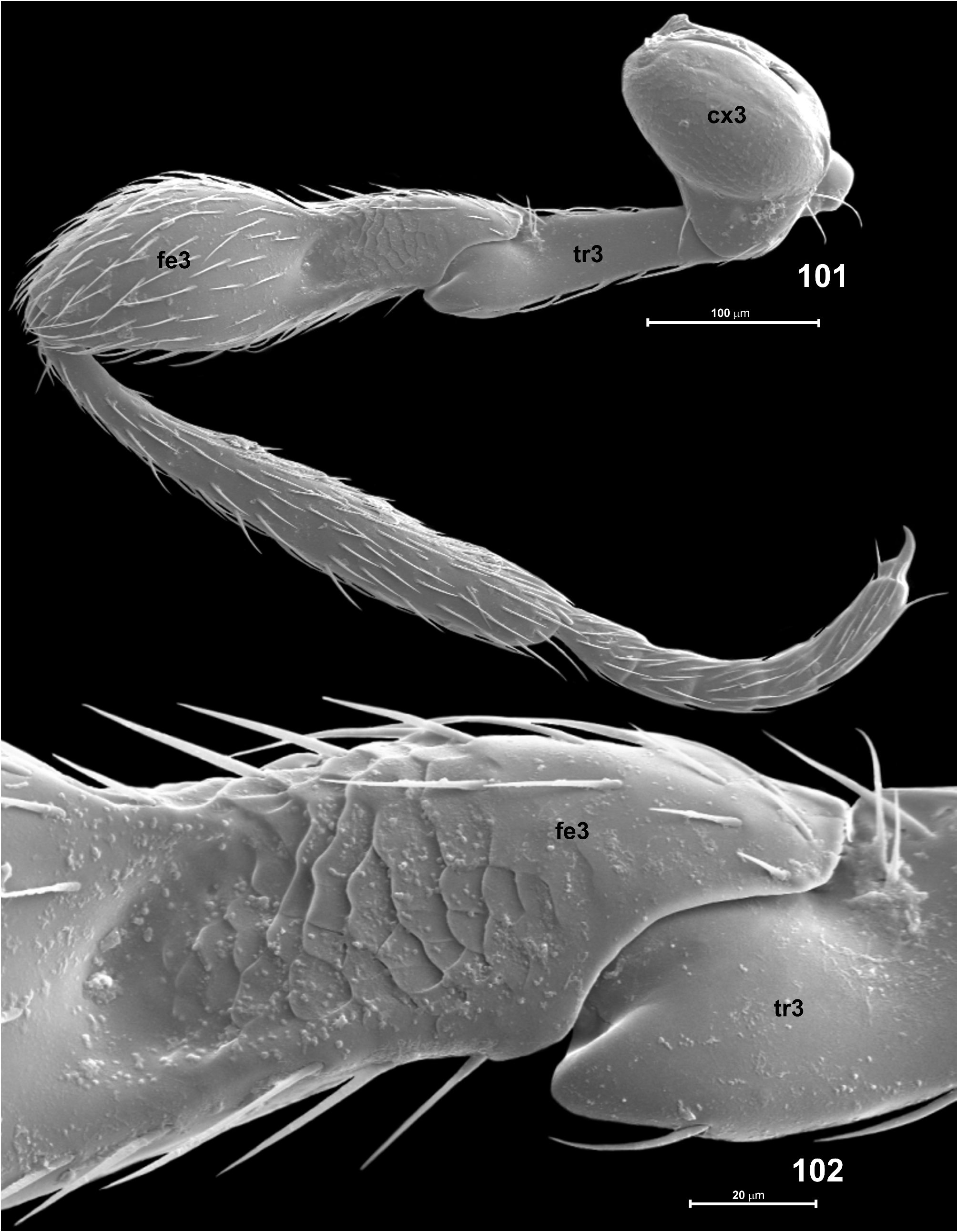
Revised diagnosis. Each elytral apex in male with small patch of long light setae surrounding small asetose impression ( Fig. 100 View FIGURES 97–100 ); metafemoral pit ( Fig. 102 View FIGURES 101–102 ) large and deep, with proximal margin diffuse; aedeagus ( Figs 105–108 View FIGURES 103–108 ) with distal broadened region indistinctly demarcated from narrow proximal portion, not capitate, weak constriction situated near middle of median lobe.
Redescription. Body of male ( Figs 103–104 View FIGURES 103–108 ) slightly flattened, elongate and slender, BL 1.30–1.35 mm; pigmentation uniformly light brown with umbra-reddish hue, appendages indistinctly lighter; cuticle moderately glossy, covered with vestiture of setae slightly lighter than body.
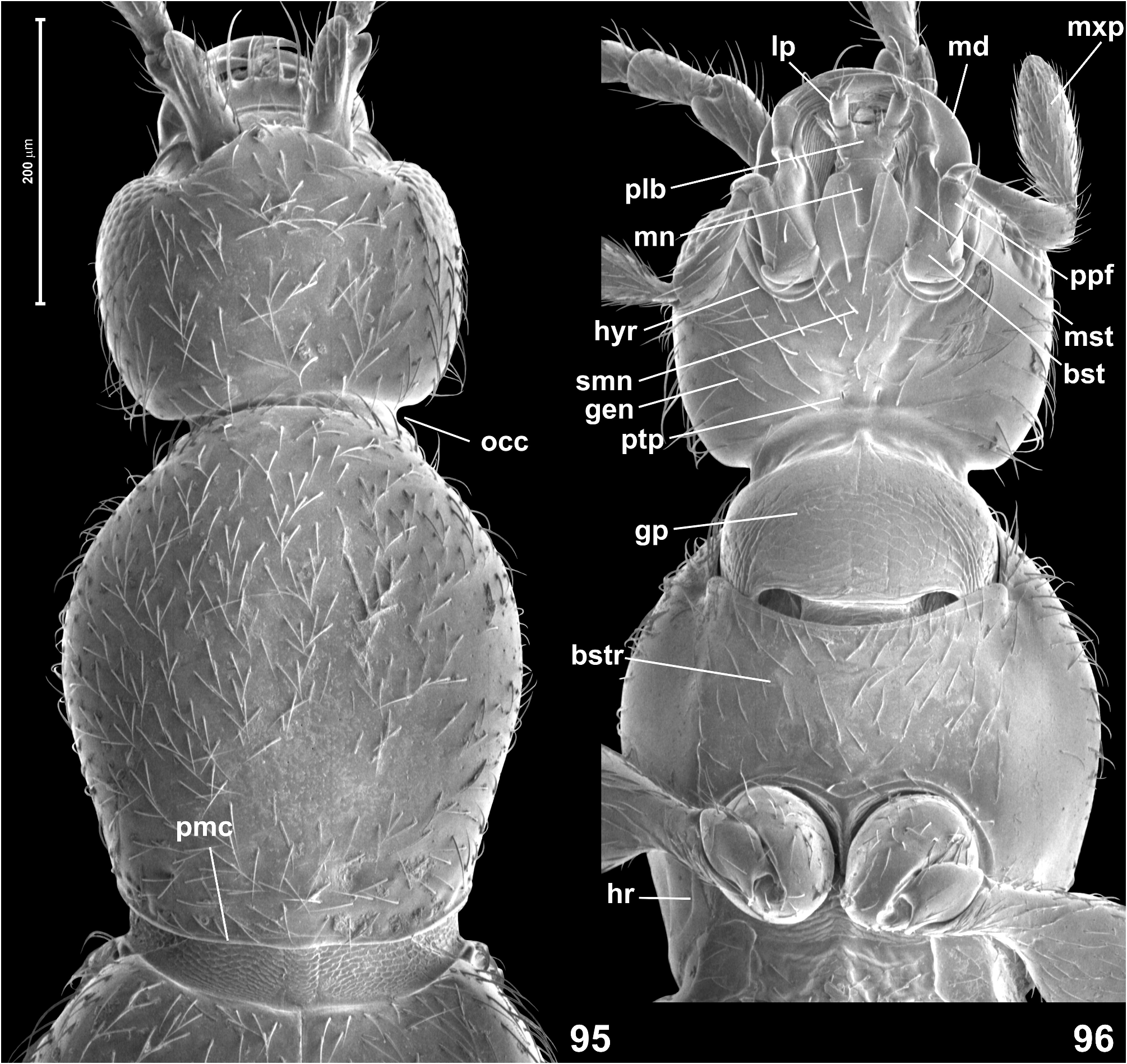
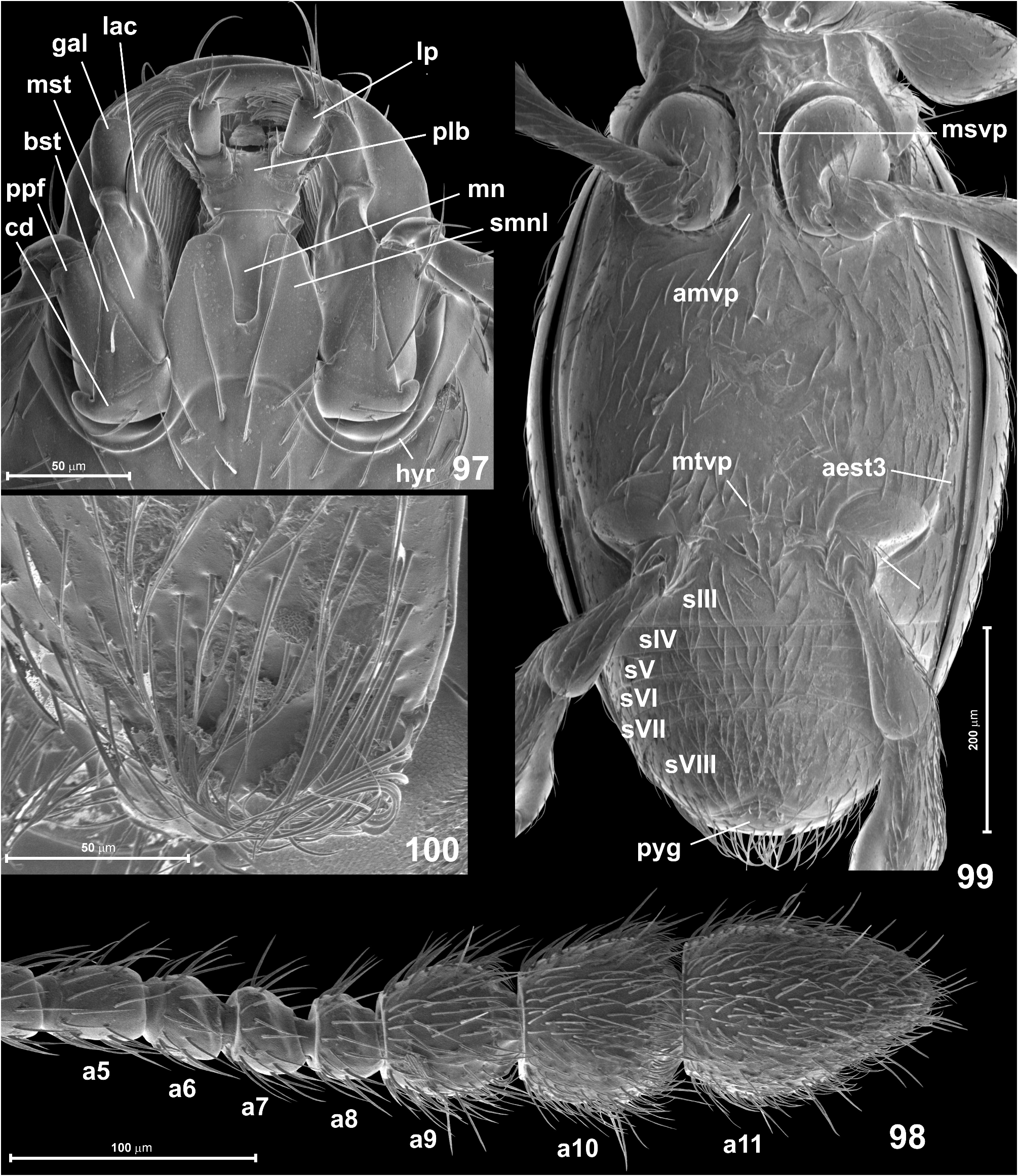
Head ( Figs 95–96 View FIGURES 95–96 ) in dorsal view transversely subrectangular, broadest at eyes, HL 0.23 mm, HW 0.25–0.28 mm; vertex and frons confluent and weakly convex, posterior margin of vertex weakly concave, evenly arcuate; tempora as long as about 1.5 × length of eye in dorsal view; supraantennal tubercles indistinct; frons over antennal fossae broadly subtriangular and with rounded anterior margin. Eyes small, nearly semicircular in shape, indistinctly emarginate posteriorly and oblique in relation to long axis of head. Punctures on frons and vertex fine, inconspicuous; setae short, sparse, suberect, those on tempora as sparse as those on head dorsum. Genae ( Fig. 96 View FIGURES 95–96 ) as sparsely setose as frons and vertex. Anterior (exposed) region of head capsule demarcated from neck region by broad abrupt impression around occipital constriction, anterior margin of gular plate on neck region accentuated by small rounded anteriorly-directed projection. Submentum ( Fig. 97 View FIGURES 97–100 ) with pair of large submental lobes, each elongate and tapering anteriorly, with truncate anterior margin, gap between lobes narrower than width of each lobe, so that only narrow median region of mentum is exposed between lobes; hypostomal ridges ( Fig. 97 View FIGURES 97–100 ) extend mesally and anteriorly to connect at middle behind submental lobe, their median transverse portion slightly diffuse. Antennae ( Figs 98 View FIGURES 97–100 , 103–104 View FIGURES 103–108 ) moderately long, slender, AnL 0.60–0.65 mm; three terminal antennomeres forming moderately sharply delimited club; scape slightly more than 3 times as long as broad, distinctly broadening distally; pedicel twice as long as broad; antennomeres 3–5 each 1.5–1.8 × as long as broad, 6 and 7 each indistinctly elongate and distinctly asymmetrical, 8 about as long as broad and distinctly asymmetrical, 9 indistinctly elongate, 10 about as long as broad, 11 1.6 × as long as broad, slightly shorter than 9–10 together, indistinctly asymmetrical, with blunt apex; proximal and middle antennomeres with sparse long suberect setae, antennomeres 9–11 each covered with short, dense and weakly suberect setae and with less numerous long erect setae.
Pronotum in dorsal view ( Fig. 95 View FIGURES 95–96 ) weakly elongate, broadest near anterior third, PL 0.40–0.43 mm, PW 0.33– 0.35 mm; anterior and lateral margins confluent and rounded; posterior corners obtuse-angled and blunt; posterior margin weakly arcuate; base with narrow and indistinct posterior marginal carina and with two pairs of indistinct, shallow and diffuse small antebasal pits. Pronotal disc covered with fine and inconspicuous punctures; setae similar to those on frons and vertex, short, moderately dense, suberect. Ventrally ( Fig. 96 View FIGURES 95–96 ) prothorax with nearly asetose and impunctate hypomera and basisternal region much longer than procoxal rests, sparsely covered with short recumbent setae, with short anterior ‘collar’ and distinct vestiges of notosternal sutures visible as notches on sides of anterior prothoracic margin; hypomeral ridges distinct and complete, demarcating narrow inner (adcoxal) region of each hypomeron, anteriorly running along procoxal rests and connecting at middle to form biarcuate anteprocoxal carina demarcating basisternal region posteriorly.
Elytra ( Figs 100 View FIGURES 97–100 , 103–104 View FIGURES 103–108 ) slightly rhomboidal, broadest slightly in front of middle, EL 0.68–0.70 mm, EW 0.45–0.50 mm, EI 1.40–1.50. Humeral calli present but weakly elevated; basal impression on each elytron barely marked; basal elytral foveae lacking; apices ( Fig. 100 View FIGURES 97–100 ) separately rounded, each slightly flattened and bearing conspicuous patch of dense long yellowish setae directed posteriorly and outwards, surrounding small impressed asetose area. Punctures fine and inconspicuous; setae (except those on apical portions) similar to those on pronotum. Hind wings fully developed.
Mesoventrite ( Fig. 99 View FIGURES 97–100 ) with broad, parallel-sided subrectangular mesoventral intermesocoxal process posteriorly touching short and equally broad anterior metaventral process, border between processes distinctly marked by transverse fissure. Metanepisterna ( Fig. 99 View FIGURES 97–100 ) narrow and completely demarcated from metaventrite. Metaventral intermetacoxal process ( Fig. 99 View FIGURES 97–100 ) broad and short, with distinctly concave posterior margin, so that its lateral portions form short subtriangular processes projecting posteriorly; distance between metacoxae subequal to half width of metaventrite at its posterior margin and wider than width of metacoxa. Metaventrite ( Fig. 99 View FIGURES 97–100 ) weakly convex, slightly flattened at middle, unmodified and evenly covered with sparse, short, nearly recumbent setae.
Legs ( Figs 96 View FIGURES 95–96 , 99 View FIGURES 97–100 , 101–102 View FIGURES 101–102 ) moderately long and slender; unmodified, except for flattened and impressed proximal portion of metafemur ( Figs 101–102 View FIGURES 101–102 ) which bears large and deep pit with diffuse proximal margin, where surface of femur is covered with scale-like microsculpture contrasting with smooth surrounding areas; protarsi nearly cylindrical, narrow, lacking tenent setae. Protarsomeres 1–4 each indistinctly elongate, 5 only twice as long as broad; meso- and metatarsi of equal length, both distinctly longer that protarsi, each with tarsomere 1 nearly twice as long as broad, tarsomeres 2–4 about 1.5 × as long as broad, and tarsomere 5 2.5 × as long as broad.
Aedeagus ( Figs 105–108 View FIGURES 103–108 ) elongate but not very slender, AeL 0.28 mm, in dorsal view median lobe broadest near distal 1/4, apical region suboval, delimited from narrow proximal region by shallow and broad constriction, apical margin broadly rounded; ostium situated in distal 1/4 of median lobe; flagellum with broad loops just proximal to ostium in resting position; median lobe lacking setae.
Female. Unknown
Distribution. Northern (N Northern Territory) and eastern (SE Queensland) Australia.
Remarks. The two known localities where S. australiensis have been collected are over 2,800 km apart in a straight line. The northern one is located within the western tropical zone, while the southeastern site is in the northernmost area of the temperate zone. The holotype from the Northern Territory ( Fig. 103 View FIGURES 103–108 ) is slightly stouter than males from Queensland ( Fig. 104 View FIGURES 103–108 ), but no differences in the aedeagi was found between all available specimens, and the apical elytral modification is also identical (although in the holotype some of the subapical setae are broken off). It is possible that this species occurs in the entire forested area along the eastern and northern coast, but it seems to be one of the rarest Australian species of Scydmaenus .
Subgenus Parallomicrus Franz
Parallomicrus Franz , 1998 (in Newton & Franz, 1998): 153. New name for Allomicrus Franz, 1975.
Allomicrus Franz, 1971a: 101 (as subgenus of Scydmaenus ; not as new; nomen nudum, no description). Note: Franz referred Allomicrus to a work published later ( Franz 1975).
Allomicrus Franz, 1975: 284 (as subgenus of Scydmaenus ). Preoccupied, not Gahan (1893) ( Coleoptera : Cerambycidae ). Type species: Scydmaenus rufus Müller & Kunze, 1822 (des. orig.).
Diagnosis. Franz (1975) defined Parallomicrus (as Allomicrus) by the following characters: (1) lack of secondary sexual characters on the head and antennae; (2) the pronotum without antebasal pits; (3) elytra lacking basal impressions and humeral folds; (4) the metaventrite fused with metanepisterna, and (5) weakly broadened protarsi in males.
Remarks. All the above-mentioned characters are present in the type species of the subgenus and in two Australian species placed in Parallomicrus , except that Franz (1975) placed in Parallomicrus also S. inflatitibia , which has two pairs of distinct antebasal pronotal pits, and therefore does not match the Franz’s diagnosis of the subgenus. However, most of the diagnostic characters of Parallomicrus are reductions that can also be found in other subgenera. The only feature that is not a reduction (metanepisterna and metaventrite fused together) also occurs in other subgenera. Moreover, the type species differs from the two Australian species placed by Franz in Parallomicrus in the structure of the aedeagus. The status and diagnosis of Parallomicrus are here treated as unclear, and no attempt is made to redefine this subgenus. It is, however, necessary to redescribe the Australian species to facilitate identifications and to document their morphological structures, which will facilitate a future reclassification of Scydmaenus .
Composition, distribution and biology. Parallomicrus includes six species, but placement of most of them requires verification. The best studied is the Western Palaearctic type species of the subgenus, S. rufus Müller & Kunze, 1822 , whose larva, feeding behavior and prey preferences were described in Jałoszyński (2015) and Jałoszyński & Olszanowski (2015). Species of Parallomicrus are distributed in Africa: Algeria, Tunisia; Asia: Lebanon, Turkey; Australia: continental Australia and Tasmania; Europe: Austria, Belgium, Bosnia & Herzegovina, Czech Rep., Denmark, Estonia, Finland, France, Great Britain, Germany, Greece, Hungary, Italy, Latvia, Netherlands, Norway, Poland, Portugal (Azores), Romania, European Russia, Slovakia, Spain, Sweden, Switzerland, Ukraine; Georgia; and Pacific islands: Fiji (Vanu Levu), and New Caledonia.
In Australia two species occur, S. inflatitibia Franz and S. myrmecobius Csiki. The latter has been collected in colonies of the ant Amblyopone australis Erichson.
Identification key to males of Australian Scydmaenus (Parallomicrus) View in CoL
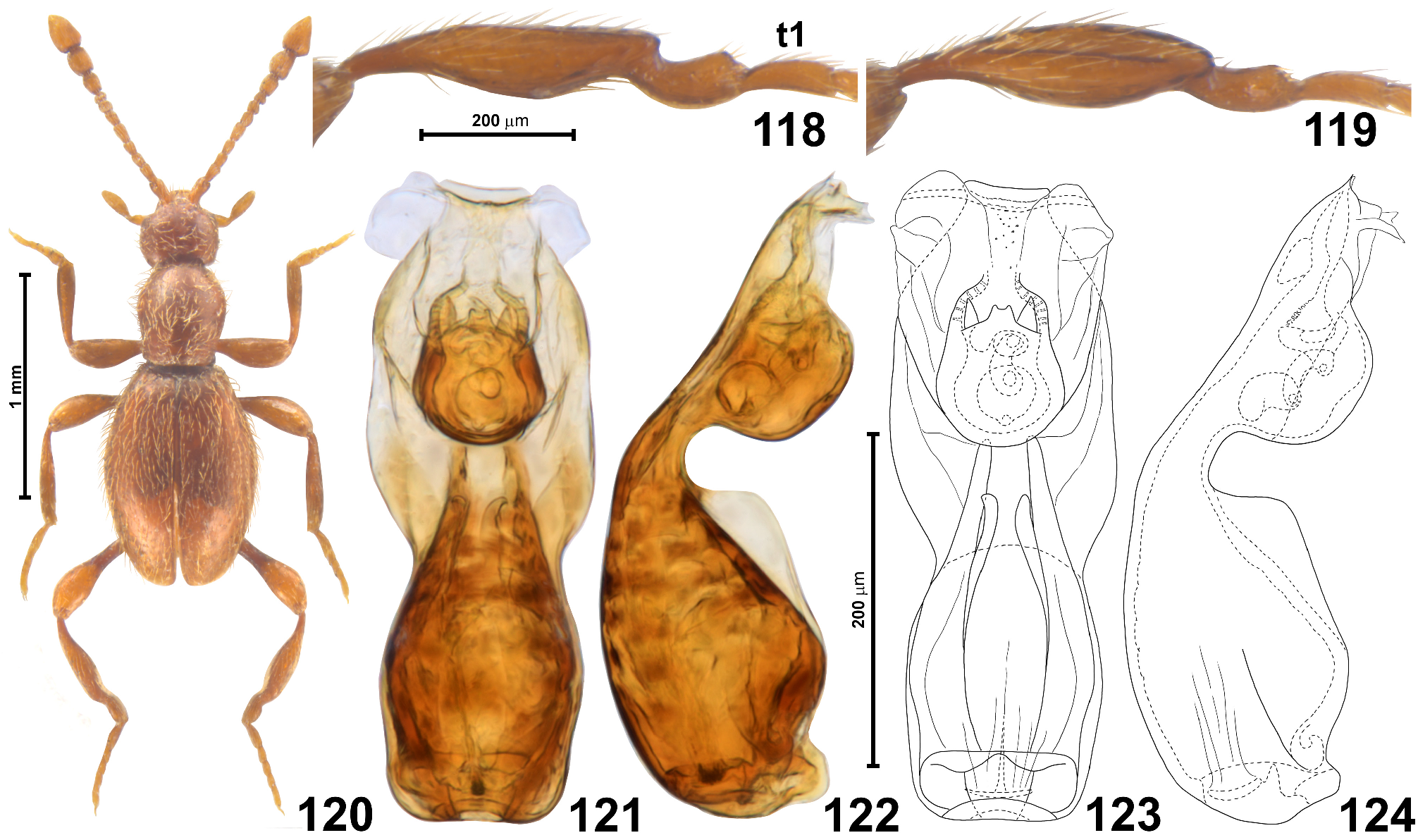
1 Pronotum with two pairs of antebasal pits; hind tibiae strongly modified, each with abrupt lateral elongate concavity on outer region and with subapical constriction demarcating short curved distal portion ( Figs 118–119 View FIGURES 118–124 ); antennomere 5 longer than 4 and 6; antennomere 11 less than twice as long as broad, with lateral outer margin bearing proximal angulate expansion ( Fig. 120 View FIGURES 118–124 , right antenna); aedeagus with lateral subapical lobes broadly subtriangular, small ( Fig. 121 View FIGURES 118–124 )..................................................................................................... Scydmaenus inflatitibia Franz View in CoL
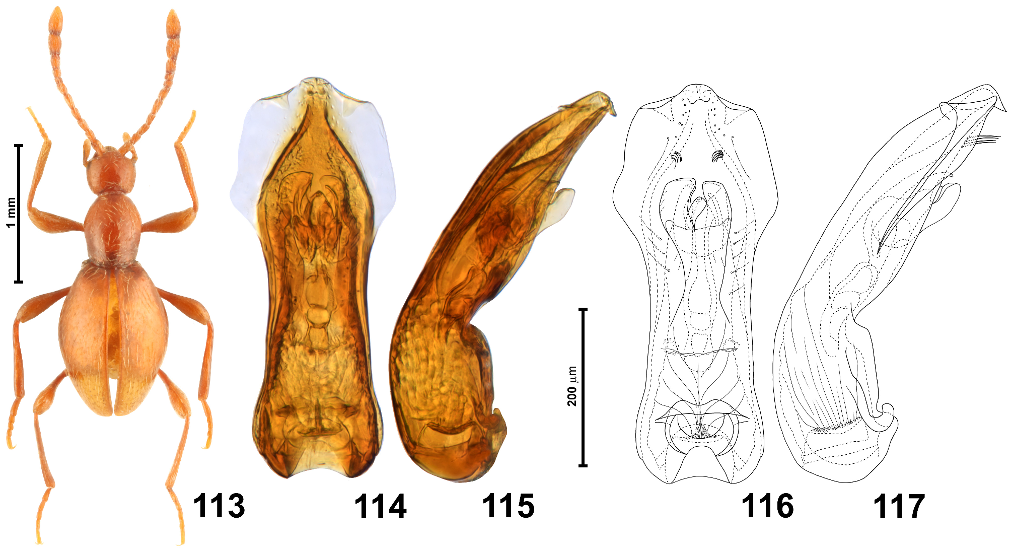
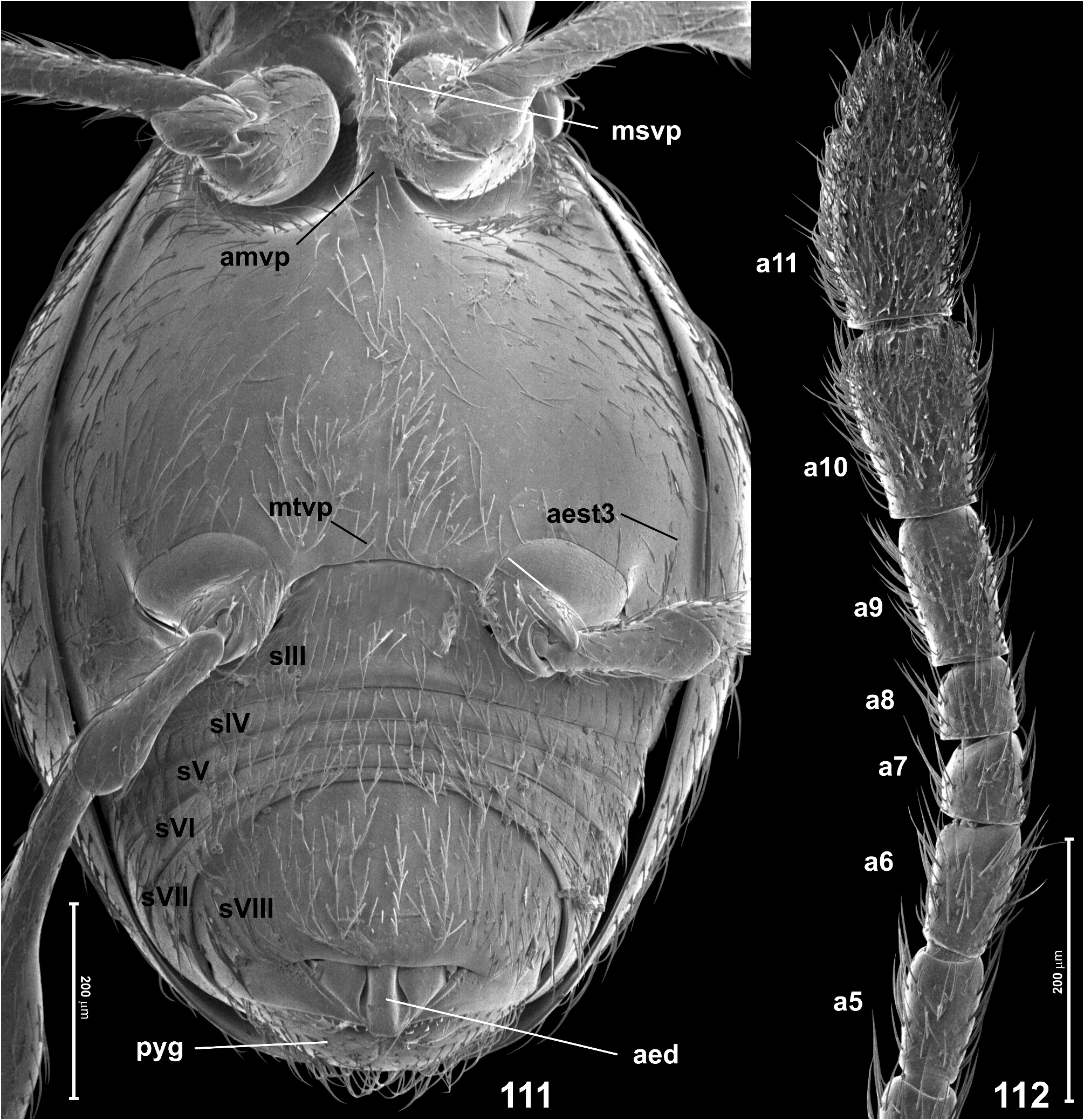
- Pronotum lacking antebasal pits; hind tibiae unmodified, slender ( Fig. 113 View FIGURES 113–117 ); antennomere 5 subequal to 4 and 6; antennomere 11 more than twice as long as broad, with lateral outer margin rounded ( Fig. 112 View FIGURES 111–112 ); aedeagus with lateral subapical lobes elongate subtrapezoidal, conspicuously long ( Fig. 114 View FIGURES 113–117 )................................ Scydmaenus myrmecobius Csiki View in CoL

No known copyright restrictions apply. See Agosti, D., Egloff, W., 2009. Taxonomic information exchange and copyright: the Plazi approach. BMC Research Notes 2009, 2:53 for further explanation.
|
Kingdom |
|
|
Phylum |
|
|
Class |
|
|
Order |
|
|
Family |
|
|
Genus |
Scydmaenus (Mascarensia) australiensis Franz
| Jałoszyński, Paweł 2023 |
Scydmaenus (Mascarensia) australiensis
| Franz, H. 1975: 282 |