
Scydmaenus (Parallomicrus) myrmecobius Csiki
|
publication ID |
https://doi.org/ 10.11646/zootaxa.5371.1.1 |
|
publication LSID |
lsid:zoobank.org:pub:D60B50D1-280B-4403-9E5B-25C0704A43A1 |
|
DOI |
https://doi.org/10.5281/zenodo.10249303 |
|
persistent identifier |
https://treatment.plazi.org/id/3E380C57-FFDE-4A6D-27AC-B4BCFDA3E737 |
|
treatment provided by |
Plazi |
|
scientific name |
Scydmaenus (Parallomicrus) myrmecobius Csiki |
| status |
|
Scydmaenus (Parallomicrus) myrmecobius Csiki View in CoL
Scydmaenus (Allomicrus) myrmecobius Csiki, 1919: 73 View in CoL . New name for Heterognathus myrmecophilus Lea, 1912: 60 View in CoL .
Heterognathus myrmecophilus Lea, 1912: 60 View in CoL . Preoccupied, not Scydmaenus myrmecophilus Aubé, 1861: 197 View in CoL (secondary homonymy in Scydmaenus View in CoL ); now not congeneric but replaced before 1961 and replacement name in use. ( Figs 113–117 View FIGURES 113–117 , 198 View FIGURES 189–205 )
Type material studied. LECTOTYPE (here designated) ( AUSTRALIA: TASMANIA): ♂ ( Fig. 113 View FIGURES 113–117 ), with labels illustrated in Fig. 198 View FIGURES 189–205 : “Tasm.H Vic.” [white, handwritten], “ Heterognathus / myrmecophilus / Lea” [white, handwritten], “ Scydmaenus / myrmecobius Lea ” / det. H.Franz ” [white, handwritten and printed], “Typus” [red, handwritten], “SAMA Database / No. 25-037031” [white, printed], “SAMA Digital Image / 21. Aug 2013 ” [green, printed and handwritten] ( SAMA). Paralectotypes: TASMANIA: 2 exx. mounted on one card, with additional card with an ant Amblyopone australis Erichson , “myrmecophilus / Lea TYPE / Marrawah ” [white, handwritten], “15065 / Heterognathus / myrmecophilus / Lea / Tas : H vic.” [white, handwritten, with red “TYPE” along right margin], “SAMA- 25 43058” [white, printed] ( SAMA) ; VICTORIA: 1 ex. mounted on one card with Amblyopone australis , “ Victoria ” [white, printed], “18396 / Heterognathus / myrmecophilus / Lea / Victoria ” [white, handwritten, with red “TYPE” along right margin], “SAMA- 25 43057” [white, printed] ( SAMA) .
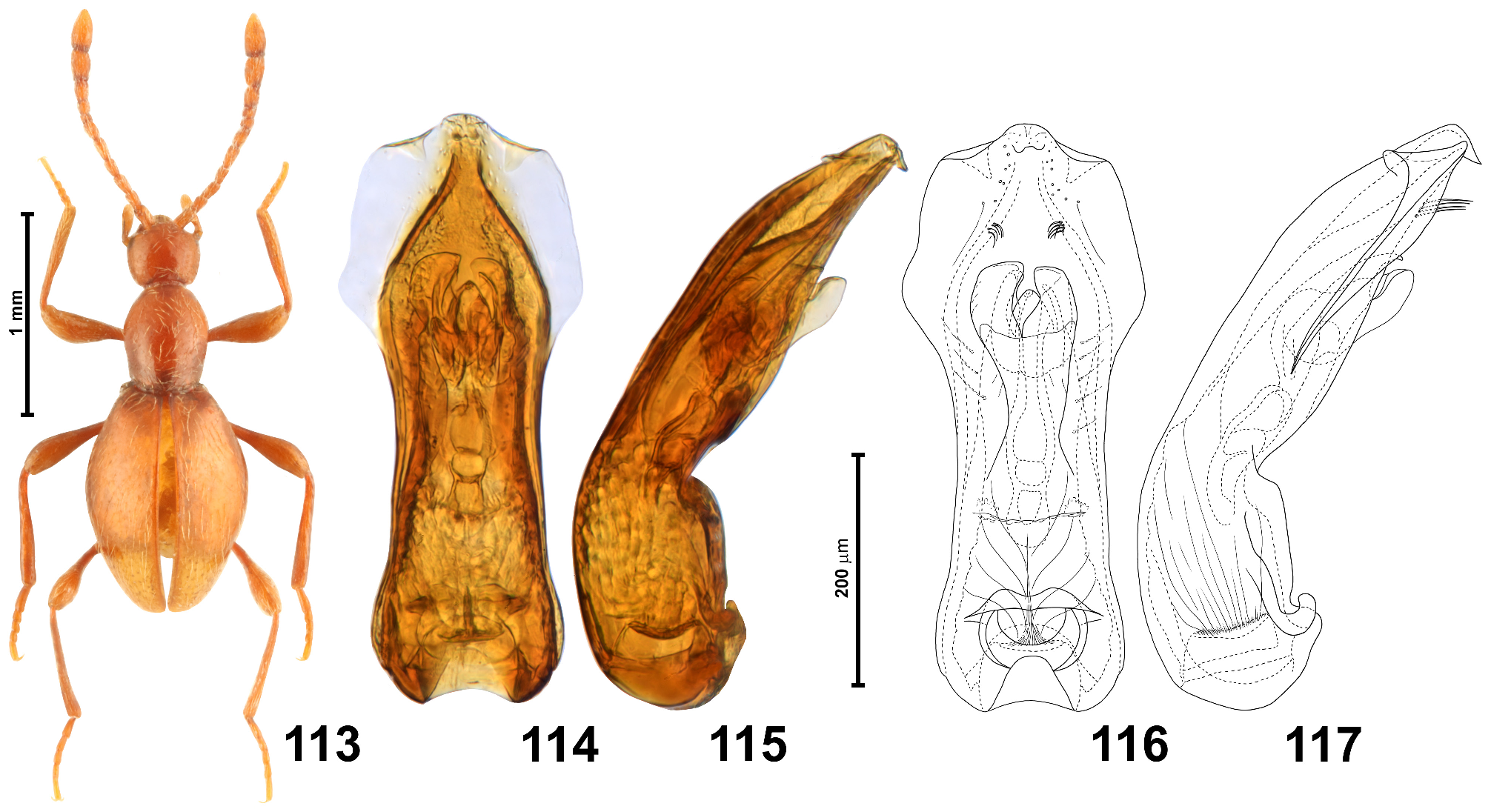
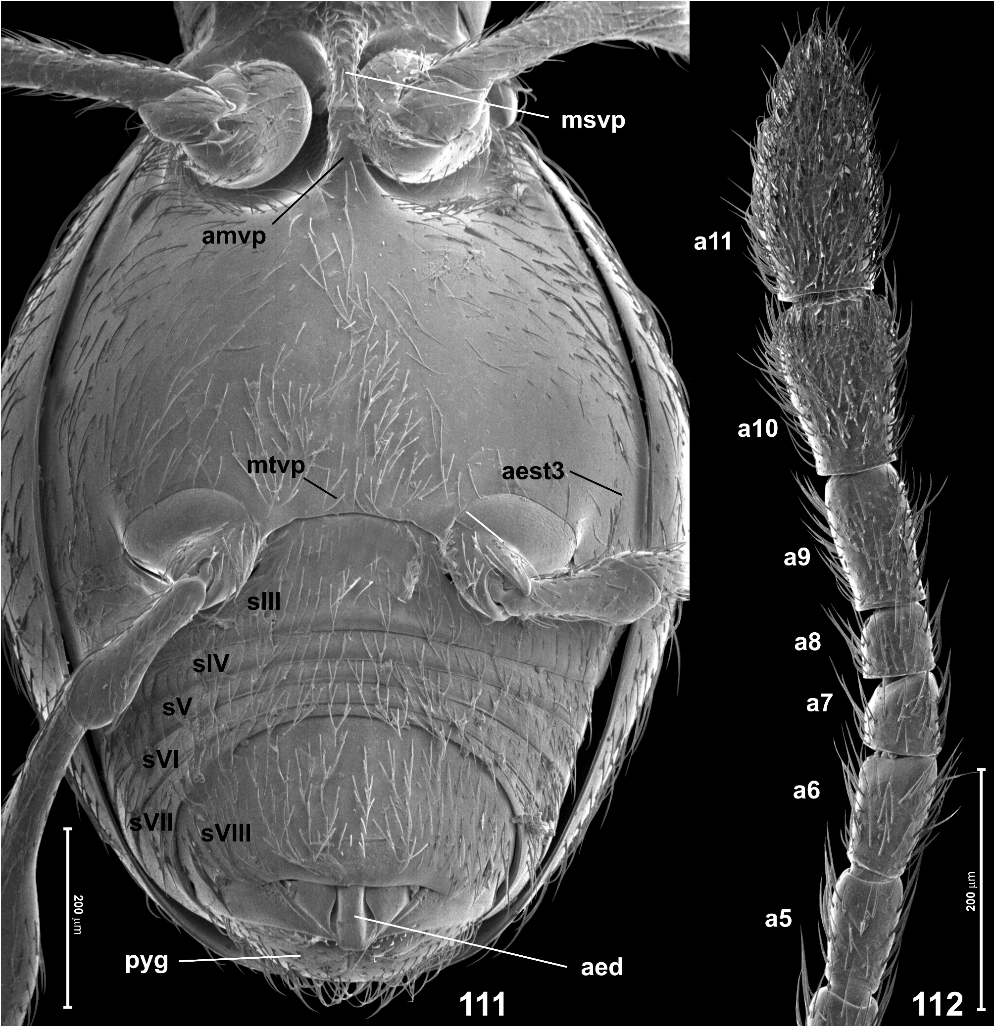
Revised diagnosis. In male antennomeres 3–6 each strongly elongate, about twice as long as broad, antennomeres 9 and 10 each twice as long as broad, and antennomere 11 more than twice as long as broad, with rounded lateral margins ( Fig. 112 View FIGURES 111–112 ); metatibiae unmodified ( Fig. 113 View FIGURES 113–117 ); pronotum lacking antebasal pits; aedeagus in dorsal view ( Figs 114, 116 View FIGURES 113–117 ) with conspicuously narrow apex of median lobe, only as broad as 1/5 of widest (sub-basal) site of median lobe, flanked by strongly elongate, subtrapezoidal and weakly sclerotized lateral subapical lobes, each with small distal subtriangular projection directed dorsally, and with two small subapical groups of long setae, also directed dorsally.
Redescription. Body in male ( Fig. 113 View FIGURES 113–117 ) strongly convex, elongate and relatively slender, BL 1.88 mm; pigmentation uniformly light brown (including appendages); cuticle moderately glossy, covered with vestiture of setae slightly lighter than body.
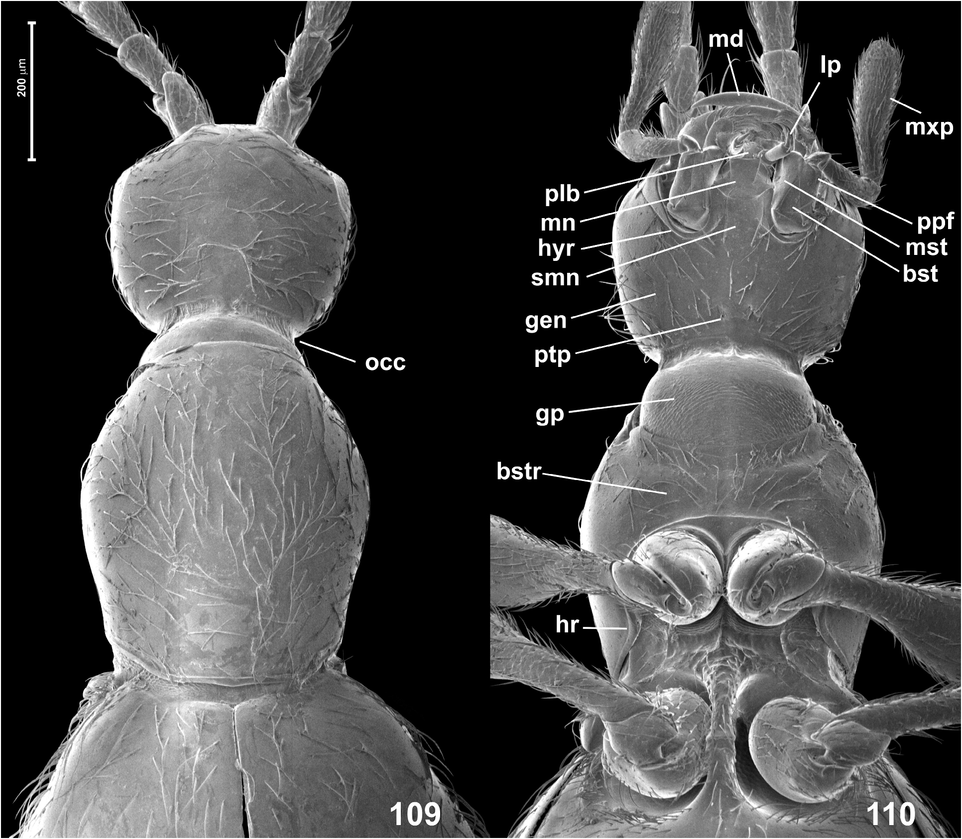
Head ( Figs 109–110 View FIGURES 109–110 ) in dorsal view indistinctly transverse and somewhat subhexagonal, broadest at eyes, HL 0.30 mm, HW 0.33 mm; vertex and frons confluent and weakly convex, posterior margin of vertex evenly arcuate and anteriorly convex; tempora about 2.5 × as long as length of eye in dorsal view; supraantennal tubercles indistinct; frons over antennal fossae broadly subtriangular and with rounded anterior margin. Eyes small, weakly oval, not emarginate posteriorly and oblique in relation to long axis of head. Punctures on frons and vertex fine, inconspicuous; setae (including those on tempora) short, sparse, nearly recumbent. Genae ( Fig. 110 View FIGURES 109–110 ) as sparsely setose as frons and vertex. Anterior (exposed) region of head capsule demarcated from neck region by short abrupt impression around occipital constriction, anterior margin of gular plate on neck region with indistinct, narrow anteriorly-directed projection. Submentum ( Fig. 110 View FIGURES 109–110 ) without submental lobes; hypostomal ridges ( Fig. 110 View FIGURES 109–110 ) extend mesally and anteriorly, but not connected at middle behind anterior margin of submentum. Antennae ( Figs 112–113 View FIGURES 111–112 View FIGURES 113–117 ) long and slender, AnL 1.18 mm; three terminal antennomeres forming indistinctly delimited club; scape 2.5 × as long as broad, distinctly broadening distally; pedicel 1.8 × as long as broad; antennomeres 3–6 each strongly elongate, about twice as long as broad, 7 and 8 each distinctly asymmetrical and about as long as broad, 9 twice as long as broad and nearly cylindrical, 10 twice as long as broad and distinctly broadening distally, 11 much shorter than 9 and 10 combined, about 2.2 × as long as broad, indistinctly asymmetrical.
Pronotum in dorsal view ( Fig. 109 View FIGURES 109–110 ) distinctly elongate, broadest between middle and anterior third, PL 0.50 mm, PW 0.40 mm; anterior margin arcuate and laterally confluent with rounded lateral margins, so that anterior corners are not marked; posterior corners obtuse-angled and blunt; posterior margin nearly straight at middle and bent anteriorly at sides; base with very narrow and indistinct posterior marginal carina, lacking pits. Pronotal disc covered with fine and shallow, unremarkable sparse punctures; setae similar to those on head, moderately dense and long, suberect. Ventrally ( Fig. 110 View FIGURES 109–110 ) prothorax with nearly asetose and impunctate hypomera and basisternal region only slightly longer than procoxal rests, sparsely covered with moderately long recumbent setae, with short but distinct and slightly impressed anterior ‘collar’, and with distinct vestiges of notosternal sutures visible as notches on sides of anterior prothoracic margin; hypomeral ridges distinct and complete, demarcating narrow inner (adcoxal) region of each hypomeron, anteriorly running along procoxal rests and connecting at middle to form biarcuate anteprocoxal carina demarcating basisternal region posteriorly.
Elytra ( Figs 109 View FIGURES 109–110 , 113 View FIGURES 113–117 ) slightly rhomboidal, broadest near middle, EL 1.08 mm, EW 0.73 mm, EI 1.48. Humeral calli small but distinctly elevated and each mesally demarcated by shallow and transverse basal elytral impression; basal elytral foveae lacking; apices separately rounded. Elytral punctures fine and inconspicuous; setae similar to those on pronotum, moderately dense. Hind wings fully developed.
Mesoventrite (partly visible in Fig. 111 View FIGURES 111–112 ) with carinate subrectangular mesoventral intermesocoxal process posteriorly fused with elongate and posteriorly broadening anterior metaventral process, fusion site distinctly marked on surface by transverse ridge. Metanepisterna ( Fig. 111 View FIGURES 111–112 ) fused with metaventrite. Metaventral intermetacoxal process ( Fig. 111 View FIGURES 111–112 ) broad and short, with strongly concave posterior margin, its lateral portions form short subtriangular processes weakly projecting posteriorly; distance between metacoxae subequal to 1/3 width of metaventrite at its posterior margin (excluding metanepisterna) and slightly wider than one metacoxa. Metaventrite ( Fig. 111 View FIGURES 111–112 ) weakly convex, unmodified and covered with sparse, short, slightly suberect setae, slightly denser on posteromedian area than those on sides.
Legs ( Figs 110 View FIGURES 109–110 , 111 View FIGURES 111–112 , 113 View FIGURES 113–117 ) long and slender; unmodified, except for slightly broadened proximal region of protarsi bearing tenent setae on tarsomeres 1–3. Protarsomere 1 nearly twice as long as broad, 2–4 each indistinctly elongate, 5 about 3 × as long as broad; mesotarsi longer than protarsi, mesotarsomere 1 about 3 × as long as broad, tarsomeres 2–4 each weakly elongate, tarsomere 5 about 2.5 × as long as broad; metatarsi slightly longer than mesotarsi, metatarsomere 1 nearly 3 × as long as broad, tarsomeres 2–4 each nearly twice as long as broad, tarsomere 5 about 2.5 × as long as broad.
Aedeagus ( Figs 114–117 View FIGURES 113–117 ) elongate but not very slender, AeL 0.50 mm, in dorsal view median lobe (excluding lateral subapical lobes) broadest near base, narrowing distally, then broadening in subapical region, to become strongly tapered in apical area, with apical margin weakly arcuate and as narrow as only 1/5 of median lobe at broadest site; lateral subapical lobes conspicuously large, each subtrapezoidal and strongly elongate, with additional subtriangular projection near apex, directed dorsally; flagellum broadened in proximal region to form three consecutive symmetrical chambers; median lobe with two small groups of long setae in subapical dorsal region directed dorsally; ostium situated in distal third of median lobe, far from its apex.
Female. Unknown.
Distribution. Known only from NW (Marrawah) and NC (Latrobe) Tasmania, and from CS Victoria (Lal Lal) (localities after Lea (2012)).
Remarks. Franz (1975) stated that the type material preserved at SAMA was composed of three specimens mounted on one pin, and farther in the text he refers to a holotype. However, the type series found in SAMA is composed of four syntypes, and a lectotype is designated here. Franz remounted one syntype (i.e., he removed one specimen from the original Lea’s card) and now it is on a separate pin, with handwritten copies of the original labels.
Scydmaenus myrmecobius has a relatively unremarkable external morphology; although its elytra are stouter than those in other similar species (mostly placed by Franz (1975) in Scydmaenus s. str.), it is easy to confuse this species with other congeners. Examination of the uniquely shaped aedeagus is the only unambiguous way to confirm identification.
Lea (1912) mentioned five studied specimens, “probably all males”; and he described “The middle trochanters each have a small acute tooth, projecting inwards and slightly forwards, but owing to its position it is not easily seen.” Such a tooth was not found in the studied syntypes, and I do not know of any species of world Scydmaenus with modified mesotrochanters. It is possible that Lea meant the distoventral corner of middle trochanters, which is subtriangular and can possibly be described as forming a tooth. However, this feature is common in most species of Scydmaenus and is not sexually dimorphic.

| SAMA |
South Australia Museum |
No known copyright restrictions apply. See Agosti, D., Egloff, W., 2009. Taxonomic information exchange and copyright: the Plazi approach. BMC Research Notes 2009, 2:53 for further explanation.
|
Kingdom |
|
|
Phylum |
|
|
Class |
|
|
Order |
|
|
Family |
|
|
Genus |
Scydmaenus (Parallomicrus) myrmecobius Csiki
| Jałoszyński, Paweł 2023 |
Scydmaenus (Allomicrus) myrmecobius
| Csiki, E. 1919: 73 |
| Lea, A. M. 1912: 60 |
Heterognathus myrmecophilus
| Lea, A. M. 1912: 60 |
| Aube, C. 1861: 197 |