
Trimma meityae, Winterbottom & Erdmann, 2018
|
publication ID |
https://doi.org/ 10.11646/zootaxa.4444.4.7 |
|
publication LSID |
lsid:zoobank.org:pub:D943FB45-6B57-430D-9B77-3B0FB0F647DE |
|
DOI |
https://doi.org/10.5281/zenodo.5997057 |
|
persistent identifier |
https://treatment.plazi.org/id/7A218DC5-BADD-46F5-AF4B-699D538BF4D3 |
|
taxon LSID |
lsid:zoobank.org:act:7A218DC5-BADD-46F5-AF4B-699D538BF4D3 |
|
treatment provided by |
Plazi |
|
scientific name |
Trimma meityae |
| status |
sp. nov. |
Trimma meityae new species
Meity’s Pygmygoby
Figs. 4–7 View FIGURE 4 View FIGURE 5 View FIGURE 6 View FIGURE 7 .
No published names pertain to this species.
Material examined. Holotype. ROM 106350 View Materials , 18.6 View Materials mm SL male, Indonesia, West Papua, Pulau Purup, Cendrawasih Bay , 02° 03.188' S, 134° 09.318' E, 56 m, 4 Aug., 2017, M.V. Erdmann. GoogleMaps
Paratypes: MZB 24599, 2 View Materials (17.1–17.2), collected with the holotype. ROM T21296, (15.5 mm SL female), Indonesia, West Papua, Cendrawasih Bay, Pulau Purup , 02° 03.188' S, 134° 09.318' E, 52 m, 24 Oct., 2016, field # MVE-16-070, M.V. Erdmann GoogleMaps . ROM 106364, 3(17.3–17.7), collected with the holotype. ROM T25134, (16.1), & T25135, (16.8), collected with the holotype.
Diagnosis. A species of Trimma with scales present on cheeks and opercle, 8–10 scales in predorsal midline, 17–18 unbranched pectoral fin rays, unbranched 5th pelvic fin ray 40–47% length of 4th ray, 21–22 total gill rakers, a broad interorbital (56–79% pupil width) with narrow crease-like postorbital groove, ending at posteriormost papilla in row p, 8 papillae in row p (with 2 papillae ventral to papilla row n), and nasal apparatus small and situated on anterior one-third of snout with posterior nares forming posterodorsal margin of nasal sac. When live or freshly collected, dorsal surface of eye light blue; preserved specimens with melanophores on dorsal surface of snout mostly adjacent to nasal capsules.
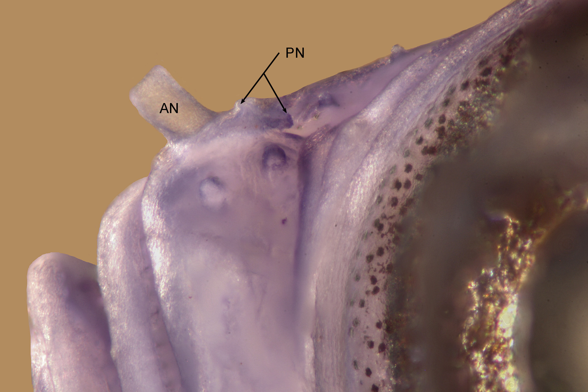
Description. The description is based on the holotype and 5 paratypes. Dorsal fin VI + I 8, second spine elongated, reaching posteriorly when adpressed to between base of 5th ray to 1 st scale behind last dorsal fin ray ( Fig. 5 View FIGURE 5 , mean = base of 7th ray, holotype to base ray 8), first ray of second dorsal fin branched (unbranched in two), remaining fin rays branched except for posterior element of last ray, fin reaches posteriorly 46– 57 –61% (54%) distance between base of last ray and first exposed dorsal procurrent caudal fin ray; anal fin I 6– 8 (7.7, single specimen with 6 rays, clearly deformed or due to injury), first ray unbranched, fin reaches posteriorly 31– 33 –42% (34%) distance between base of last ray and first exposed ventral procurrent caudal fin ray; pectoral fin 17 –18 (17.7), all rays unbranched, fin reaching posteriorly to region above urogenital papilla to anal spine; pelvic fin I 5, fifth ray unbranched and 40– 46 –47% (44%) length of fourth ray, which reaches posteriorly to between bases of anal spine to 2nd anal ray, pelvic rays 1–4 with single sequential branch point; basal membrane forming fold across midline above last pre-pelvic scale; no fraenum. Lateral scales 23; anterior transverse scales 8; posterior transverse scales 7; cheek with two rows cycloid scales, upper row of 1 –2 and lower row with 6– 7 (means = 1.5 and 6.8 respectively, midline of predorsal with 8– 9 –10 (9.0) scales, anterior 1–4 rows may be cycloid, otherwise ctenoid; anteriormost scales on sides (may be ctenoid or cycloid) and top of nape reaching anteriorly almost to posterior margin of eye; opercle fully scaled above papilla row oi in 4 horizontal rows, dorsalmost row of 4 –5, then 2– 3, 3 and 1 –2 mostly cycloid scales, although scales in mid-upper region may be ctenoid; 3 vertical rows of cycloid scales on pectoral fin base with 1 –2 in anteriormost row, 2– 3 in second row and 5 in outer row; 7 –8 (7.2) cycloid scales in midline anterior to pelvic fin base; area between pelvic spine and ventral margin of pectoral fin base with cycloid (smaller specimens) or ctenoid (larger specimens) scales; anterior few rows of scales in midline of belly cycloid; circumpeduncular scales 12, scales rows in midline between base of last anal ray and first ventral procurrent caudal fin ray 8– 9 –10 (9.2). Upper jaw with outer row of spaced, enlarged curved canines which decrease slightly in height and reach posteriorly 4/5ths of length of premaxilla, two irregular inner rows of small conical teeth (about half height of outer teeth) becoming reduced to single row at bend of premaxilla and continuing posteriorly to 3/5ths length of premaxilla. Lower jaw with short row of about 5 enlarged, spaced, curved canines from symphysis almost to bend of dentary, about 2 irregular rows of slightly curved smaller (2/3rds height of outer teeth) teeth at symphysis, grading to single row, decreasing slightly in size posteriorly, and reaching almost to tip of coronoid process of dentary. Tongue broadly rounded to spatulate with small central tip. Gill opening extending anteroventrally to below mid to posterior pupil; gill rakers 4 –5 + 16– 18 = 21– 22 (4.7 + 17.0 = 21.7). Nasal apparatus small, situated on anterior one-third of snout, anterior naris short tapering tube reaching anteriorly to above anterior margin of upper lip, posterior opening a large, pore-like opening with slightly raised rim covering posterodorsal width of nasal sac, transverse width of pore about 65% length of nasal capsule, posterior margin of posterior naris separated from bony front of orbit by 3.5 –4.0 times its transverse width (mean = 3.9), nasal sac only very slightly raised above surrounding area of snout ( Fig. 6 View FIGURE 6 ). Bony interorbital width 56– 70 –79.0% (65.4) pupil diameter; profile of snout gently convex, with shallow concave depression between eyes (between 4th and 5th papillae of row p); epaxialis reaching anteriorly in midline to vertical above posterior margin of pupil; no narrow ridge of skin in midline of nape extending anteriorly from origin of first dorsal fin. Caudal peduncle depth as percentage caudal peduncle length 34.4– 42.2 (38.4); head length as percentage SL 32.9– 33.2 –35.2 (33.7); as percentage head length: horizontal eye diameter 34.6 –38.9 (36.9); snout length 21.2– 22.1 –22.8 (22.2); cheek depth 16.8– 20.9 –21.2 (19.1). Cephalic sensory papillae as in Fig. 7 View FIGURE 7 . Number of papillae in each row: a = 6; b = 4– 5 –6 (5.0); c = 6; cp = 1; d = 6– 7 (6.5); dʹ = 6– 7 –8 (7.3); e-anterior = 12– 13 –14 (12.8); e-posterior = 12– 15 –17 (14.5); i-anterior = 6 –7 (6.7); i-posterior = 7; p = 8, with 2 papillae just medial to posterior naris and 2 papillae ventral to row n; r = 2; f = 3 –4 (3.2); cs" = 3; g = apparently absent, but may be represented by zig-zag line of 7 papillae in one specimen; n = 1; x = 6– 7 –8 (7.2, n = 5); u = 4– 5 (4.3); z = 5– 7 (6.3, n = 4); ot = 11– 13 (12.3, n = 3); os = 7– 8 (7.7, n = 3); oi = 4– 7 (5.5, n = 4). Abdominal/caudal vertebral transition not examined.
Colour pattern. Live, based on 12 images from Cendrawasih Bay, West Papua, Indonesia. Specimens ( Fig. 5 View FIGURE 5 , A, B) almost entirely translucent light pink or off-white with yellow spinal cord, much lighter and brighter blue over dorsomedial surface of eye than T. blematium , brown iris ring relatively indistinct and white inner ring around pupil wider and about half iris width. Fin rays pinkish, no apparent pigmentation in fin membranes. Other photographed specimens from this locality with darker pink background and blue over eyes less well developed. Freshly collected, ( Fig. 5 C View FIGURE 5 ). Upper and lower midlines reddish, body semi-translucent greenish with scale margins diffusely outlined with dark red, posterior region of cranium dull yellow, snout reddish, distal half of upper lip and region of snout below anterior margin of eye orange-red. Dorsal fins with basal stripe of whitish chromatophores, stripe half pupil-width anteriorly to full pupil-width posteriorly, fin elements pink above this; caudal with bluepurple margins and 3 equidistant similar streaks in body of fin, the middle medial in position, rest of fin greenishyellow; membranes of pelvic fin with some iridocytes, rays off-white, membranes of pectoral fin hyaline with rays light pink. Preserved. Holotype pale off-white with scale margins in predorsal region and dorsal-most scale row along entire dorsum outlined with brown melanophores; a thin line of melanophores in midline from end of anal fin base almost to first ventral procurrent ray, a few melanophores medial to posterior nares ( Fig. 7 B View FIGURE 7 ), on snout below anteroventral margin of eye, and from upper operculum anterior to posterior margin of eye ( Fig. 7 A View FIGURE 7 ). Paratypes similar, but lack melanophores anteroventral to the eye, and may lack those over operculum.
Etymology. The species is name “ meityae ” in honour of Meity Mongdong, one of Indonesia’s foremost marine conservationists, who has dedicated the past several decades of her career towards expanding and improving the management of marine protected areas in West Papua, including the Cendrawasih Bay National Park where this species is found.
Distribution and habitat. This species is currently known only from Cendrawasih Bay (Pulau Purup), West Papua, Indonesia. It was observed and collected from a nearshore reef with almost no exposure to waves or currents, in the 50-60 m depth zone only, on a silty sand and rubble slope.
Comparisons. There are 9 other described species of Trimma which possess predorsal scales in the midline, scales on the cheek, unbranched pectoral fin rays, an unbranched 5th pelvic fin ray, and a bony interorbital width that is ~50% or more of the pupil width. These are: T. abyssum Allen, 2015 , T. burridgeae Winterbottom, 2016 , T. caudomaculatum Yoshino & Araga, 1975 , T. corerefum Winterbottom, 2016 , T. habrum Winterbottom, 2011 , T. helenae Winterbottom et al., 2014a , T. hollemani Winterbottom, 2016 , T. kitrinum Winterbottom & Hoese, 2015 and T. tevegae Cohen & Davies, 1969 . None of these species has 17 or more pectoral fin rays as found in T. meityae . Trimma abyssum has 16 such rays (the others have 15 or fewer), but has a second dorsal spine reaching posteriorly only as far as the bases of the first or second ray of the second dorsal fin (vs. to between base of 5th ray to 1 st scale behind last dorsal fin ray in T. meityae ). Only three of these species have been recorded with as many as 21 total gill rakers ( T. burridgeae with 19-21 (mean = 19.6), T. caudomaculatum with 17–21 (18.9) and T. kitrinum 20–22 (21.3), versus 21–22 (21.7) in T. meityae . In these species, the bony interorbital is 70–100% of pupil width (vs. 56–79.0%, mean = 65.4). A dark caudal spot is present in all the above listed species except T. meityae , T. habrum , T. helenae and T. kitrinum , where it is absent. See T. blematium under the Comparisons section for the similarities and differences between that species and T. meityae .
Discussion. See T. blematium under the Discussion section for comments.


No known copyright restrictions apply. See Agosti, D., Egloff, W., 2009. Taxonomic information exchange and copyright: the Plazi approach. BMC Research Notes 2009, 2:53 for further explanation.
|
Kingdom |
|
|
Phylum |
|
|
Class |
|
|
Order |
|
|
Family |
|
|
Genus |