
Lutetialestes uniformis, Greenwalt, Dale E. & Bechly, Günter, 2014
|
publication ID |
https://doi.org/ 10.11646/zootaxa.3887.2.2 |
|
publication LSID |
lsid:zoobank.org:pub:D9275063-BD74-498E-B504-1421B50114A5 |
|
DOI |
https://doi.org/10.5281/zenodo.6141530 |
|
persistent identifier |
https://treatment.plazi.org/id/40446A46-FFB2-670F-FF4C-043A8AF9FE11 |
|
treatment provided by |
Plazi |
|
scientific name |
Lutetialestes uniformis |
| status |
sp. nov. |
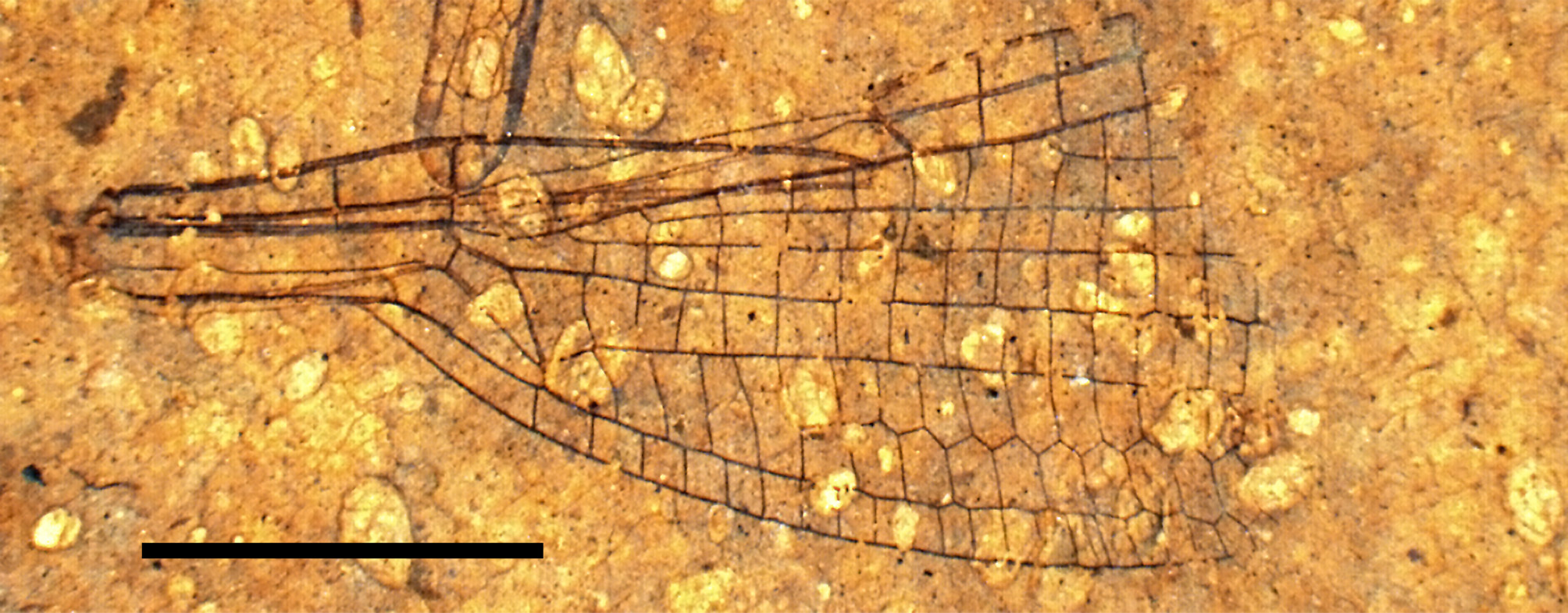
Lutetialestes uniformis n. sp.
( Figures 8 View FIGURE 8 , 9 View FIGURE 9 A, 10)
Holotype. USNM 559048, National Museum of Natural History, Washington, D.C.
Type locality and stratum. Disbrow Creek site, Middle Fork of the Flathead River, Pinnacle, Montana, USA. Coal Creek Member of the Kishenehn Formation, early Middle Eocene, 46.2±0.4 or 43.5±4.9 mya ( Constenius, 1996).
Etymology. Species name uniformis from the Latin word uniformis (uniform, consistent), an indication of the very uniform shape of the cells in the cubital field.
Diagnosis. 1) MA slightly zigzagged only distal of RP 2 origin; 2) Ax2 opposite anterior arcular crossvein; 3) Discoidal cell closed; 4) RP separates from RA+ RP distal of anterior arcular crossvein so that there is no RP +MA vein; 5) IR2 and RP 3/4 origin closer to arculus than nodus; 6) A relatively short petiole; 7) Postnodal crossveins aligned; 8) A broad cubital field consisting of five basal cells longer than wide followed by a single supplementary longitudinal sector that defines two rows of cells with the shape of isosceles right pentagons.
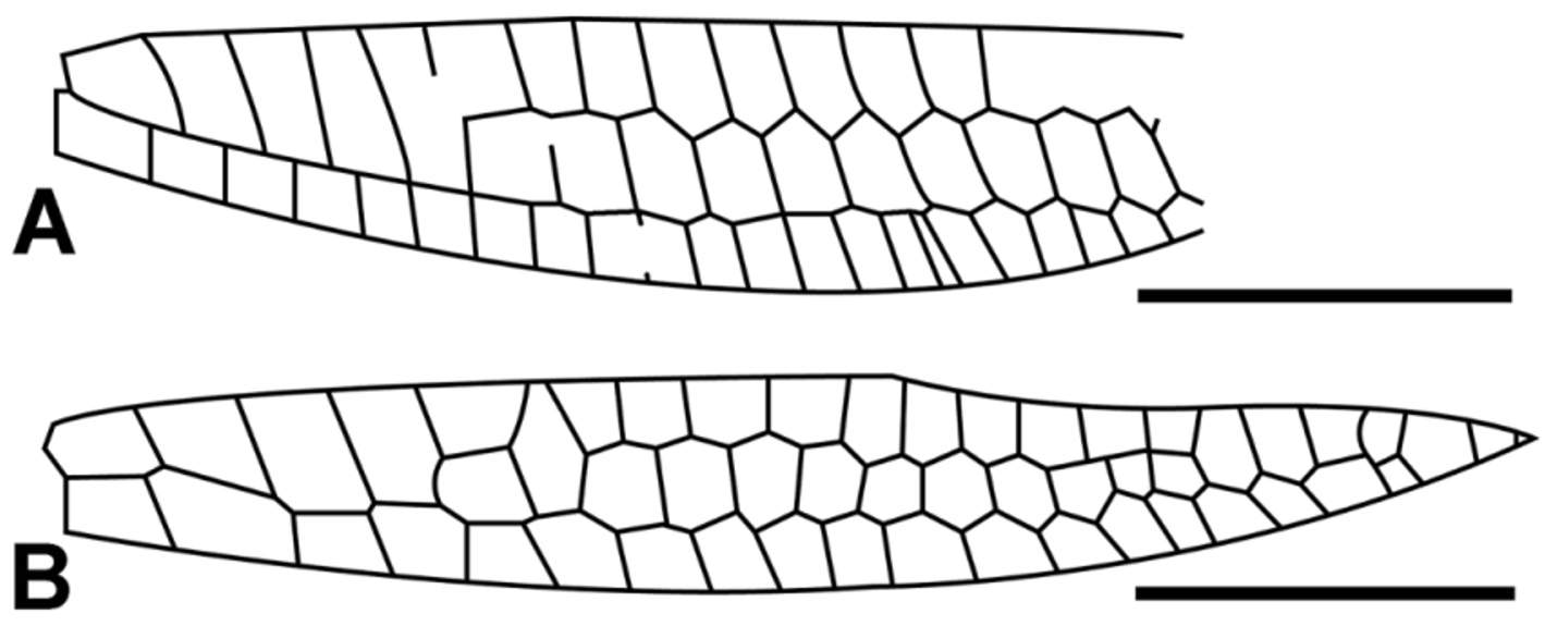
Description. Basal half of a fossil damselfly wing (anterior veins CA & CP & ScA and ScP have broken apart from nodus, moved posteriorly approximately 1 mm and, in so doing, have caused a buckling of RP 1 just distal of arculus.) ( Fig. 8 View FIGURE 8 ); wing hyaline, 14.7 mm long (from base to a point four cells distal of RP 2 origin) and 5.59 mm and 6.80 mm wide at nodus and widest point respectively; distance between nodus and arculus and arculus and base 5.41 and 4.62 mm respectively; Petiole length relative to distance from petiole to nodus = 0.50; two antenodal and two antesubnodal crossveins. Ax1 0.2 mm basal of separation of AA’ & AA’’ and separated from Ax2 by 1.45 mm; Ax2 opposite arcular crossvein and basal of RP origin; supplementary antenodal crossveins absent; posterior arcular crossvein separating from discoidal cell 0.19 mm below RA; nodal and subnodal crossveins oblique; subnodal bracket apparently not thickened; all three (first three) postnodal crossveins present exactly aligned with postsubnodal crossveins; base of RP 2 three cells and 1.64 mm distal of subnodus and seven cells and 5.04 mm distal of origin of IR2; IR2 and PR3/4 originate much closer to arculus than nodus. IR2 arched abruptly toward RP at base with crossvein opposite, one cell and 0.93 mm from origin of RP 3/4; RP 3/4 originates one cell and 1.45 mm from arculus; MA straight proximal of origin of RP 2 and slightly zigzagged distal to RP 2 origin; MP with no or very little arch as it leaves discoidal cell at angles of 92 and 74 degrees from MAb and vertical respectively; CuA a prominent vein that leaves subdiscoidal cell 0.29 mm below MP and transitions to a zigzagged pattern between levels of subnodus and origin of RP 2; CuA underlies a broad cubital field 1.73 mm in height that starts with a single cell below base of MP and continues through four cells 1.8 to 2.8 times as high as wide, at which point a supplementary longitudinal vein forms and delineates two rows of cells, ten cells in length ( Fig. 9 View FIGURE 9 A). Supplementary longitudinal sector highly and very uniformly zigzagged and a defining characteristic of this specimen. Except for the very first cell, cubito-anal field cells are square in shape transitioning to higher than wide at level of subnodus; cubito-anal field 2.41 mm in height; CuP origin 0.49 mm distal of separation of AA’ & AA’’, approximately halfway between Ax1 and Ax2; petiole well defined, 3.44 mm long (from base to separation of AA’ and AA’’); discoidal cell closed basally, 1.80 mm long (end of posterior arcular vein to origin of MP) and 0.79 mm wide (origin of arcular vein to origin of MAb) with an acute posterior internal angle of 26 degrees; distal side of discoidal cell (MAb) 18 degrees from vertical relative to RA and 1.24 mm in length; anterior, posterior and basal (posterior arculus) sides 0.70 mm, 1.67 mm and 0.35 mm long; ratio of lengths of anterior and posterior sides of discoidal cell = 0.42; RP separates from RA+ RP distal of anterior arcular crossvein so that vein RP +MA does not exist ( Fig. 10 View FIGURE 10 ); subdiscoidal cell elongate with no fusion of CuP & AA’ to posterior wing margin distal of CuP. The same slab also contains fossil ostracods and the wing of a hemipteran.

| USNM |
Smithsonian Institution, National Museum of Natural History |
No known copyright restrictions apply. See Agosti, D., Egloff, W., 2009. Taxonomic information exchange and copyright: the Plazi approach. BMC Research Notes 2009, 2:53 for further explanation.
|
Kingdom |
|
|
Phylum |
|
|
Class |
|
|
Order |
|
|
Family |
|
|
Genus |