
Eolestes syntheticus Cockerell, 1940
|
publication ID |
https://doi.org/ 10.11646/zootaxa.3887.2.2 |
|
publication LSID |
lsid:zoobank.org:pub:D9275063-BD74-498E-B504-1421B50114A5 |
|
DOI |
https://doi.org/10.5281/zenodo.6141524 |
|
persistent identifier |
https://treatment.plazi.org/id/40446A46-FFBA-6706-FF4C-01EE8D69F85A |
|
treatment provided by |
Plazi |
|
scientific name |
Eolestes syntheticus Cockerell, 1940 |
| status |
|
Eolestes syntheticus Cockerell, 1940
( Figures 1 View FIGURE 1 , 2 View FIGURE 2 , 3 View FIGURE 3 , 4 View FIGURE 4 , 5 View FIGURE 5 A, 5B, 6A, 6B)
Synonomy.
v. 1940 Eolestes synthetica Cockerell 1940 ; p. 105, figs. 1, 2. v. 1974 Eolestes syntheticus Fisher 1974 ; p. 218.
Range. Eocene of Northwestern United States (Green River Formation near DeBeque, Colorado, and Kishenehn Formation, Pinnacle, Montana).
Holotype ( UCM 19170) ( Figures 1 View FIGURE 1 , 2 View FIGURE 2 , 3 View FIGURE 3 , 4 View FIGURE 4 , 5 View FIGURE 5 A, 6A)
Type locality and stratum. 39.5° N, 108.4, Roan Creek near de Begue, Garfield County, Colorado, USA. Green River Formation, Early Eocene, 53.5–48.5 mya ( Smith et al. 2003).
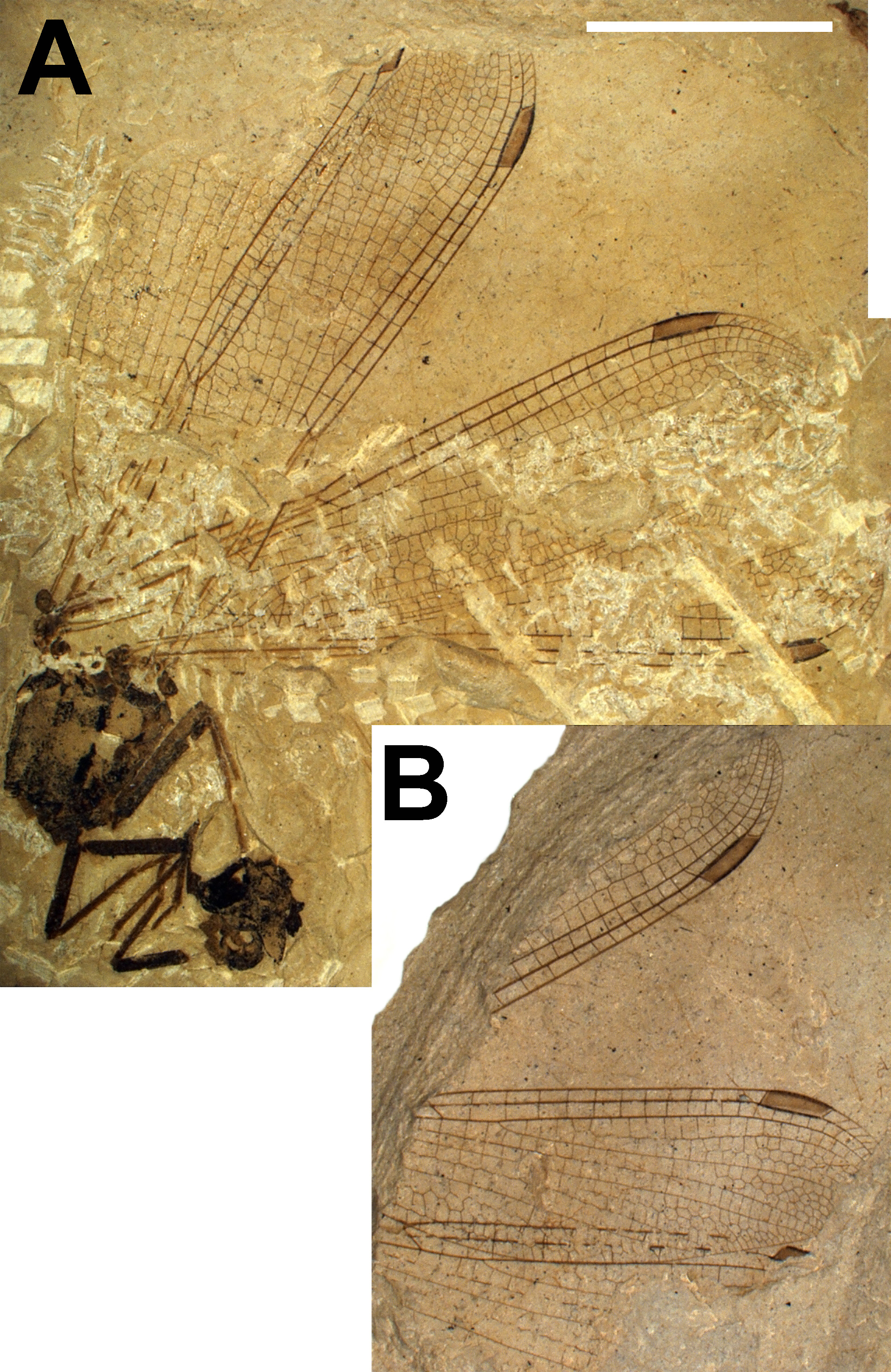
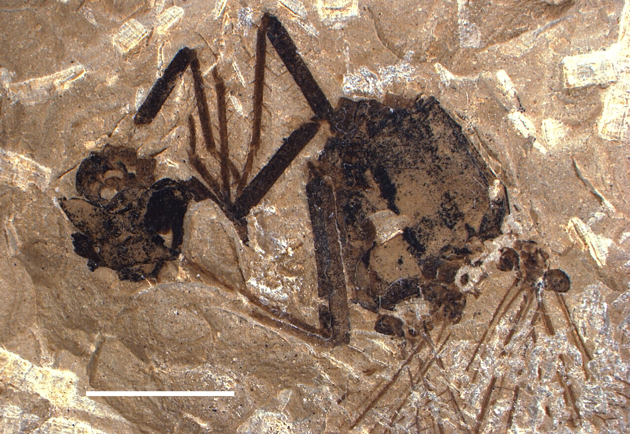
Re-description. Part and counterpart of a fossil damselfly with all or most of four wings, five legs, the dorsal aspect of the thorax and a poorly preserved head ( Fig. 1 View FIGURE 1 ); pterothorax square, about as long as high (lateral aspect), approximately 4.8 mm long at the mid-dorsal stripe of the mesanepisternum and 3.0 mm wide (between the humeral stripes) ( Fig. 2 View FIGURE 2 ); pro-, meso- and metafemora 3.10, 4.15 and 5.47 mm in length; pro- and mesotibiae 3.36 and 4.17 mm in length; tarsi 3-segmented with pro- and mesotarsi 1.37 and 1.98 mm in length respectively; pro- and mesofemora and pro- and mesotibiae with spines, mesotibial spines gradually tapering distally with a maximum length of 0.6 mm; pro- and mesotibial apical spurs 0.32 and 0.35 mm respectively; protarsal claw 0.36 mm in length.
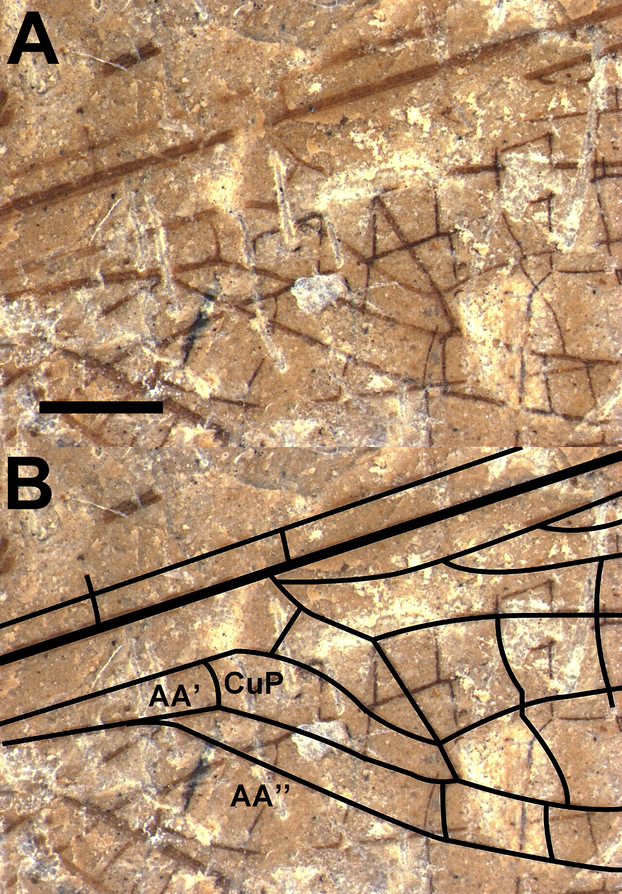
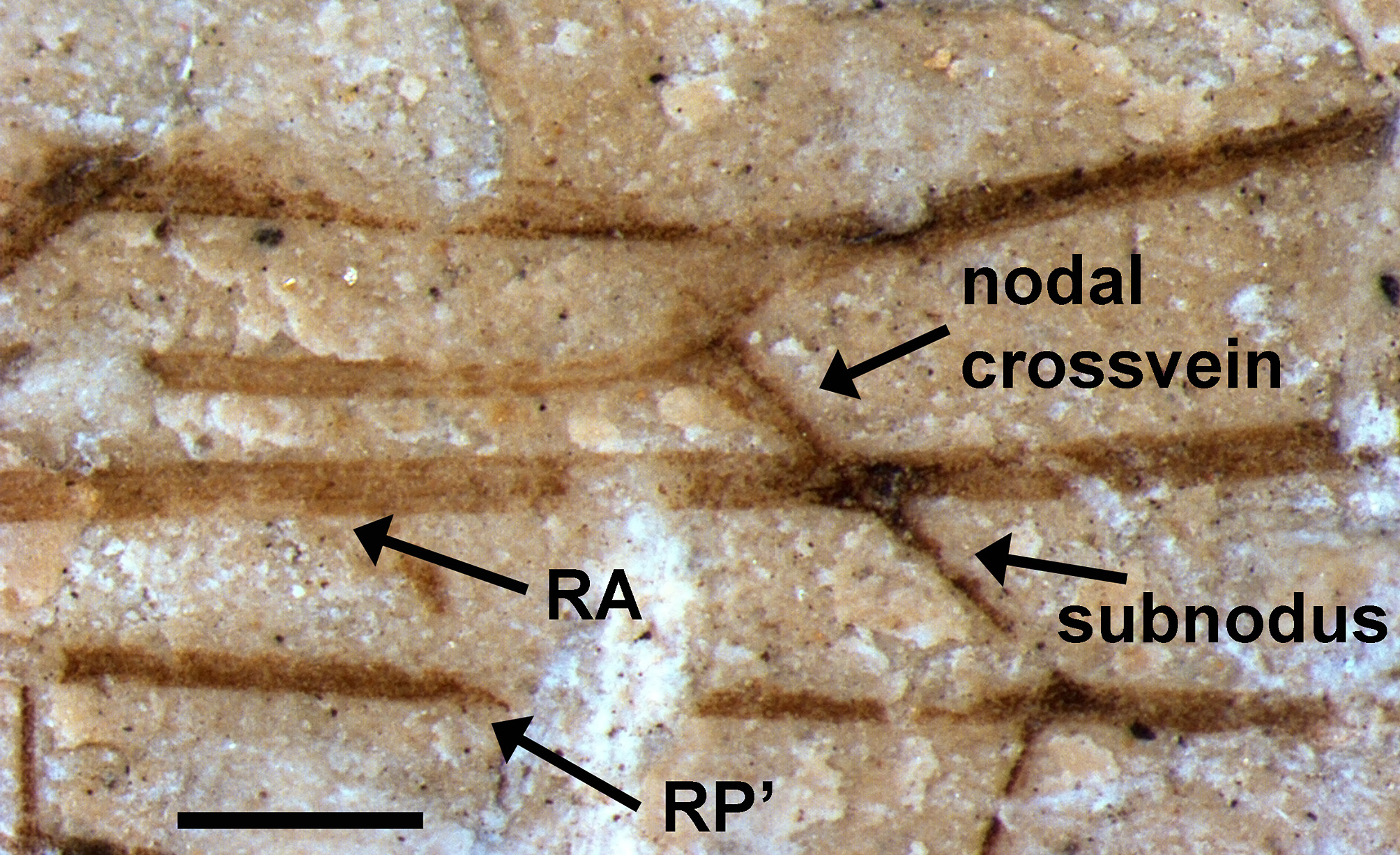
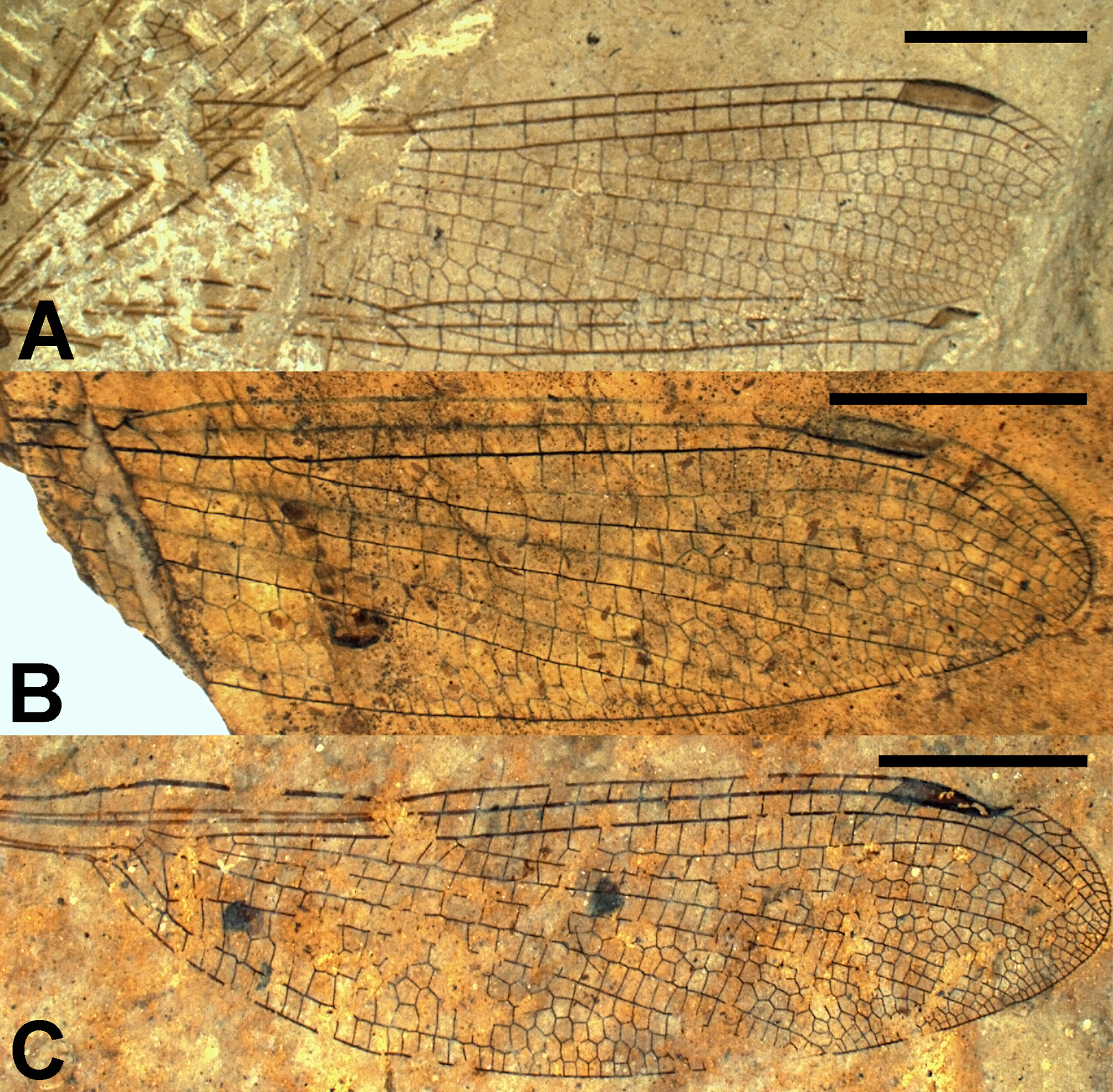
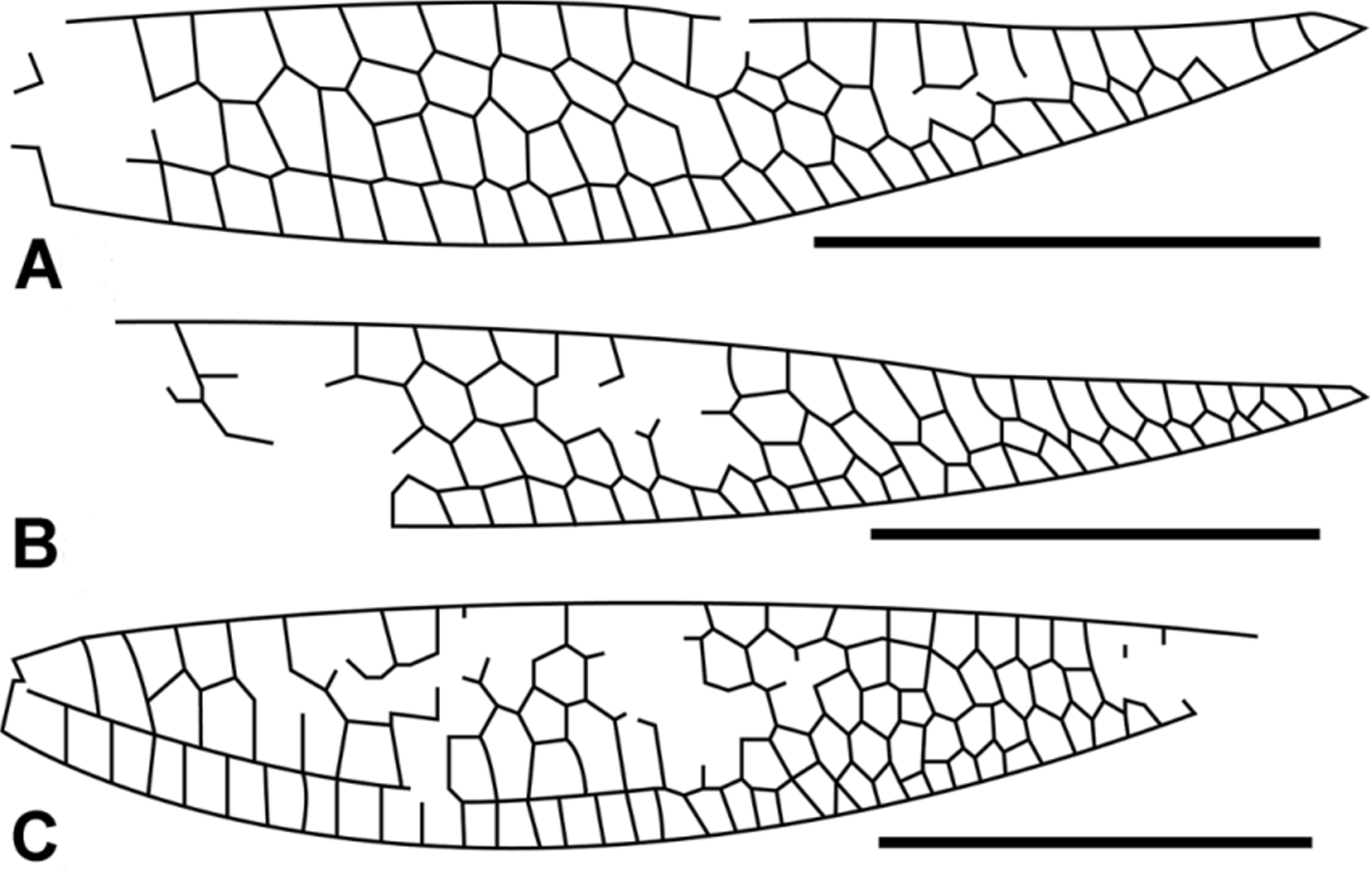
Forewing: Intact, hyaline, 30.62 mm long and 5.34 mm and 7.01 mm wide at nodus and widest point respectively; pterostigma four and a half cells and 2.52 mm long, 0.70 mm wide; pterostigmal brace oblique; distance between wing apex and pterostigma, pterostigma and nodus, nodus and arculus and arculus and base 3.03, 14.17, 5.74 and 5.23 mm respectively; the post-pterostigmal cell area does not appear to contain a supplementary sector; two antenodal and two antesubnodal crossveins with Ax1 basal of the separation of AA’ & AA’’ and separated from Ax2 by 1.61 mm ( Fig. 3 View FIGURE 3 ); Ax2 distal of anterior arcular crossvein; supplementary antenodal crossveins absent; posterior arcular crossvein separating from discoidal cell 0.39 mm below RA; RP separates from RP +MA just below RA+ RP so that RP +MA very short; nodal and subnodal crossveins oblique; dorsal subnodal bracket thickened ( Fig. 4 View FIGURE 4 ); 13 postnodal crossveins most of which are aligned with postsubnodal crossveins; ‘‘lestine’’ oblique vein ‘‘O’’ present 3.5 cells distal of base of RP 2 between RP 2 and IR2; base of IR1 four cells and 3.28 mm distal of RP 2 origin; IR1 underlies a single row of cells that, proximal to pterostigma, are higher than they are wide and overlies a row of seven single cells that transition through three double cells and, distally, an area of reticulated cells. Apical fifth of left forewing torn/split, causing gross misalignment of veins/cells; base of RP 2 three cells and 2.41 mm distal of subnodus and at least six cells and 6.56 mm distal of origin of IR2; IR2 and PR3/ 4 originate much closer to arculus than nodus. IR2 arched abruptly toward RP at base, one half cell and 0.86 mm from origin of RP 3/4; IR2 underlies a row of single cells basally, after which a supplementary longitudinal sector originates proximal to level of pterostigma and branches to five supplementary sectors near wing margin; RP 3/4 originates 1.33 mm from arculus and underlies a single row of single cells that extends to level of pterostigma, after which it is no longer visible; MA of right forewing slightly zigzagged only distal to subnodus; MP underlies a single row of cells that is obscured between subnodus and pterostigma; CuA a prominent vein that leaves subdiscoidal cell 0.33 mm below MP and transitions to a slightly zigzagged pattern at about level of origin of subnodus. It underlies a broad cubital field 1.81 mm in height that starts with a single cell below base of MP but transitions to contain three supplementary longitudinal veins with four rows of cells; anal field consists of a single row of cells that are either square or higher than wide; terminus of CuA at a point 2/3 of the way between nodus and pterostigma; cubito-anal field 2.64 mm in height; CuP distal of separation of AA’ & AA’’ and halfway between Ax1 and Ax2; petiole well defined, 3.69 mm long (from base to separation of AA’ and AA’’) and 12.05% of wing length; Petiole length relative to distance from petiole to nodus = 0.56; discoidal cell closed basally, 1.63 mm long (end of posterior arcular vein to origin of MP) and 0.81 mm wide (origin of arcular vein to origin of MAb) with an acute posterior internal angle of 25 degrees; distal side of discoidal cell (MAb) nearly perpendicular to RA (12 degrees from vertical relative to RA), and 1.01 mm in length; anterior, posterior and basal (posterior arculus) sides of discoidal cell 0.70 mm, 1.55 mm and 0.45 mm long; ratio of lengths of anterior and posterior sides of discoidal cell = 0.45; subdiscoidal cell elongate with no fusion of CuP & AA’ to posterior wing margin distal of CuP; MP slightly arched as it leaves discoidal cell at an angle of 93 degrees between MP and MAb.
Hind wing: intact, hyaline, 28.84 mm long and 5.17 mm and 6.68 mm wide at nodus and widest point respectively ( Fig. 5 View FIGURE 5 A); pterostigma three and a half cells and 2.62 mm long, 0.72 mm wide; pterostigmal brace oblique; distance between wing apex and pterostigma, pterostigma and nodus, nodus and arculus and arculus and base 2.48, 13.06, 5.36 and 5.43 mm respectively; post-pterostigmal cell area (five cells preserved only) does not appear to contain a supplementary longitudinal sector; two antenodal and two antesubnodal crossveins with Ax2 opposite arcular crossvein and separated from Ax1 by 1.65 mm; supplementary antenodal crossveins absent; subnodal crossvein oblique, nodal crossvein slightly less so; dorsal subnodal bracket slightly thickened; 11 postnodal crossveins exactly aligned with postsubnodal crossveins with exception of last two; ‘‘lestine’’ oblique vein ‘‘O’’ present four cells distal of base of RP 2; base of IR1 four cells and 3.04 mm distal of RP 2 origin; IR1 zigzagged to level of pterostigma and underlies a single row of cells that, proximal to the pterostigma, are higher than wide and overlies a row of seven single cells that expand distally through four double cells, four triple cells via two and then three supplementary longitudinal sectors—distal edge of wing not preserved. Base of RP 2 two and a half cells and 2.23 mm distal of subnodus; IR2 and PR3/4 originate closer to arculus than nodus. IR2 underlies a row of single cells basally, followed by a gradual increase in the number of supplementary longitudinal sectors until there are three supplementary longitudinal sectors distal of pterostigma; RP 3/4 underlies a row of single cells that transitions from cells longer than high to higher than long proximal to stigma; MA zigzagged from a point two cells proximal of subnodus—basal of that point, MA not preserved; MA underlies a row of cells that transitions to double cells at level of IR1 origin and then gradually to 16 small cells at wing margin; MP, not zigzagged, underlies a single row of cells that gradually transitions from square-shaped to cells higher than wide; CuA leaves subdiscoidal cell 0.29 mm below MP, becomes zigzagged as it approaches subnodus, and underlies a broad cubital field, 1.92 mm in height, that starts with a single cell below base of MP and quickly transitions through one and then two supplementary longitudinal sectors and then back to a single row of cells at wing margin ( Fig. 6 View FIGURE 6 A); cubito-anal field (between CuA and hind margin) with a single row of cells higher than wide with the exception of the first and a terminus at a point three or four cells from terminus of MP; height of cubito-anal field 2.51 mm; CuP origin distal of separation of AA’ & AA’’ approximately halfway between Ax1 and Ax2; petiole well defined, 3.65 mm long (from base to separation of AA’ and AA’’) and 12.65 % of wing length; Petiole length relative to distance from petiole to nodus = 0.59; discoidal cell poorly preserved; distal side of discoidal cell (MAb) nearly perpendicular to RA and 1.2 mm in length; MP slightly arched as it leaves the discoidal cell.
Both part and counterpart also contain a small fossil tipulid dipteran.
New specimen ( USNM 559049) ( Figures 5 View FIGURE 5 B, 6B)
Deposition. USNM 559049, National Museum of Natural History, Washington, D.C.
Locality and stratum. Spring site, Middle Fork of the Flathead River, Pinnacle, Montana, USA. Coal Creek Member of the Kishenehn Formation, early Middle Eocene, 46.2±0.4 or 43.5±4.9 mya ( Constenius, 1996).
Description. Apical portion (≈ 70%) of a fossil damselfly wing, hyaline, 21.48 mm long and 5.13 mm and 6.35 mm wide at nodus and widest point respectively ( Fig. 5 View FIGURE 5 B); pterostigma four cells and 2.60 mm long, 0.54 mm wide; pterostigmal brace oblique; distance between wing apex and pterostigma and pterostigma and nodus 3.22 and 13.4 mm respectively; post-pterostigmal cell area consists of three single cells proximally, four smaller cells distally and five double cells in between; nodal crossvein and subnodal crossvein oblique; anterior nodal bracket apparently thickened; 12 postnodal crossveins aligned with postsubnodal crossveins except for two most distal; ‘‘lestine’’ oblique vein ‘‘O’’ present three cells distal of base of RP 2; base of IR1 four cells and 3.18 mm distal of RP 2 origin, IR1 underlies a single row of cells that, proximal to the pterostigma, are higher than wide and overlies a row of nine single cells that distally expand through three double cells and, more distally, three rows of cells; base of RP 2 three cells and 2.13 mm distal of subnodus; IR2 and PR3/4 appear to originate closer to arculus than nodus, although their origins are not preserved (IR2 extends basally to edge of fossil at which point it is slightly less than halfway (0.44) between RP 1 and RP 3/4); IR2 underlies a row of at least 18 single cells basally, followed by a gradual increase in number of secondary veins until there are eight small cells at wing margin; RP 3/4relatively straight and underlies a row of single cells, although a short two-cell-long supplementary longitudinal sector appears at level of pterostigma; MA relatively straight proximal of RP 2, only very slightly zigzagged thereafter, and underlies a row of cells that transitions to double cells just distal of IR1 origin and then gradually to 17 small cells at wing margin; MP relatively straight, underlies a single row of cells; CuA zigzagged throughout and terminates three cells short of end of MP. Broad cubital field 1.78 mm in height and contains two supplementary longitudinal sectors that transition to a single supplementary sector through six cells and then disappear ten cells prior to termination of MP ( Fig. 6 View FIGURE 6 B); cubito-anal field 2.10 mm in height and with a single row of cells.
The same slab also contains a single fossil of the dipteran family Chironomidae .
No known copyright restrictions apply. See Agosti, D., Egloff, W., 2009. Taxonomic information exchange and copyright: the Plazi approach. BMC Research Notes 2009, 2:53 for further explanation.
|
Kingdom |
|
|
Phylum |
|
|
Class |
|
|
Order |
|
|
Genus |
|
Kingdom |
|
|
Phylum |
|
|
Class |
|
|
Order |
|
|
Genus |