
Phenacoscorpius longirostris, Motomura, Hiroyuki & Last, Peter R., 2009
|
publication ID |
https://doi.org/ 10.5281/zenodo.275294 |
|
publication LSID |
lsid:zoobank.org:pub:9731F1D9-58C8-49AE-9A34-0EB63D519167 |
|
DOI |
https://doi.org/10.5281/zenodo.6224604 |
|
persistent identifier |
https://treatment.plazi.org/id/F6556223-8129-41B2-AEB5-3B1360C826BB |
|
taxon LSID |
lsid:zoobank.org:act:F6556223-8129-41B2-AEB5-3B1360C826BB |
|
treatment provided by |
Plazi |
|
scientific name |
Phenacoscorpius longirostris |
| status |
sp. nov. |
Phenacoscorpius longirostris View in CoL new species
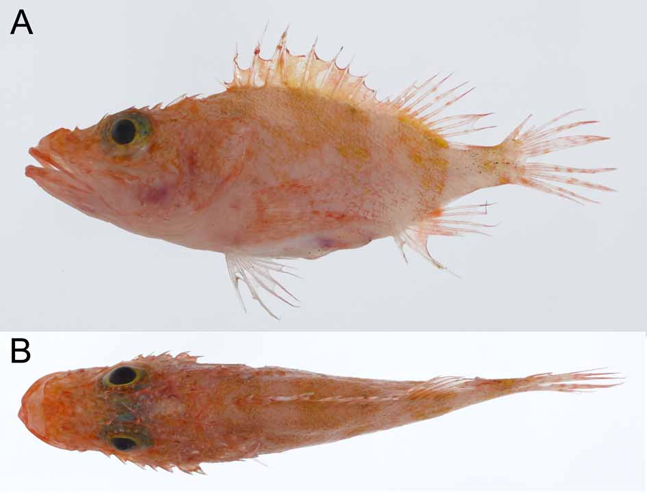
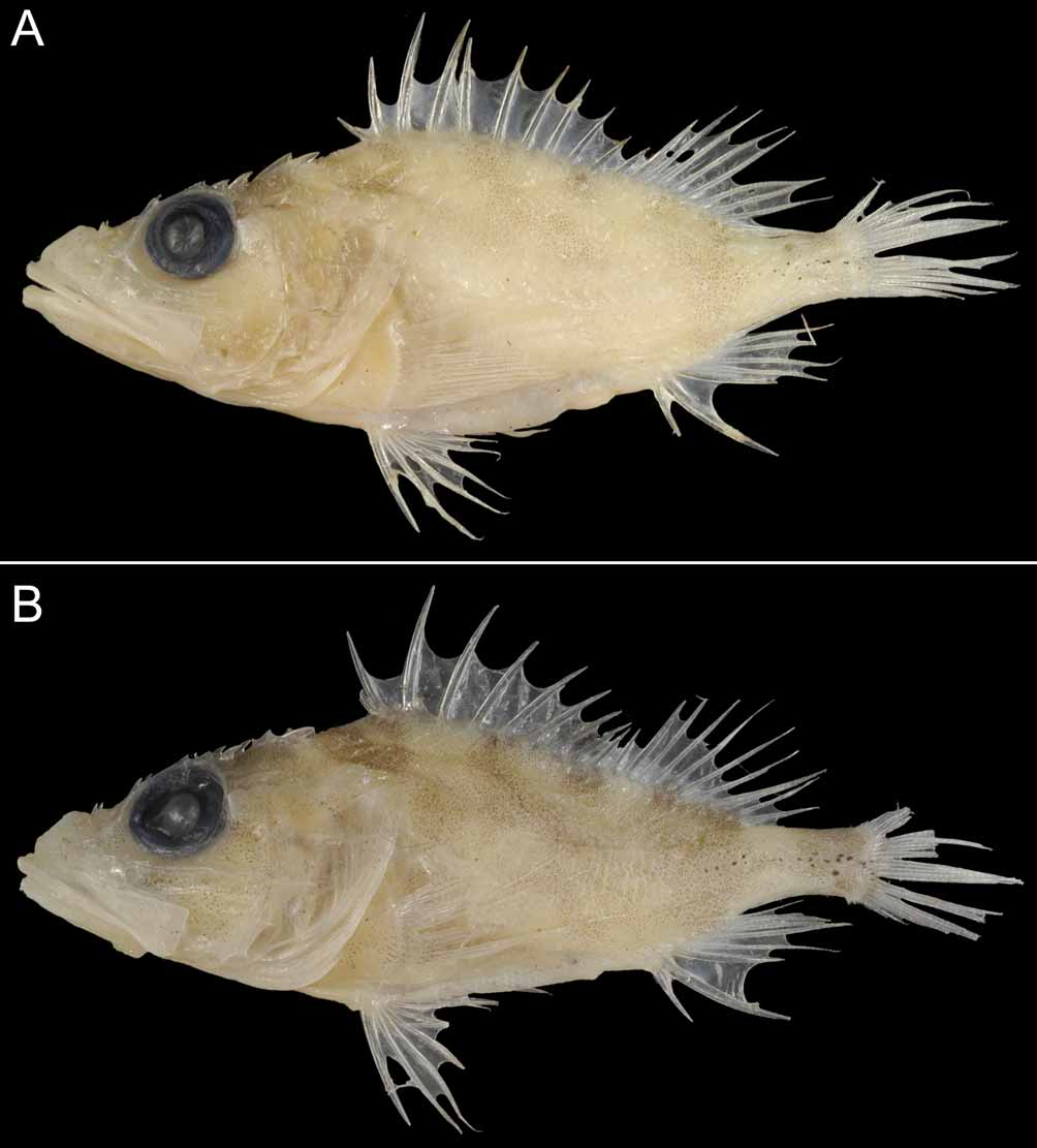
[New English name: Longsnout No-line Scorpionfish] Figures 1–2 View FIGURE 1 View FIGURE 2
Holotype. CSIRO H 6007–14, female, 63.2 mm SL, south of Norfolk Island, Norfolk Island Ridge, Tasman Sea, 29°42’S, 168°01’E, 322 m depth, coll. by FRV Tangaroa , 14 May 2003.
Paratype. CSIRO H 6024–04, male, 47.9 mm SL, Lord Howe Rise, Tasman Sea, 29°13’S, 159°00’E, 300 m depth, coll. by FRV Tangaroa , 20 May 2003.
Diagnosis. A species of Phenacoscorpius with the following combination of characters: pectoral-fin rays 17, all rays unbranched; no palatine teeth; second preopercular spine well-developed, its length longer than third or fourth spines; long snout, its length 12.8–13.1% (mean 12.9%) SL and 27.9–28.6% (mean 28.2%) HL; small eye, orbit diameter 12.3–13.2% (mean 12.8%) SL and 26.9–28.8% (27.8%) HL; narrow head, its width 27.4–28.3% (27.8%) SL; long and shallow caudal peduncle, length and depth 16.9–17.6% (17.3%) SL and 7.1–8.2% (7.7%) SL respectively; long predorsal length 42.2–43.1% (42.7%) SL; long preanal length 74.9–75.0% (74.9%) SL; long prepelvic length 43.1–43.5% (43.3%) SL.
Description. Data for the holotype are presented first, followed by paratype data (if different) in parentheses. Dorsal fin with 12 spines; length of first spine 2.1 (first spine tip broken in paratype) in second spine; third spine longest, its length subequal to pelvic-fin spine length; third to eleventh spines progressively shorter; length of eleventh spine 2.0 (1.7) in last spine; membrane of spinous portion of dorsal fin moderately notched. Dorsal fin with 9 soft rays; all rays branched, divided into 2 branches; third soft ray longest, its length slightly shorter than longest anal-fin soft ray length; posterior branch of last ray joined by membrane to caudal peduncle for approximately one-fourth its length. Anal fin with 3 spines; second spine longest and widest, its length longer than pelvic-fin spine length; first spine 2.7 (spine tip broken in paratype) in second spine, 2.0 (1.6) in third spine. Anal fin with 5 soft rays; all rays branched, divided into 2 branches; second ray longest; posterior branch of last ray not joined by membrane to caudal peduncle. Pectoral fin with 17 rays on each side of body; all rays unbranched; tenth ray longest, its length subequal to body depth; ninth and tenth rays extremely longer than other rays; lower rays not covered with thick skin; posterior margin of fin not bilobed. Pelvic fin with 1 spine and 5 soft rays; last ray unbranched, other rays branched, divided into 2 branches; second ray longest, its length longer than longest anal-fin soft ray length; last soft ray joined by membrane to abdomen for more than two-thirds its length. Posterior margin of caudal fin slightly rounded. Caudal-peduncle depth 2.1 (2.4) in caudal-peduncle length.
Pored lateral-line scales 2 and 3 in left and right sides of body respectively (3 and 2). Predorsal scale rows, between first dorsal-fin spine origin and anterior end of nuchal spine base, about 10. Gill rakers on upper limb 6, lower limb 13 (15), including 4 (5) rakers on hypobranchial; total gill rakers 19 (21); upper 4 rakers on upper limb and 4 rakers on hypobranchial rudimentary and spinous; other rakers relatively long, length of longest raker on first gill arch about four times length of gill filaments around angle of gill arch; a small slit behind lower fourth gill arch. Branchiostegal rays 7. Swimbladder absent.
Body strongly compressed anteriorly, progressively more compressed posteriorly; body width at pectoralfin bases less than upper-jaw length. Nape and anterior body not strongly arched. Body depth relatively shallow; less than head length. Very few small papillae on head; some small papillae scattered on interorbital space and occipital pit. No skin flap on outer margin of anterior and posterior nostrils. A short, slender, unbranched tentacle on posterior end of preocular spine base; its length slightly longer than nasal spine length. Tentacle on posterior end of supraocular spine base unbranched, short, slender; its length longer than length of preocular tentacle, but slightly less than pupil diameter; tip not extending beyond tip of tympanic spine when dpressed. Several small, short, unbranched tentacles on upper and anterior margin of eye membrane. No other distinct tentacles on head, including interorbital space, occipital pit, snout, lacrimal spines, cheek, lips, maxilla, underside of mandible, preopercular margin and opercle. No tentacles on fins and lateral surface of trunk. Pectoral-fin axil without skin flaps. Posterior lacrimal spine not linked posteriorly to head by skin. Anterior nostril not associated with membranous tube.
Well-exposed ctenoid scales covering interorbital space, lateral margins of occipital pit in dorsal view, an area between parietal and pterotic spine bases, and an area surrounded by posterior margin of orbit, upper preopercular margin and pterotic spine base. Well-exposed ctenoid scales covering upper opercle, including between upper and lower opercular spines; scales becoming cycloid on lower opercle; no scales on opercular margin. Well-exposed cycloid scales covering cheek. Other parts of head not covered with scales. Wellexposed ctenoid scales on lateral surface of body, scales becoming cycloid on abdomen. Body scales not extending onto rays or membranes of fins, except base of caudal fin. Exposed cycloid scales covering pectoral-fin base and ventral surface of body, including between pelvic fins.
Cephalic sensory pores prominent; 3 circular pores just below suborbital ridge, first pore largest, located just below first suborbital spine, second pore below and between second and third suborbital spines, and third pore below end of suborbital ridge. Anterior nostril circular, located just below nasal spine base, its diameter more than twice posterior nostril diameter. Posterior nostril circular, located just above end of upward low ridge on lacrimal. A pair of elliptic pores on interorbital space in dorsal view; each pore located anterior to midline through pupil and between interorbital ridge and supraocular ridge; longitudinal length of pore subequal to anterior nostril diameter. A circular pore located behind posterior margin of orbit between tympanic and sphenotic spines; its diameter smaller than anterior nostril diameter, but larger than posterior nostril diameter. An elliptic pore just behind posterior end of pterotic spine base, longitudinal length of pore larger than anterior nostril. A circular pore just behind posterior end of lower posttemporal spine base; its diameter subequal to posterior nostril diameter. Six elliptic pores along preopercular margin; each pore larger than or subequal to anterior nostril diameter; upper 2 pores between upper end of preopercular margin and first preopercular spine, third pore just below first preopercular spine base, fourth pore just above second preopercular spine base, fifth pore between second and third preopercular spine bases, and sixth pore between third and fourth preopercular spine bases. Underside of dentary with 3 sensory pores on each side in ventral view; first pore below anterior edge of lacrimal bone, second pore below and between tips of anterior and posterior lacrimal spines, third pore located on posterior margin of dentary. A pore behind symphysial knob of lower jaw on each side; an indistinct pore each side of symphysial knob.
Mouth large, slightly oblique, forming an angle of about 20 degrees to horizontal axis of head and body. Posterior margin of maxilla just reaching a vertical through posterior margin of pupil. Upper edge of posterior maxilla slightly swollen laterally; anterior part of maxilla slightly convex, central part nearly flat. Lower jaw with a symphysial knob. Width of symphysial gap separating premaxillary teeth bands slightly greater than (or approximately equal to) width of each band. Upper jaw with a band of short, incurved, conical teeth, tips of teeth not strongly pointed. Tooth band in about 8 tooth at front of upper jaw, narrowing posteriorly; band of upper jaw wider than band of lower jaw. Lower jaw with a band of short, incurved, conical teeth; lengths of most teeth subequal to those of upper jaw. Three rows of small teeth at front of vomer, forming 1 or 2 rows posteriorly, forming a V-shaped patch. No palatine teeth. Underside of lower jaw smooth without ridges.
Dorsal profile of snout elevated slightly, forming an angle of about 20 degrees to horizontal axis of head and body. Nasal spine simple, sharp, directed dorsally, its length equal to (less than) anterior nostril diameter. Ascending process of premaxilla not intruding into interorbital space, its posterior margin just reaching (extending beyond) level of anterior margin of anterior nostril in dorsal view. Median interorbital ridge absent. Interorbital ridges poorly developed, separated by a very shallow furrow, beginning posterior to nasal spines and joining tympanic spine bases; anterior angular edge of occiput indistinct; interorbital ridges diverging anteriorly and posteriorly in dorsal view, distance between interorbital ridges narrowest slightly anterior to a vertical midline through eye. Interorbital space relatively shallow, about one-tenth of orbit diameter, extending above dorsal profile of head. Preocular spine simple, directed dorsoposteriorly; tip of spine extending beyond level of upper margin of pupil in lateral view. Supraocular spine simple, its tip extending slightly beyond a vertical midline through pupil in lateral view; spine shorter than preocular, postocular and tympanic spines. Postocular spines simple, slightly canted laterally. Tympanic spine simple, strongly pointed, directed dorsoposteriorly. Interorbital, coronal and pretympanic spines absent. Indistinct transverse ridge anterior to occiput slightly curved posteromedially in dorsal view. Occiput nearly flat, lacking pit. No transverse ridge in rear of occiput between bases of parietal and nuchal spines. Nuchal and parietal spines joined at base. Sphenotic with 2 small spines. Postorbital bone absent. Pterotic spine simple, located below parietal spine. No ridge on area surrounded by parietal, nuchal, pterotic and lower posttemporal spines. Upper posttemporal spine absent. Lower posttemporal spine simple, its base length less than that of pterotic spine. Supracleithral spine simple, not strongly pointed. Cleithral spine flattened, pointed with a low median ridge.
Lateral lacrimal spine present. Anterior lacrimal spine triangular, simple, not strongly pointed, directed ventrally, its tip not reaching dorsal margin of upper lip. Posterior lacrimal spine simple, pointed, directed ventroposteriorly, its tip not reaching upper lip; posterior lacrimal spine much greater than anterior spine. Suborbital ridge with 6 (5) spines, last spine vestigial and located basally behind posteriormost welldeveloped spine (last spine absent); first to third spines below orbit, fourth to sixth (fifth) behind orbit. Space between ventral margin of eye and suborbital ridge relatively narrow. Suborbital pit absent. Preopercle with 5 spines; uppermost spine largest with a supplemental preopercular spine on its base; second to fifth spines without a distinct median ridge; second spine well developed, longer than third and fourth spines; fifth spine minute. Preopercle, between uppermost preopercular spine and upper end of preopercle, smooth without serrae or spines. Upper opercular spine simple without a distinct median ridge. Lower opercular spine simple with a distinct median ridge. Space between upper and lower opercular spines without ridges. Posterior tips of upper and lower opercular spines not reaching opercular margin (tip of lower opercular spine just reaching opercular margin).
Origin of first dorsal-fin spine above supracleithral spine. Posterior margin of opercular membrane reaching a vertical through third (second) dorsal-fin spine base. Posterior tip of pectoral fin extending beyond a vertical through third anal-fin spine base. Origin of pelvic-fin spine slightly anterior to origin of pectoral fin. Posterior tip of depressed pelvic fin not reaching anus. Origin of first anal-fin spine slightly posterior to origin of last dorsal-fin spine.
Morphometrics. Expressed as percentage of SL: Body depth 34.7% (33.3%); body width 18.8 (16.7); head length 45.9 (45.8); snout length 13.1 (12.8); orbit diameter 12.3 (13.2); interorbital width at middle of eye 4.9 (4.2); interorbital width between supraocular spine bases 4.6 (3.6); head width 13.0 (12.6); upper-jaw length 22.8 (22.2); maxillary depth 7.6 (7.1); suborbital space 1.9 (1.5); postorbital length 20.9; between tips of opercular spines 5.4 (5.9); predorsal length 42.2 (43.1); preanal length 75.0 (74.9); prepelvic length 43.5 (43.1); first dorsal-fin spine length 4.5 (spine damaged); second dorsal-fin spine length 9.3 (11.1); third dorsal-fin spine length 15.0 (16.3); fourth dorsal-fin spine length 13.9 (spine damaged); fifth dorsal-fin spine length 11.9 (spine damaged); sixth dorsal-fin spine length 11.7 (14.0); seventh dorsal-fin spine length 11.2 (13.0); eighth dorsal-fin spine length 10.3 (spine damaged); ninth dorsal-fin spine length 8.5 (9.2); tenth dorsal-fin spine length 6.6 (7.5); eleventh dorsal-fin spine length 5.9 (6.9); twelfth dorsal-fin spine length 11.5 (11.9); longest dorsal-fin soft ray (third) length 16.6 (18.6); first anal-fin spine length 6.6 (8.8); second analfin spine length 17.5 (spine damaged); third anal-fin spine length 13.1 (14.0); longest anal-fin soft ray (second) length 18.4 (18.6); longest pectoral-fin ray (tenth) length 34.5 (33.7); pelvic-fin spine length 15.0 (15.9); longest pelvic-fin soft ray (second) length 20.4 (21.8); caudal-fin length 22.6 (fin damaged); caudalpeduncle length 17.6 (16.9); caudal-peduncle depth 8.2 (7.1).
Color when fresh. Based on color photographs of holotype ( Fig. 1 View FIGURE 1 ) and paratype. Body pinkish (reddish), with four, indistinct orange or yellowish saddles; first saddle on nape, second extending from fifth to eleventh dorsal-fin spine bases to middle of body, third extending from soft-rayed portion of dorsal-fin base to anal fin, fourth on caudal peduncle. Spinous portion of dorsal fin pinkish, without distinct marking (reddish, with dark red blotch on membrane between seventh and tenth dorsal-fin spines in paratype). Soft-rayed portion of dorsal fin, pectoral fin and caudal fin with indistinct reddish spots. Pelvic fin whitish. Anal fin whitish with an indistinct reddish band basally. Small, but distinct black spots scattered on posterior portion of caudal peduncle.
Color of preserved specimens. Body and all fins whitish, with small black spots scattered on caudal peduncle. ( Fig. 2 View FIGURE 2 ). In paratype, body with four indistinct brownish saddles; blackish blotch on membrane between seventh and tenth dorsal-fin spines still evident.
| CSIRO |
Australian National Fish Collection |
No known copyright restrictions apply. See Agosti, D., Egloff, W., 2009. Taxonomic information exchange and copyright: the Plazi approach. BMC Research Notes 2009, 2:53 for further explanation.