
Neocephalosphaera carinae, Ramos-Pastrana & Marques & Rafael, 2022
|
publication ID |
https://doi.org/ 10.11646/zootaxa.5178.4.1 |
|
publication LSID |
lsid:zoobank.org:pub:6E706C39-8F42-4050-8792-0423F4267D2B |
|
DOI |
https://doi.org/10.5281/zenodo.7037013 |
|
persistent identifier |
https://treatment.plazi.org/id/41113E13-FF86-FFD8-FF12-90CE4F20B5FF |
|
treatment provided by |
Plazi |
|
scientific name |
Neocephalosphaera carinae |
| status |
sp. nov. |
Neocephalosphaera carinae View in CoL sp. nov.
Figs 14–26 View FIGURES 14–26 , 129 View FIGURE 129
Type material. HOLOTYPE ♂: COLOMBIA, Boyacá, SFF[Santuario de Fauna y Flora] Iguaque, Cab [aña] Mamarramos , 0525’N/7325’W, 2855 m [eters], 23.Sep[ix].–11.Oct[x].2000, 752 (1♂, IAvH) (photographed specimen) . PARATYPE idem, 25.May[v].–08.Jun[vi].2000 (1♂, LEUA) . Holotype with left wing mounted on microslide with Canada balsam. Left antenna and terminalia placed in a microvial with glycerin, both pinned along the specimen.
Diagnosis. Trochanters light brown on the anterior face, dark brown posterodorsally; femora light brown with a dark brown spot on proximal third, dark brown dorsally. Tergites 2–5 velvety black anteriorly, brown pruinose posteriorly. Surstyli subsymmetrical, both with carinae on inner edges. Apex of phallic guide stout, long, with rigid and distinct submedian lobe dorsally, bearing a row of thin and clear apical setae ventrally and a hooked tip. Phallus with ejaculatory ducts completely spiralized.
Description. MALE (holotype). Body length 4.6 mm. Head ( Figs 14–15 View FIGURES 14–26 ). Eyes contiguous for 21 facets. F, EM, V (mm) = 0.4, 0.5, 0.2. Frons brown pruinose. Postcranium brown, laterally and ventrally gray pruinose, dorsally brown pruinose. Antenna ( Fig. 16 View FIGURES 14–26 ) with scape and pedicel dark brown, pedicel with three setae dorsally and three longer setae ventrally; postpedicel light brown, with long acuminate apex. LPP/WPP = 3.3. Thorax ( Figs 14–15, 17 View FIGURES 14–26 ). Postpronotal lobe brown. Scutum black, brown pruinose. Notopleuron concolorous with the scutum, brown pruinose. Scutellum black, brown pruinose, with 14 conspicuous setae along posterior margin. Mesopleuron and mediotergite brown, gray-brown pruinose. Wing ( Fig. 18 View FIGURES 14–26 ). Length 5.7 mm. LW/MWW = 3.5; LTC/LFC = 0.6. Membrane faintly brown infuscated; third section costal shorter than the length of fourth; vein r-m located after the basal third of upper section of cell dm; vein M 2 short; dm-m/M 2 = 5.3; section between cell dm and vein M 2 greater than vein dm-m; vein dm-m slightly curved. Halter dark brown except stem beige. Legs ( Figs 14–15 View FIGURES 14–26 ). Coxae dark brown; trochanters light brown on the anterior face, dark brown posterodorsally; femora light brown with a dark brown spot on proximal third, dark brown dorsally, with a row of long and fine yellow setae posterolaterally; tibiae dark yellow, hind tibia with brown spots and posterior erect setae medially; tarsomeres 1–4 yellow, 5 brown; pulvilli yellow. Abdomen ( Figs 14–15, 19 View FIGURES 14–26 ). Ground color dark, tergite 1 sparsely gray-brown pruinose, with seven stout black setae laterally; tergites 2–5 velvety black anteriorly, brown pruinose posteriorly; tergites 1–3 gray pruinose laterally; tergites 4–5 brown pruinose laterally; tergites and sternites 6 and 7 as in Fig. 20 View FIGURES 14–26 . Syntergosternite 8 brown, brown pruinose, shorter than tergite 5 length, with membranous area large, dividing the syntergosternite 8 ventrally and reaching epandrium ( Figs 19, 21 View FIGURES 14–26 ). Terminalia ( Figs 20–26 View FIGURES 14–26 ). Epandrium and surstyli dark brown ( Figs 21–22 View FIGURES 14–26 ). Surstyli ( Figs 21–24 View FIGURES 14–26 ) subsymmetrical, shorter than the length of epandrium, completely setose; both surstyli with carinae on apical inner edges ( Fig. 22 View FIGURES 14–26 ); left surstylus slightly shorter and thinner than right, both surstyli with tips downward directed when seen in lateral view ( Figs 23–24 View FIGURES 14–26 ). Apex of phallic guide stout, long, with rigid and distinct submedian lobe dorsally; bearing a row of thin clear setae ventrally and hooked tip ( Fig. 25 View FIGURES 14–26 ). Ejaculatory apodeme funnel-shaped, with one margin somewhat straight ( Fig. 26 View FIGURES 14–26 ). Phallus trifid, with ejaculatory ducts completely spiralized ( Fig. 25 View FIGURES 14–26 ).
FEMALE. Unknown.
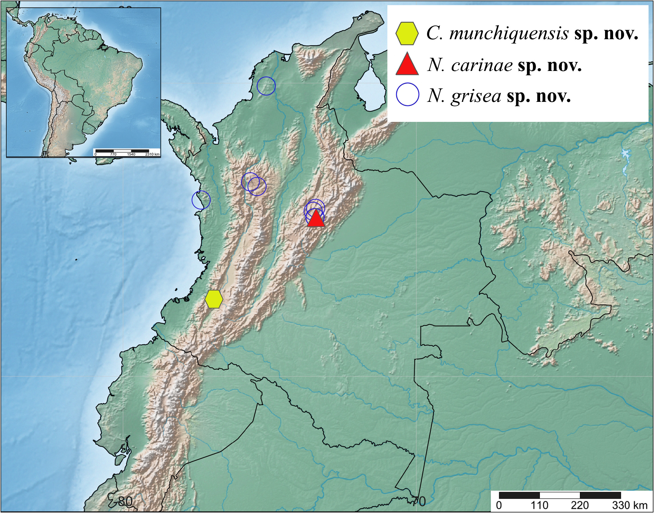
Geographical distribution. Colombia (Boyacá) ( Fig. 129 View FIGURE 129 ).
Etymology. The specific epithet refers to the type locality, Boyacá, Colombia.
Habitat. The specimens were collected with Malaise traps at ground level. The vegetation of the collection site is composed of cloud Andean forests of the Eastern Cordillera of Northeastern region of Colombia.
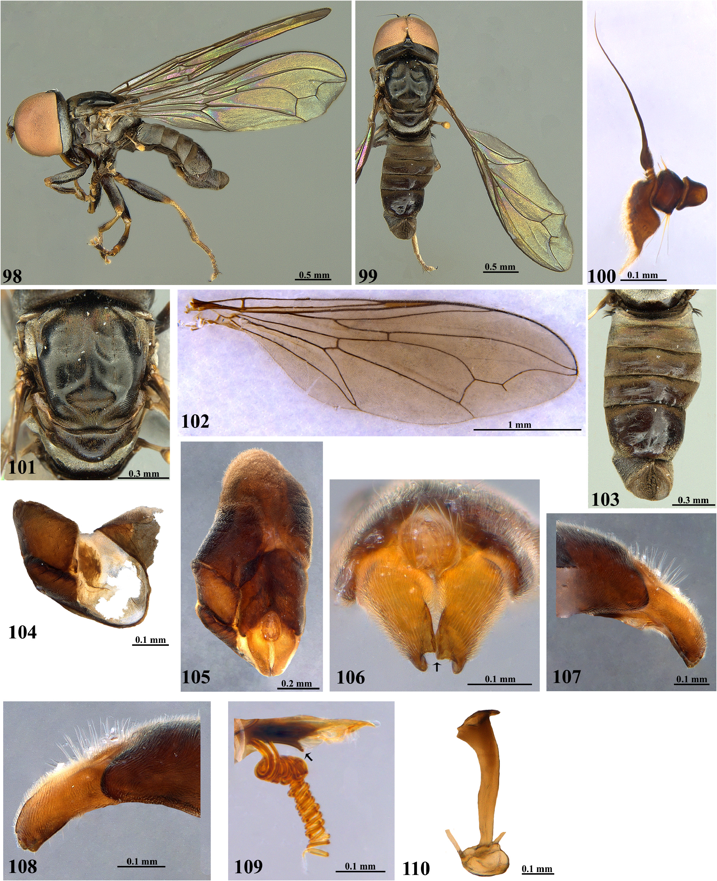
Taxonomic notes. Neocephalosphaera carinae sp. nov. runs to N. jamaicensis Johnson, 1919 in the couplet 18 of the key presented by Souza & Ale-Rocha (2009). It differs from N. jamaicensis in having the antenna with postpedicel long, with acuminate apex ( Fig. 16 View FIGURES 14–26 ) [versus antenna with postpedicel short, with the acuminate apex in N. jamaicensis , figure 42.6, presented by Rafael (1992)]; femora light brown with a dark brown spot on proximal third, dark brown dorsally ( Figs 14–15 View FIGURES 14–26 ) (versus femora dark brown to black, with bases and apices yellow); trochanters light brown on the anterior face, dark brown posterodorsally ( Fig. 14 View FIGURES 14–26 ) (versus trochanters yellow); apex of phallic guide with rigid and distinct submedian lobe dorsally and hooked tip ( Fig. 25 View FIGURES 14–26 ) [versus apex of phallic guide with straight margins dorsally and ventrally and straight tip, figure 3.I, presented by Souza & Ale-Rocha (2009)]; phallus with ejaculatory ducts completely spiralized ( Fig. 25 View FIGURES 14–26 ) [versus phallus with ejaculatory ducts spiralized only in distal 3/4, figure 3.I, presented by Souza & Ale-Rocha (2009)]. Based on the male specimen and due to the shape of the surstyli, N. carinae sp. nov. is also related to N. iguaquensis sp. nov. ( Figs 50–51 View FIGURES 43–55 ), N. muisca sp. nov. ( Figs 66–67 View FIGURES 59–71 ), and N. spiralis sp. nov. ( Figs 105–106 View FIGURES 98–110 ). It differs from N. iguaquensis sp. nov. in having apex of phallic guide with rigid and distinct submedian lobe dorsally and hooked tip ( Fig. 25 View FIGURES 14–26 ) [versus apex of phallic guide with distinct submedian truncated lobe ventrally, slightly straight dorsally, tip slightly downward directed with a small translucent lobe dorsally in N. iguaquensis sp. nov. ( Fig. 54 View FIGURES 43–55 )]; phallus with ejaculatory ducts completely spiralized ( Fig. 25 View FIGURES 14–26 ) [versus phallus trifid, with ejaculatory ducts spiralized, only in distal 3/4 ( Fig. 54 View FIGURES 43–55 )]; ejaculatory apodeme funnel-shaped, with one margin somewhat straight ( Fig. 26 View FIGURES 14–26 ) [versus ejaculatory apodeme somewhat nail-shaped ( Fig. 55 View FIGURES 43–55 )]. It differs from N. muisca sp. nov. in having syntergosternite 8 shorter than tergite 5 length, without crestlike membranous area ( Figs 19, 21 View FIGURES 14–26 ) [versus syntergosternite 8 larger than tergite 5, with crestlike membranous area in N. muisca sp. nov. ( Figs 64, 66 View FIGURES 59–71 )]; phallus with ejaculatory ducts completely spiralized ( Fig. 25 View FIGURES 14–26 ) [versus phallus trifid, with ejaculatory ducts spiralized, only in distal 3/4 ( Fig. 70 View FIGURES 59–71 )]. It differs from N. spiralis sp. nov. in having section between cell dm and vein M 2 greater than vein dm-m ( Fig. 18 View FIGURES 14–26 ) [versus section between cell dm and vein M 2 equal than vein dm-m in N. spiralis sp. nov. ( Fig. 102 View FIGURES 98–110 )]; apex of phallic guide with rigid and distinct submedian lobe dorsally and hooked tip ( Fig. 25 View FIGURES 14–26 ) [versus apex of phallic guide with distinct submedian acute lobe ventrally, which is downward directed; tip slightly downward directed ( Fig. 109 View FIGURES 98–110 )]; ejaculatory apodeme funnel-shaped, with one margin somewhat straight ( Fig. 26 View FIGURES 14–26 ) [versus ejaculatory apodeme somewhat funnel-shaped narrowed, slightly inclined ( Fig. 110 View FIGURES 98–110 )].


No known copyright restrictions apply. See Agosti, D., Egloff, W., 2009. Taxonomic information exchange and copyright: the Plazi approach. BMC Research Notes 2009, 2:53 for further explanation.