
Hypselodoris rositoi, Gosliner & Johnson, 2018
|
publication ID |
https://doi.org/10.1093/zoolinnean/zly048 |
|
publication LSID |
urn:lsid:zoobank.org:pub:F0065FD2-417C |
|
DOI |
https://doi.org/10.5281/zenodo.5980694 |
|
persistent identifier |
https://treatment.plazi.org/id/411BF606-FFC9-FF93-FC74-FB78FAAC6BE3 |
|
treatment provided by |
Plazi |
|
scientific name |
Hypselodoris rositoi |
| status |
sp. nov. |
HYPSELODORIS ROSITOI GOSLINER & JOHNSON View in CoL SP. NOV.
(FIGS 2N, 13J, 25A, B, 26)
LSID: urn:lsid:zoobank.org:act:
Type material
Holotype: NMP 0 41283 (formerly CASIZ 186099 ), s u b s a m p l e d f o r m o l e c u l a r s t u d y, d i s s e c t e d, Malajibomanoc ( Chicken Feather Island ), 13.628°N, 120.966°E, 30 m depth, Batangas Bay, Batangas Province, Luzon, Philippines, 16 May 2011. GoogleMaps
Paratype: CASIZ 220675 , one specimen, Cavite Province, Luzon , Philippines, May 2009, specimen provided by Denis Ty of Aquascapes Philippines Company .
Geographical distribution
Known only from the Batangas and Cavite provinces of Luzon, Philippines (present study).
Etymology
The name rositoi comes from the Latin for rose, referring to the distinctive rose-pink body colour that is predominant in this species and also honors Kumataro Ito, the artist on the 1908 Albatross Expedition who first illustrated this species ( Fig. 25B View Figure 25 ).
Description
External morphology: Living animals ( Fig. 25A View Figure 25 ) of moderately large size, reaching 50–60 mm in length. Entire dorsal surface deep rose pink, with thick white band encircling the margin of notum. Sides of body lighter pink, fading to white margin of foot. Five unipinnate gill branches on notum. Holotype with four large gill branches and five smaller ones, possibly indicating regrowth from damage. Paratype with nine unipinnate gill branches. Gill pocket well elevated. Gill branches elongate, deep orange, with lighter pigment on inner rachis of each branch. Base of gill pocket deep pink. Bulb of rhinophores bright orange with redder apex. Bulb with ~25–32 densely packed lamellae. Base of rhinophore sheath deep pink.
Mantle glands: Small subcutaneous mantle glands present along entire mantle margin, somewhat enlarged anteriorly and posteriorly ( Fig. 2N View Figure 2 ).
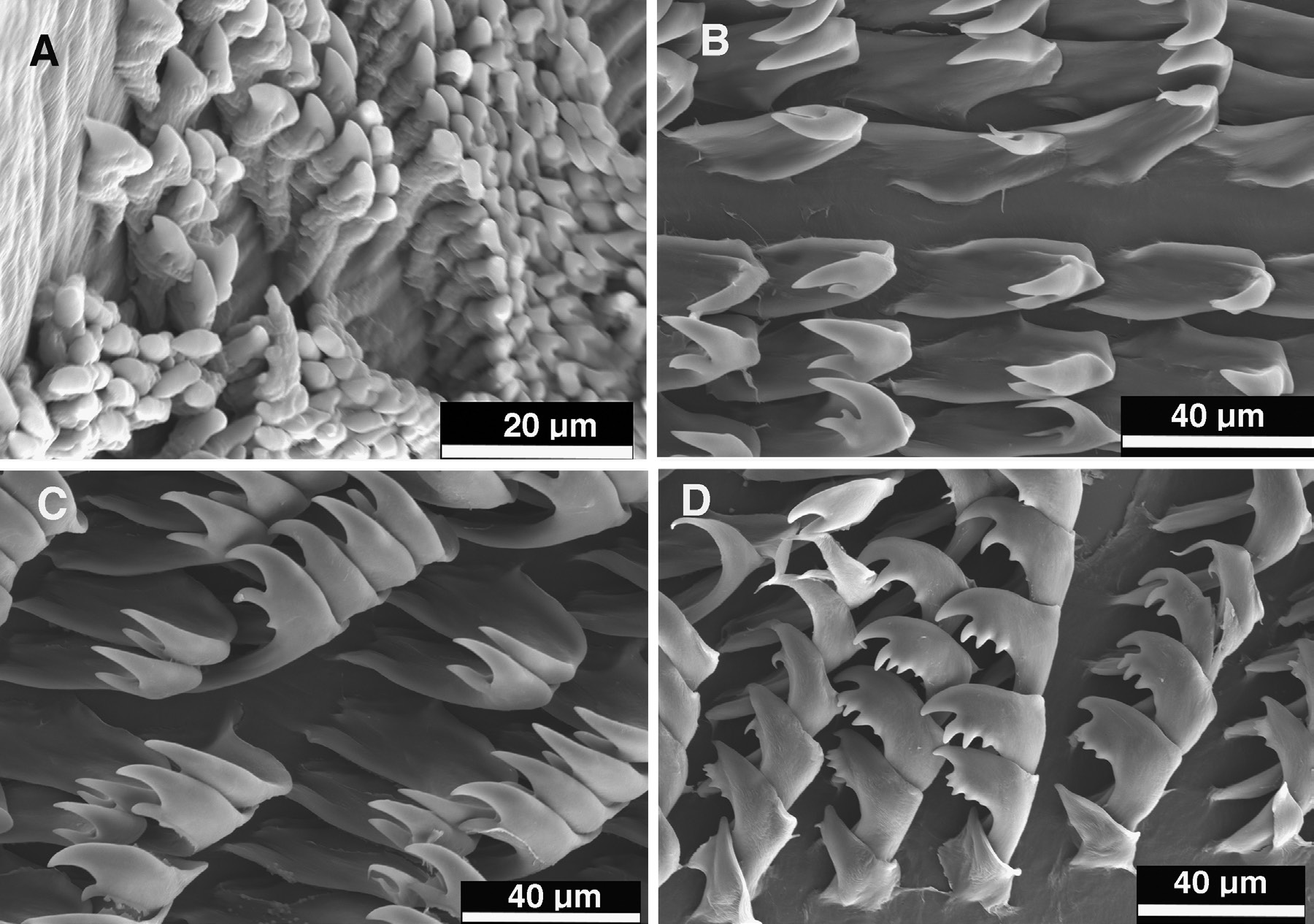
Buccal armature: Muscular portion of buccal mass slightly larger than oral tube. Chitinous labial cuticle found at anterior end of muscular portion of the buccal mass bearing numerous jaw rodlets ( Fig. 26A View Figure 26 ). Rodlets narrow and short with long base and evenly curved, with single, acutely pointed apex. Radular formula of holotype (CASIZ 186099) 66 × 46.0.46. Rachidian row of teeth absent ( Fig. 26B View Figure 26 ). Innermost lateral teeth having one short denticle on inner side of bifid primary cusp, lacking outer denticles. Next several laterals lacking inner and outer denticles, possessing only primary bifid cusps. Midlateral teeth ( Fig. 26C View Figure 26 ) all lacking inner and outer denticles, with exception that single outer denticle may be present on some teeth. Outermost teeth having a narrower base and lacking denticles. Three to four teeth inside outermost teeth, with one to four denticles on outer side of bifid cusp ( Fig. 26D View Figure 26 ).
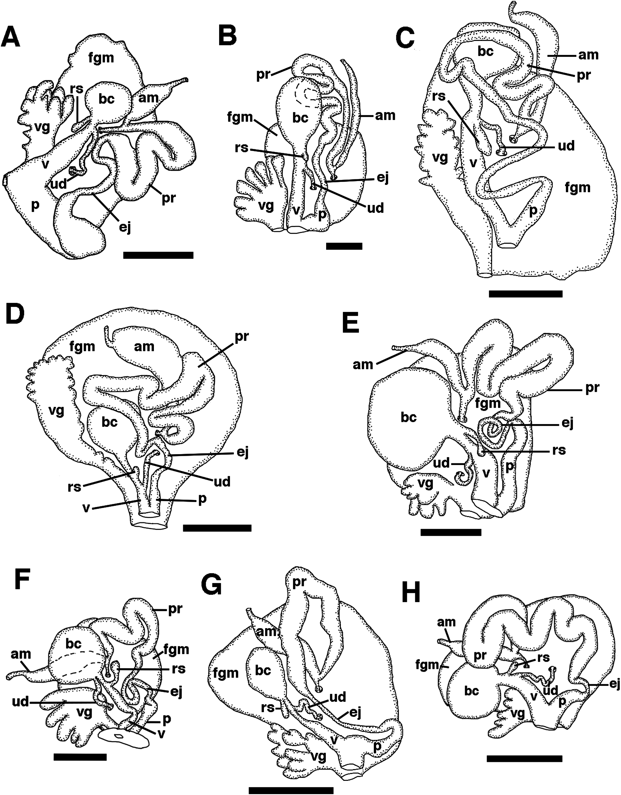
Reproductive system: Reproductive organs of the holotype fully mature ( Fig. 13J View Figure 13 ). Ampulla thick, tubular and slightly curved, narrowing somewhat before bifurcating into oviduct and vas deferens. Short oviduct entering female gland mass near albumen gland. Prostatic proximal portion of vas deferens convoluted, curved and thick and narrowing abruptly as it transitions into muscular ejaculatory portion. Ejaculatory portion short, curved, narrow, entering short, wider penial bulb. Penial bulb adjacent to straight, moderately wide vaginal duct at common gonopore. Distal end of vas deferens devoid of penial hooks. Female gland mass consisting of large mucous gland and small membrane and albumen glands. Large, lobate vestibular gland situated near exit of mucous gland. Relatively long vagina leading to small, straight receptaculum seminis and larger spherical, thin-walled bursa copulatrix. Receptaculum seminis appressed against vagina in distal half of vaginal length. Moderately short uterine duct emerging from vagina close to receptaculum and entering female gland mass near the albumen gland.
Remarks
In our phylogenetic analyses, H. rositoi is sister to the rest of the H. bullockii clade. In the ABGD analysis, H.rositoi is indicated to represent a distinct species and is> 13% different in its COI gene compared with any other members of the H. bullockii clade. Hypselodoris rositoi is unique among members of the H. bullockii clade in having a bright rose-pink body colour. All other members of the clade are light pink to purple. Hypselodoris rositoi , H. violacea sp. nov. and H. variobranchia sp. nov. are the only members of the H. bullockii clade with a broad, solid white marginal band. Hypselodoris rositoi is also the only member of the H. bullockii clade with obvious mantle glands. Internally, H. rositoi has a radula with fewer teeth per half row (46) than found in other members of the H. bullockii clade. It is also unique in lacking outer denticles on the vast majority of its middle lateral teeth. All the remaining members of the H. bullockii clade have numerous prominent denticles on their middle lateral teeth.
The reproductive system of members of the H. bullockii clade has slight variation. In H. bullockii ( Fig. 13F View Figure 13 ) and H. apolegma ( Fig. 4D View Figure 4 ), the vaginal duct is relatively short, whereas in H. rositoi , H. violacea , H. variobranchia , H. brycei and H. melanesica the vagina is more elongate. In H. rositoi , H. violacea , H. brycei and H. melanesica , the uterine duct branches from the middle of the vagina near the receptaculum seminis, whereas in H. variobranchia , the uterine duct branches near the base of the vagina. In H. rositoi , the ejaculatory portion of the vas deferens is relatively short, whereas it is much longer in H. violacea , H. variobranchia , H. brycei and H. melanesica .


| NMP |
National Museum (Prague) |
No known copyright restrictions apply. See Agosti, D., Egloff, W., 2009. Taxonomic information exchange and copyright: the Plazi approach. BMC Research Notes 2009, 2:53 for further explanation.
|
Kingdom |
|
|
Phylum |
|
|
Class |
|
|
SubClass |
Heterobranchia |
|
Order |
|
|
Family |
|
|
Genus |