
Hypselodoris variobranchia, Gosliner & Johnson, 2018
|
publication ID |
https://doi.org/ 10.1093/zoolinnean/zly048 |
|
publication LSID |
urn:lsid:zoobank.org:pub:F0065FD2-417C |
|
DOI |
https://doi.org/10.5281/zenodo.5980698 |
|
persistent identifier |
https://treatment.plazi.org/id/411BF606-FFD2-FF8E-FC35-FBD1FB68685F |
|
treatment provided by |
Plazi |
|
scientific name |
Hypselodoris variobranchia |
| status |
sp. nov. |
HYPSELODORIS VARIOBRANCHIA GOSLINER & JOHNSON View in CoL SP. NOV.
(FIGS 25C–F, 29, 30A)
LSID: urn:lsid:zoobank.org:act:723905BD-FADE-4891-B2A5-3F8546935808
H y p s e l o d o r i s b u l l o ck i, m i s i d e n t i f i c a t i o n, n o t H. bullockii ( Collingwood, 1881) View in CoL ; Rudman, 1999a: photograph E; Izumi, 2003; Adams, 2004; Brauchli, 2004; Lau, 2006; Turker, 2006; Debelius & Kuiter, 2007: 116: middle right photograph; Tanke 2008.
Hypselodoris View in CoL sp. Coleman, 2001: 82, middle photograph, fourth row.
Hypselodoris View in CoL sp. Coleman, 2001: 82, lower right photograph.
Hypselodoris View in CoL cf. bullocki -1 Debelius & Kuiter, 2007: 116: lower two rows of photographs.
Hypselodoris View in CoL sp. 6 Gosliner et al., 2008: 268, top photograph.
Hypselodoris View in CoL sp. 1 Humann & DeLoach, 2010: 339, upper right photograph.
Hypselodoris View in CoL sp. 15 Gosliner et al., 2015: 262, middle left photograph.
Type material
Holotype: NMP 0 41285 (formerly CASIZ 177455 ), subsampled for molecular study, dissected, Aphol’s Rock , 13.6586°N, 120.90129°E, 30 m depth, Maricaban Island , Tingloy, Batangas Province, Luzon, Philippines, 17 May 2008, Peri Paleracio. GoogleMaps
Paratypes: CASIZ 0 85901, one specimen, Liuay Rock , Dakak, Zamboanga del Norte, Mindanao, Philippines, 29 March 1993, T. Gosliner . CASIZ 0 96279, one specimen, Sepok Point , 13.68806°N, 120.82678°E, Maricaban Island , Tingloy, Batangas, Luzon, Philippines, 14 March 1994, Mike Severns GoogleMaps . CASIZ 177618 , subsampled for molecular study, dissected, Aphol’s Rock , 13.6586°N, 120.90129°E, 30 m depth, 17 April 2008, Peri Paleracio GoogleMaps . CASIZ 182841 , one specimen, Devil’s Point , 13.65083°N, 120.84127°E, Tingloy , Maricaban Island, Batangas Province, Luzon, Philippines, 21 May 2010, T. Gosliner GoogleMaps . CASIZ 208189 , one specimen, subsampled for molecular study, La Laguna, 13.525953°N, 120.970160°E, Puerto Galera , Mindoro Oriental, Philippines, 26 April 2015, T. Gosliner GoogleMaps . CASIZ 217246 , Bonito Island , 13.6305°N, 120.94763°E, Maricaban Island , Tingloy, Batangas, Luzon, Philippines, 21 April 2016, Brenna Green GoogleMaps . CASIZ 217389 , Sepok Wall , 13.68806°N, 120.82678°E Maricaban Island , Tingloy, Batangas, Luzon, Philippines, 15 April 2016, Brenna Green GoogleMaps . CASIZ 217273 , 13.6880278°N, 120.8971833E °, Calumpan Peninsula , Mabini , Batangas, Luzon Island, Philippines, Bubbles, 22 April 2016, T. Gosliner GoogleMaps . CASIZ 104704 , one specimen, 69 m depth, Horseshoe Cliffs , (26.5000°N, 127.854307°E, 1 km WNW of Onna Village, Okinawa, Japan, R. Bolland. GoogleMaps
Geographical distribution
Known from Queensland, Australia ( Rudman, 1999a), Malaysia ( Lau, 2006), Indonesia, Okinawa, Japan and the Philippines (present study).
Etymology
The name variobranchia comes from Latin for variable gills, referring to the gill, which may be either orange or bright purple.
Description
External morphology: Living animals ( Fig. 25 View Figure 25 C–F) of moderately large size, reaching 50 mm in length. Entire dorsal surface deep purple, with thick, solid opaque white band encircling the margin of notum. Sides of body and margin of foot same colour as notum. Five to nine unipinnate gill branches on notum. Gill branches deep orange or deep purple. In specimens with orange gill branches, common base often with purple. Base of gill pocket well elevated, deep purple. Bulb of rhinophores bright orange throughout. Bulb with 19–23 densely packed lamellae. Base of rhinophore sheath deep purple.
Mantle glands: Mantle glands entirely absent from mantle margin.
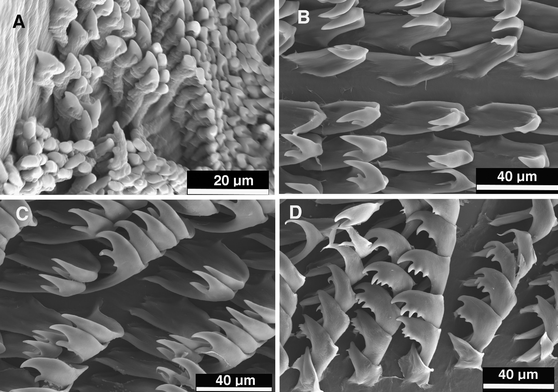
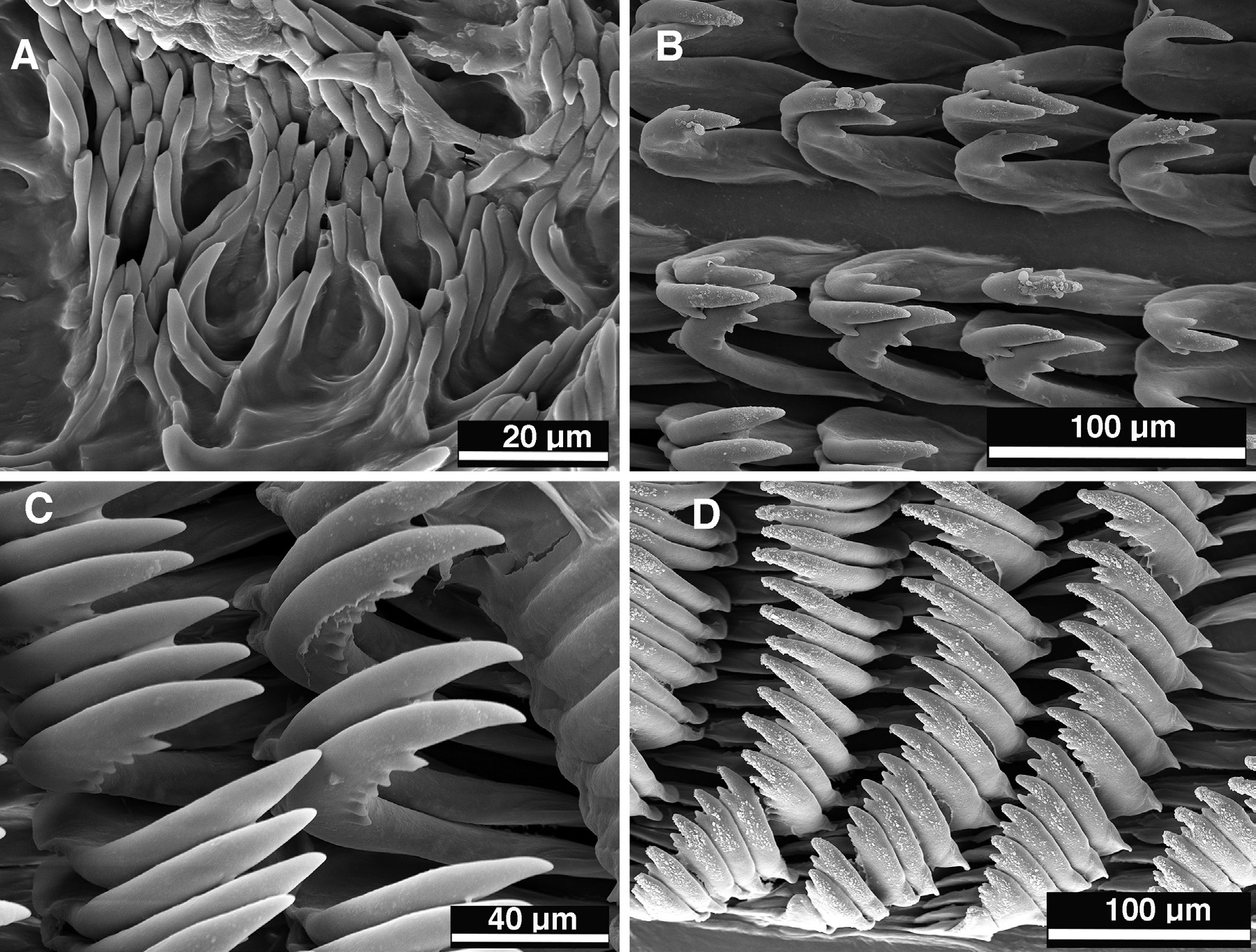
Buccal armature: Muscular portion of buccal mass much shorter than oral tube. Chitinous labial cuticle found at anterior end of muscular portion of the buccal mass bearing numerous jaw rodlets ( Fig. 29A View Figure 29 ). Rodlets narrow and short with long base and evenly curved, with single, acutely pointed apex. Radular formula of holotype (CASIZ 177455) 71 × 98.0.98, and 68 × 106.0.106 (CASIZ 177618) in one paratype. Rachidian row of teeth absent ( Fig. 29B View Figure 29 ). Innermost lateral teeth having one short denticle on inner and outer sides of bifid primary cusps. Outer cusp of bifid cusps much shorter than inner one. Next several laterals lacking inner denticles possessing primary bifid cusps and two or three outer denticles. Midlateral teeth ( Fig. 29C View Figure 29 ) all lacking inner denticles, but with five or six prominent outer denticles. Outermost teeth having a narrower base and having three or four outer denticles ( Fig. 29D View Figure 29 ).
Reproductive system: Reproductive organs of the holotype fully mature ( Fig. 30A View Figure 30 ). Ampulla thick, tubular and straight, narrowing somewhat before bifurcating into oviduct and vas deferens. Short oviduct entering female gland mass near albumen gland. Prostatic proximal portion of vas deferens long, convoluted, curved and thick and narrowing abruptly as it transitions into muscular ejaculatory portion. Ejaculatory portion elongate, convoluted, narrow, entering moderately long, wider penial bulb. Penial bulb adjacent to straight, moderately wide vaginal duct at common gonopore. Distal end of vas deferens devoid of penial hooks. Female gland mass consisting of large mucous gland and small membrane and albumen glands. Large, lobate vestibular gland situated near exit of mucous gland. Relatively long vagina leading to small, straight receptaculum seminis and larger spherical, thin-walled bursa copulatrix. Receptaculum seminis appressed against vagina in distal half of vaginal length. Moderately short uterine duct emerging from vagina proximally to receptaculum and entering female gland mass near the albumen gland.
Remarks
In our phylogenetic analyses, H. variobranchia is sister to the clade containing H. bullockii , H. melanesica , H. brycei and H. apolegma . Externally, it is most similar to H. rositoi , H. violacea and some colour morphs of H. iba , which is a member of a separate clade. All of these species have a wide, solid opaque white marginal band. Externally, H. rositoi has a pink rather than purple body colour, and H. violacea has purple rhinophores in contrast to the orange rhinophores of H. iba and H. variobranchia . Both H. iba and H. rositoi have mantle glands that are absent in H. variobranchia and H. violacea . Also, H. iba has a higher body profile than that of H. variobranchia .
The radula formula of H. iba , H. violacea and H. variobranchia is similar, with almost 100 teeth per half row, whereas H. rositoi has only 46 teeth per half row. In H. iba , only the innermost radular teeth have denticles other than the two primary cusps, whereas in H. variobranchia and H. violacea the majority of teeth have numerous outer denticles. In H. rositoi , only the outer teeth have prominent outer denticles, with the exception of some of the middle lateral teeth, which may have a single denticles ( Fig. 26C View Figure 26 ). The radula is very similar in H. variobranchia and H. violacea , but in H. violacea the innermost teeth have a longer, more acutely pointed inner cusp of the bifid cusps. This difference in inner cusp length and sharpness is also found in the middle lateral teeth. The reproductive systems of H. variobranchia and H. violacea differ in a couple of key regards. In H. variobranchia , the ejaculatory portion of the vas deferens is far more elongate than in H. violacea . In H. variobranchia , the uterine duct emerges from the proximal portion of the vagina, whereas in H.violacea the uterine duct emerges from the distal third of the vagina. In the ABGD analysis, H. variobranchia and H. violacea are clearly indicated as distinct species. The three specimens of H. variobranchia are only 0.15–0.3% different in their COI gene from each other, whereas all three specimens are 8.8–9.0% different from H. violacea .


| NMP |
National Museum (Prague) |
No known copyright restrictions apply. See Agosti, D., Egloff, W., 2009. Taxonomic information exchange and copyright: the Plazi approach. BMC Research Notes 2009, 2:53 for further explanation.
|
Kingdom |
|
|
Phylum |
|
|
Class |
|
|
Order |
|
|
Family |
|
|
Genus |
Hypselodoris variobranchia
| Epstein, Hannah E., Hallas, Joshua M., Johnson, Rebecca Fay, Lopez, Alessandra & Gosliner, Terrence M. 2018 |
Hypselodoris
| Stimpson 1855 |
Hypselodoris
| Stimpson 1855 |
Hypselodoris
| Stimpson 1855 |
Hypselodoris
| Stimpson 1855 |
Hypselodoris
| Stimpson 1855 |
Hypselodoris
| Stimpson 1855 |